




Abstract
Kiwifruit (Actinidia deliciosa) is a high-acidity fruit, with two varieties available on the market. One is the green-fleshed, fuzzy, sweet but tangy-tasting kiwifruit, and the other is the yellow-fleshed variety called “golden” kiwifruit. While the whole kiwifruit is sold at room temperature at grocery stores, sliced kiwifruit is usually sold as a part of fruit salad in the refrigerated section. The survival of a five-strain Escherichia coli O157:H7 cocktail, a five-strain Salmonella cocktail, and a five-strain Listeria monocytogenes cocktail was evaluated on whole and sliced green and golden kiwifruit. Two inoculation levels were tested (∼7 and ∼4 Log colony-forming unit (CFU)/kiwi). A significantly higher amount of wet inoculum was attached to the green kiwifruit than to the golden kiwifruit (p < 0.05). The scanning electron microscope examination showed that pathogens can attach on both the surface and the hair structure of green kiwi skin. At room temperature, all three pathogens survived for 30 days on whole kiwifruit. Although the pH of sliced kiwifruit was low (∼3.5), all three pathogens survived on sliced kiwifruit for 7 days when stored at 4°C. These results highlight the importance of preventing contamination of fresh fruit during the preharvest and processing stages.
Introduction
M
Listeria monocytogenes is another major foodborne pathogen that causes about 1600 illnesses annually in the United States (Scallan et al., 2011). Increasing numbers of L. monocytogenes outbreaks have been associated with fresh produce. For example, the 2011 cantaloupe outbreak associated with L. monocytogenes resulted in 143 hospitalizations and 33 deaths (CDC, 2011). The 2014–2015 caramel apple outbreak related to L. monocytogenes caused 35 illnesses in 12 states and 7 deaths (CDC, 2015). These outbreaks highlighted the importance of having additional interventions to control microbial contamination risks associated with fresh fruits and vegetables.
Kiwifruit (Actinidia deliciosa) is a low-acidity fruit that was introduced to the United States and New Zealand early in the 20th century. While the green-fleshed, fuzzy, sweet but tangy tasting “traditional” kiwifruit is the most common one, the yellow-fleshed variety called “golden” kiwifruit is also available on the market. The golden kiwifruit has a sweeter taste and smoother skin compared to the traditional green kiwifruit. Kiwifruits are low-pH fruits, with pH values of ∼3.5. The major kiwifruit production area in the United States is in California, producing around 98% of the kiwifruit grown in the United States. Harvest in California usually begins in late September, with most of the fruits being harvested in October and early November (California kiwifruit, 2017). In addition to the U.S. grown kiwifruits, 176 million pounds of kiwi were imported for the production year of 2015–2016, valued at approximately $128 million, to meet the needs of the market (USDA ERS, 2017).
Kiwifruit is usually sold whole at room temperature in the grocery store. When they are sliced, they are stored at refrigerated temperatures as a part of fruit salads. Although there has not been a recall or an outbreak associated with kiwifruit, survey studies conducted at the kiwifruit orchards and processing plants had identified pathogenic E. coli isolates (eae gene or stx2 gene positive) from the fruit and environmental samples (Feng et al., 2014, 2015). Thus, the objective of this study was to investigate the survival of E. coli O157:H7, Salmonella spp., and L. monocytogenes on both whole and sliced kiwifruit. Two kiwifruit varieties were tested, one being the traditional green kiwifruit and the other being the golden kiwifruit.
Materials and Methods
Culture and inoculum preparation
Three sets of five-strain cocktails were used for this study. E. coli O157:H7 ATCC 35150, ATCC 43894, 505B (a beef outbreak isolate), and AU strains 301 and 305 (culture collection at Auburn University) were used to prepare the E. coli O157:H7 cocktail. Salmonella Typhimurium ATCC 14028, Salmonella Enteritidis (ATCC BAA-1045), Salmonella Newington (AU-SN1), Salmonella Stanley (AU-SS1), and Salmonella Thompson (AU-ST2) were used to prepare the Salmonella cocktail. L. monocytogenes 10403s (serotype 1/2a, obtained from UC Davis), 101M (serotype 4b, a beef and pork sausage isolate), 108M (serotype 1/2b, a hard salami isolate), ATCC19115 (serotype 4b), and ATCC 49594 (serotype 4b) were used to prepare the L. monocytogenes cocktail. Unless otherwise stated, all bacterial strains were obtained from the culture collection of the food microbiology laboratory at Auburn University. Frozen stock cultures were first revived by transferring 100 μL of thawed stock culture into 10 mL of tryptone soy broth (Difco, Becton, Dickson and company, Sparks, MD) and incubated at 37°C for 24 h. The revived cultures were transferred again on the second day and incubated overnight for preparing cultures used for inoculation. Each overnight fresh culture was harvested and washed twice using MilliQ water by centrifugation at 3000 × g for 10 min (5810R; Eppendorf, Hauppauge, NY). Washed cell pellets were resuspended in MilliQ water and mixed in equal volume to make cocktails.
Selection of kiwifruit
Two varieties of kiwifruit, green and golden, were purchased from a local supermarket (Auburn, AL) (Fig. 1). All kiwifruits were purchased within the 2 days before the experimental trials and held at room temperature before inoculation. The pH of each kiwifruit variety was measured by a pH meter (Mettler Toledo, Columbus, OH). To do so, three kiwifruits of each variety were peeled and smashed in a Whirl-Pak® filter bag (Nasco, Fort Atkinson, WI) by hand for 2 min. The probe of the pH meter was dipped into the smashed samples directly to take the pH readings.
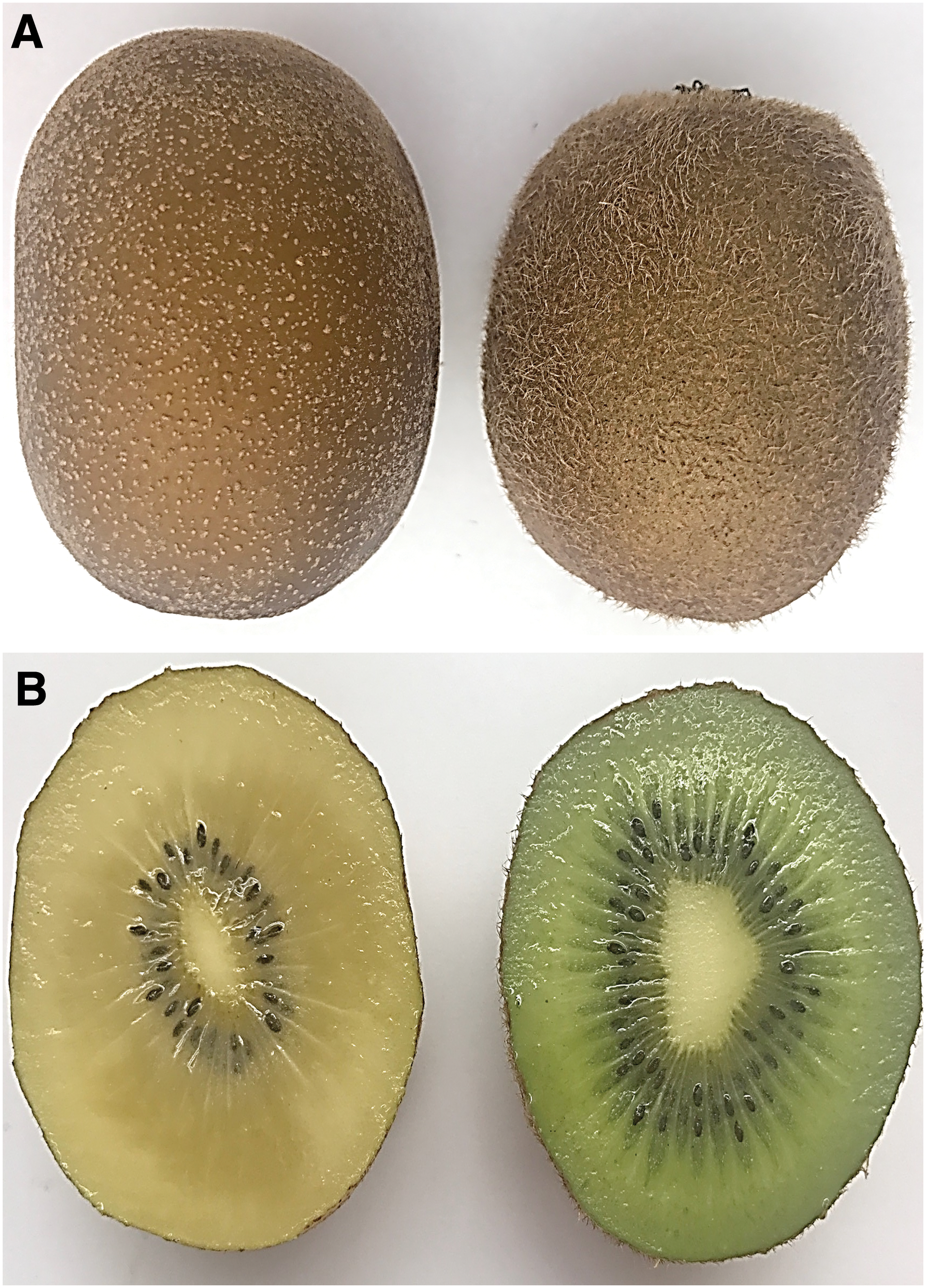
Whole and sliced green and golden kiwifruits.
Before inoculation, three kiwifruits were randomly picked from each variety and were confirmed to be negative for E. coli O157:H7, Salmonella spp., and L. monocytogenes following the FDA Bacteriological Analytical Manual (US Food and Drug Administration FDA (2017)). An additional three kiwifruits of each variety were also picked. One hundred milliliters of 0.1% peptone water (Difco, Becton, Dickson and company) was added to each fruit, and samples were homogenized by hand shaking them for 2 min. The suspension was plated onto plate count agar (PCA) (Difco, Becton, Dickson and company) to enumerate total aerobic microorganisms. PCA plates were incubated at 37°C for 24 h and enumerated.
Kiwifruit inoculation and storage
Whole kiwifruits were inoculated by dipping the entire fruit into cocktail broth (150 mL for each cocktail) for 30 s at room temperature. The inoculated kiwifruits were air dried in a biosafety cabinet (Labconco®, Kansas City, MO) for 1 h with the airflow on at room temperature. The dried kiwifruits were then placed back into their original containers and stored at room temperature (23°C ± 2°C) for 30 days. Three kiwifruits of each variety inoculated with every cocktail were randomly collected and analyzed every 5 days during the storage.
Sliced kiwifruit was prepared by first peeling off the skin using a 6-inch “Y” shaped vegetable peeler with a serrated stainless steel blade and then cutting the fruit into 5 mm-thick slices (∼11 g per slice) with a 3.5 inch stainless steel knife under aseptic conditions. The sliced kiwifruit was inoculated by dipping each slice into each cocktail for 30 s and then air drying the slices on aluminum foil for 10 min in a biosafety cabinet with the airflow on at room temperature. The inoculated kiwi slices were stored at 4°C for 7 days. Surviving pathogens were enumerated by taking three slices out and plating them separately every 6 h for the first 48 h and on days 3, 5, and 7.
Pathogen enumeration
At each sampling time, every randomly picked sample (one whole kiwifruit or a slice of kiwi) was transferred into a Whirl-Pak filter bag (Nasco) and mixed with 100 mL of 0.1% peptone water for whole kiwifruit or 50 mL of D/E neutralizing broth for sliced kiwi. D/E neutralizing broth was used following protocols published by Strawn and Danyluk (2010). The whole kiwifruit mixture was shaken by hand for 2 min, while the kiwi slices were homogenized in a Smasher (BioMérieux, Marcy-l'Étoile, France) for 2 min. Serial dilutions were made, and two 100 μL-aliquots of sample suspensions were taken from every dilution tube and plated onto two selective agar plates. The surviving E. coli O157:H7 was enumerated using CHROMagar™ O157 agar (CHROMagar, Paris, France) (Franz et al., 2011), Salmonella populations were enumerated using Xylose Lysine Deoxycholate (XLD) agar, and L. monocytogenes was enumerated by plating the samples onto modified Oxford agar (Difco, Becton, Dickson and company) (FDA, 2017).
To lower the limit of enumeration, 1 mL of sample suspension was taken directly from the original stomacher bag and distributed evenly among four plates (0.25 mL/plate). By doing so, the limit of enumeration was 2 Log colony-forming unit (CFU) for each whole kiwifruit and 1.70 Log CFU for every sliced kiwi.
Enrichment
When culturable surviving pathogens approached the limit of enumeration, samples were enriched by adding100 mL (for whole kiwifruit) or 50 mL (for sliced kiwi) of 2 × (at twice the concentration recommended by the manufacturer) brain heart infusion broth to E. coli O157:H7-inoculated samples; 2 × lactose broth to Salmonella-inoculated samples, and 2 × UVM modified Listeria enrichment broth (Difco, Becton, Dickson and Company) to L. monocytogenes-inoculated samples. Samples were enriched by incubating the mixture at 37°C for 24 h. After enrichment, samples were streaked onto respective selective agar plates to check for the presence of suspect pathogens.
Scanning electron microscope
To better understand the structural difference between the golden kiwifruit and green kiwifruit and to take a closer look at the attachment of pathogens on kiwifruit skins, both inoculated and uninoculated kiwifruit skins were prepared for scanning electron microscope (SEM) evaluation. Inoculated and uninoculated kiwifruit skins were cut into 6 × 6 mm2 squares and were placed onto moist filter papers (42.5 mm Ø filter papers; Whatman, Maidstone, United Kingdom) in disposable Petri dishes. The Petri dishes were placed at an angle of ∼30°, and the filter papers with kiwi skins on top were placed on the higher part. Two milliliters of 2% osmium tetroxide (Electron Microscopy Sciences, Hatfield, PA) was added to the lower part of the Petri dishes. The covered Petri dishes were then placed in a lightproof container and stored overnight in a biosafety hood with the airflow on. On the next day, the osmium tetroxide liquid was removed and the fixed kiwi skin samples were left to air dry with the Petri dish being opened for one more day. Dried samples were then mounted onto stubs and coated with gold by using the EMS 550X sputter coating device (Electron Microscopy Sciences). Samples were examined with Zeiss EVO 50 Scanning Electron Microscope (Carl Zeiss, Oberkochen, Germany). At least three areas per sample on each stub were photographed to determine the pathogen attachment and colonization locations on the kiwifruit skin.
Statistical analysis
Three trials of experiments were done with two replicates in each trial. The means and standard deviations were calculated. At each time point, the surviving bacterial populations between the green and the golden kiwifruit were compared using a Student's t-test. For analyzing the changes of surviving pathogens during the storage, the analysis of variance was carried out by conducting single-factor ANOVA using an SPSS software package (SPSS Statistics for Windows 19.0.0; SPSS, Inc., Chicago, IL). Significant differences were determined when p values were less than 0.05 based on the Duncan's multiple-range test.
Results
pH and total aerobic plate count of kiwifruit
The pH value of the green kiwifruit meat was 3.48 ± 0.03, and the pH value of the golden kiwifruit meat was 3.51 ± 0.02. The initial total aerobic plate count of the green kiwifruit before inoculation was 3.75 ± 0.66 Log CFU/kiwi, while the total aerobic plate count of the golden kiwifruit before inoculation was 2.93 ± 0.14 Log CFU/kiwi.
Survival of pathogens on whole kiwifruit
Two inoculation levels were prepared for whole green kiwifruit and whole golden kiwifruit (∼7 and ∼4 Log CFU/kiwi). As shown in Figure 2A, significantly higher number of pathogens were attached to green kiwifruit right after inoculation than were attached to the golden kiwifruit. For example, the wet inoculation level of E. coli O157:H7 on green kiwifruit was 7.27 ± 0.23 Log CFU/kiwifruit, while the wet inoculation level of E. coli O157:H7 on golden kiwifruit was 6.65 ± 0.09 Log CFU/kiwifruit (p < 0.05). Both varieties were inoculated with the same cocktail broth; the higher wet inoculation level on green kiwifruit might be due to the differences in skin structure.
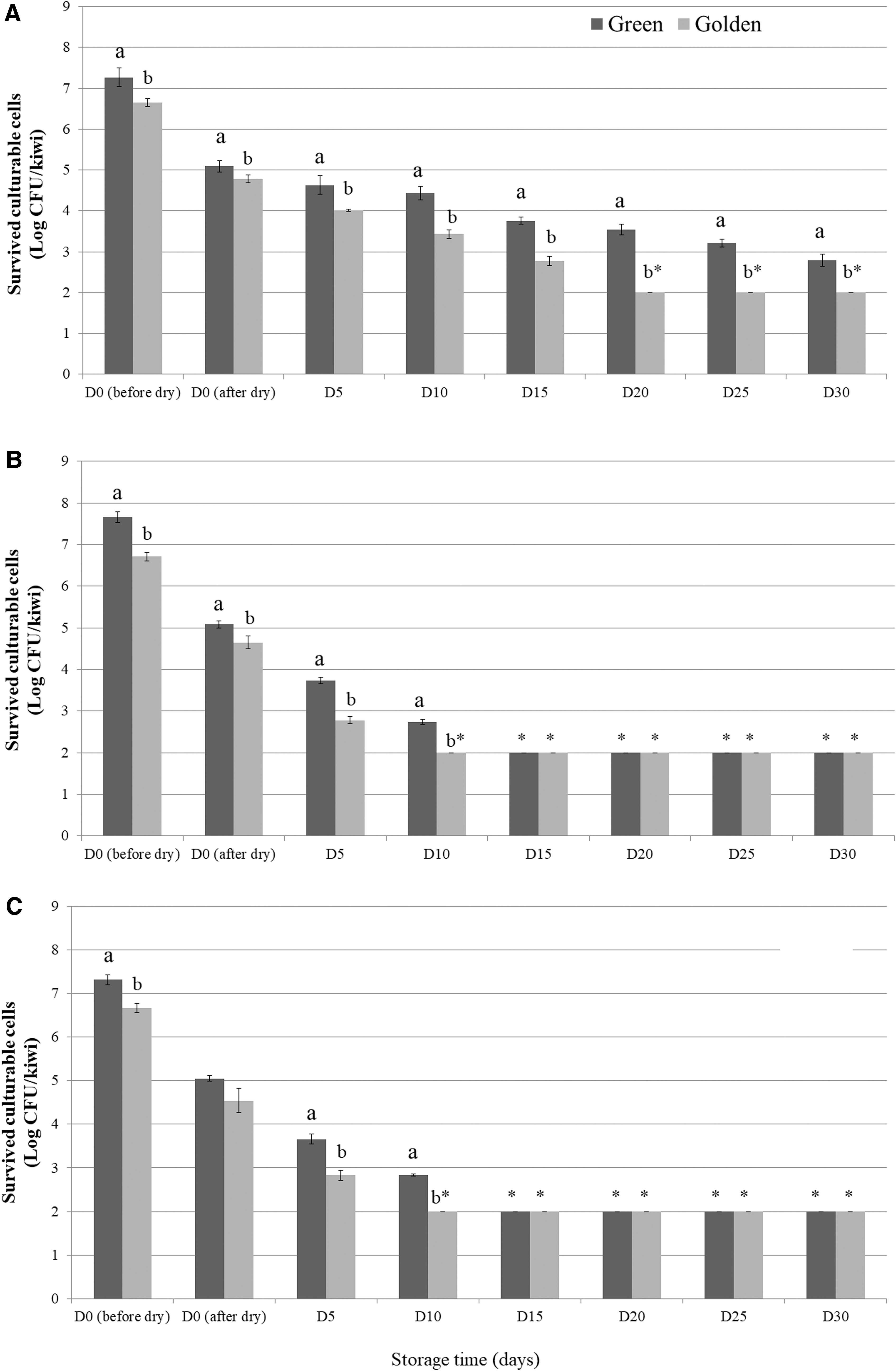
Survival of three pathogens on whole kiwifruits stored at room temperatures for 30 days (high inoculation level).
During the 1-h drying period, an ∼2 Log CFU/kiwi reduction in all pathogens was observed, regardless of the kiwifruit variety. During the 30-day storage period, the culturable E. coli O157:H7 on golden kiwifruit fell below the limit of enumeration after 20 days, while the culturable E. coli O157:H7 on green kiwifruit remained above the limit of enumeration through the entire storage time (Fig. 2A). Significant differences in culturable E. coli O157:H7 cells were observed between the green kiwifruit and golden kiwifruit from day 5 to 30. On day 30, the number of culturable E. coli O157:H7 on green kiwifruit was 2.79 ± 0.15 Log CFU/kiwi.
Culturable Salmonella cells and culturable Listeria cells fell below the limit of enumeration on day 10 on golden kiwifruit and on day 15 on green kiwifruit, respectively, as shown in Figure 2B and C. Enrichment results showed that both of these two pathogens could survive for 30 days on whole kiwifruit. On day 5 and 10, significantly higher numbers of culturable Salmonella and Listeria cells were recovered from the green kiwifruit than from the golden kiwifruit.
Figure 3 shows the survival of E. coli O157:H7, Salmonella spp., and L. monocytogenes on whole green and golden kiwifruit when inoculated at lower levels and stored at room temperature for 30 days. Higher initial wet inoculum was recovered from green kiwifruit than the golden kiwifruit, however, the difference was not significant (p > 0.05). During the storage, on day 10, the numbers of surviving E. coli O157:H7, Salmonella spp., and L. monocytogenes on green kiwifruit were significantly higher than golden kiwifruit. E. coli O157:H7 on golden kiwifruit fell below the limit of enumeration after day 10, while the surviving E. coli numbers on green kiwifruit were 3.16, 2.94, and 2.71 Log CFU/kiwi on day 10, 15, and 20 respectively.
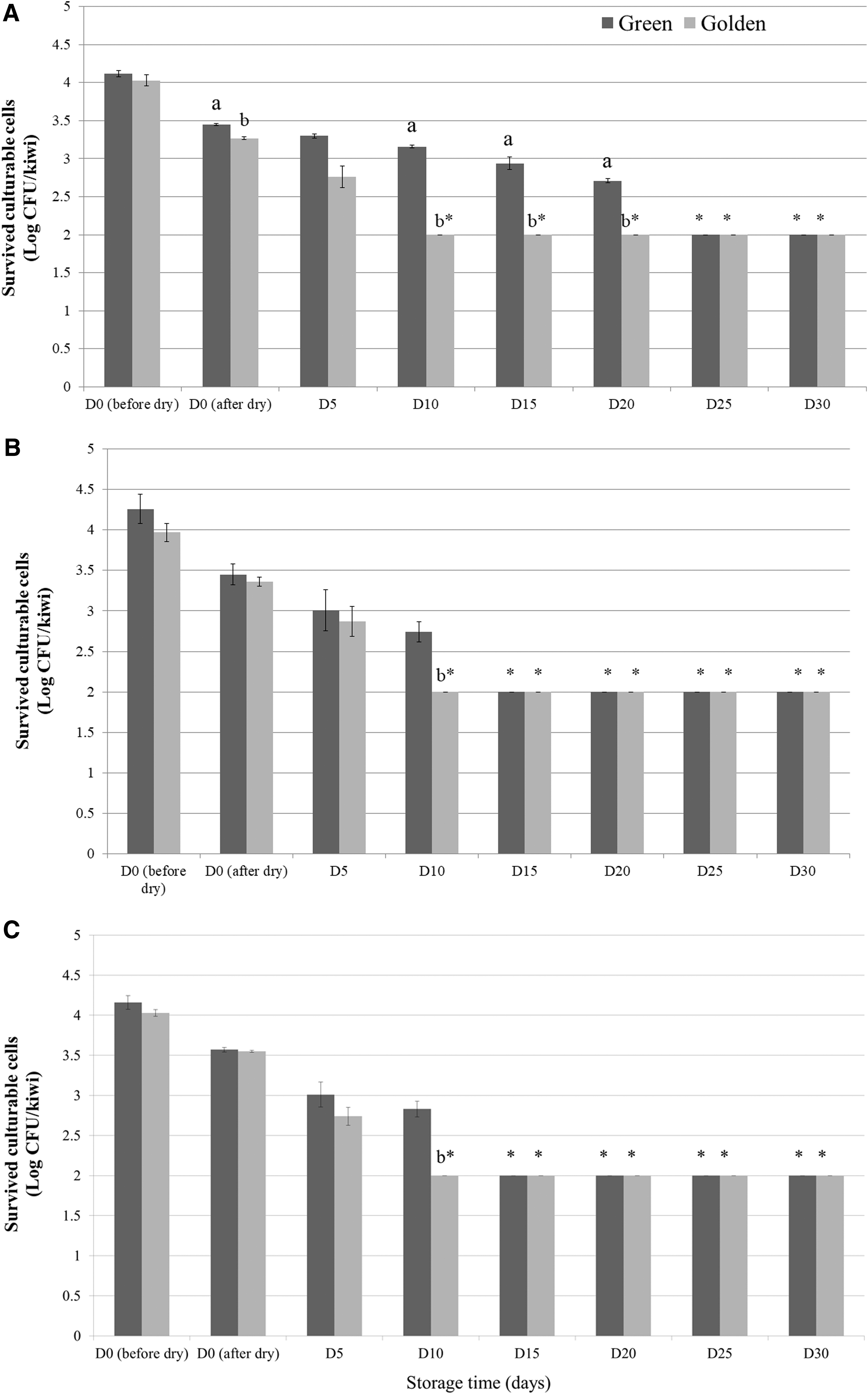
Survival of three pathogens on whole kiwifruits stored at room temperature for 30 days (low inoculation level).
SEM examination of pathogen attachment on whole kiwifruit
As shown in the whole kiwifruit study, green kiwifruit supported the attachment of the pathogen cells during inoculation and also their survival during storage. To examine the attachment of pathogen cells on two different kinds of kiwifruit skins, SEM was conducted. The hair structure of the green kiwifruit can be clearly seen in Figure 4A. Compared with the green kiwifruit, the skin of the golden kiwifruit is much smoother (Fig. 4B). Once pathogens (E. coli O157:H7) are inoculated and dried on the whole kiwifruit, the SEM examination showed that pathogens are attached to both the surfaces and the hair structure of the green kiwifruit (Fig. 4C), while the cells only are attached to the surface of the golden kiwifruit (Fig. 4D).
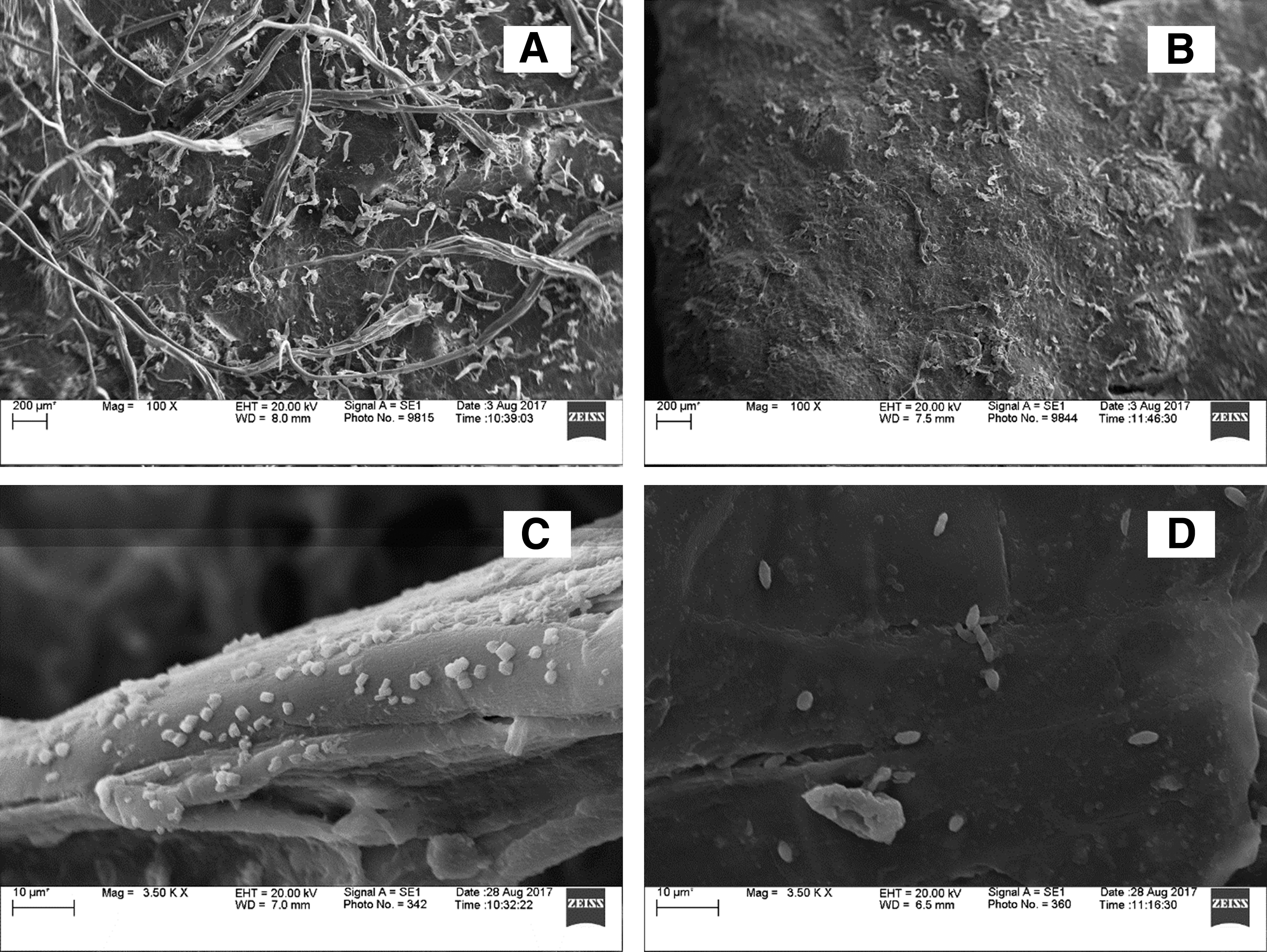
SEM examination. The skin structure of green kiwifruit
Survival of pathogens on sliced kiwifruit
The shelf life of fruit salad is usually about 2–5 days when stored properly at low temperatures (0°–5°C). Due to the high sugar content, fruit salad may start fermenting and develop a “fermented” flavor after 2–3 days. In this study, sliced golden and green kiwifruit started to develop a “fermented” smell after ∼3 days. As shown in Figure 5, at the high inoculation level, ∼3 Log reduction was observed when storing the inoculated green and golden kiwifruit at 4°C for 7 days (p < 0.05). Although the pH was low, all three pathogens survived during the refrigeration storage. Similar situation was observed at the low inoculation level (Fig. 6), all of the three inoculated pathogens survived for 7 days on sliced kiwifruit without significant decrease (p > 0.05).
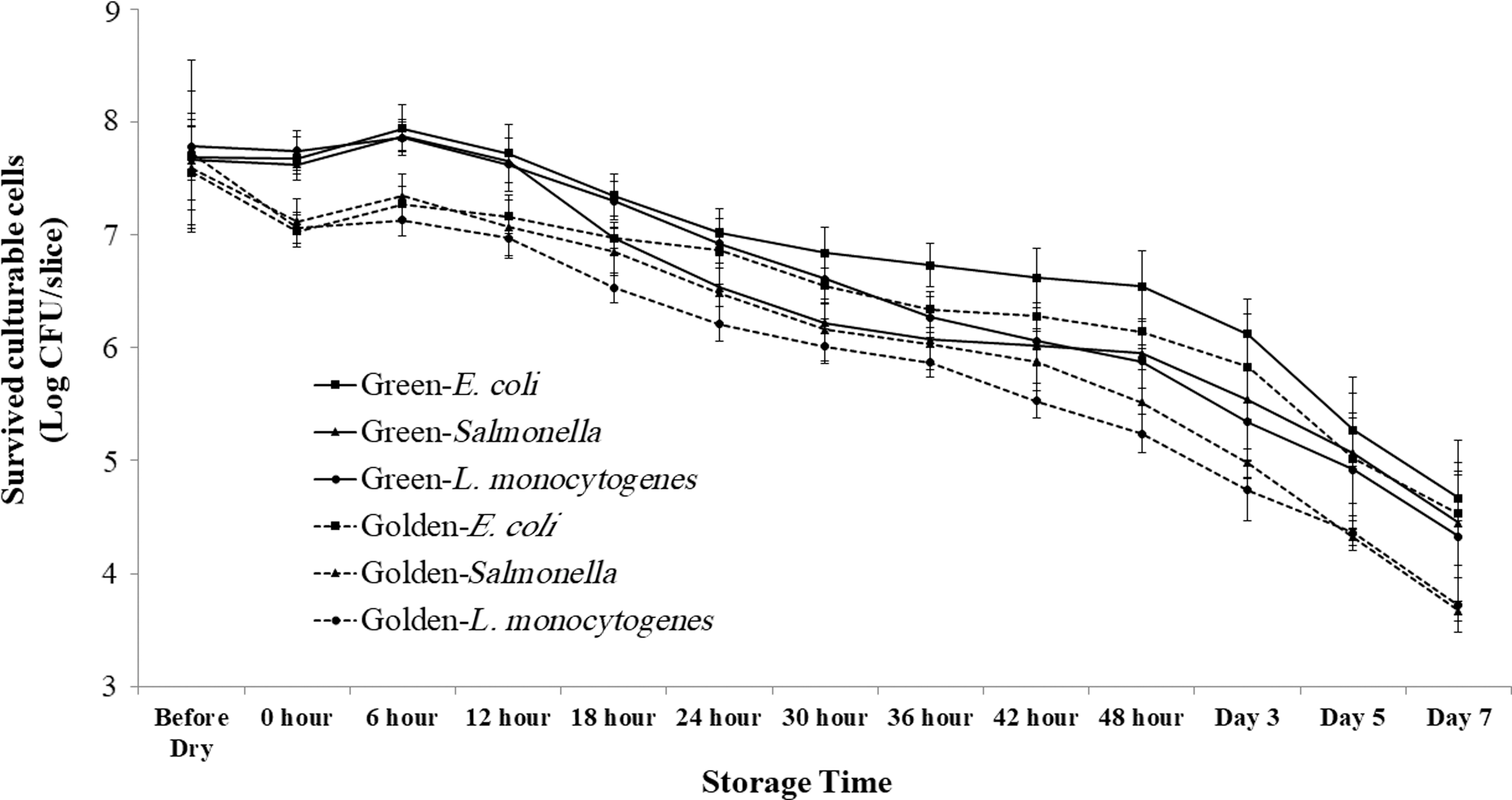
Survival of three pathogens on sliced kiwifruit at 4°C (high inoculated level). Limit of enumeration: 1.70 Log CFU/slice. CFU, colony-forming unit.
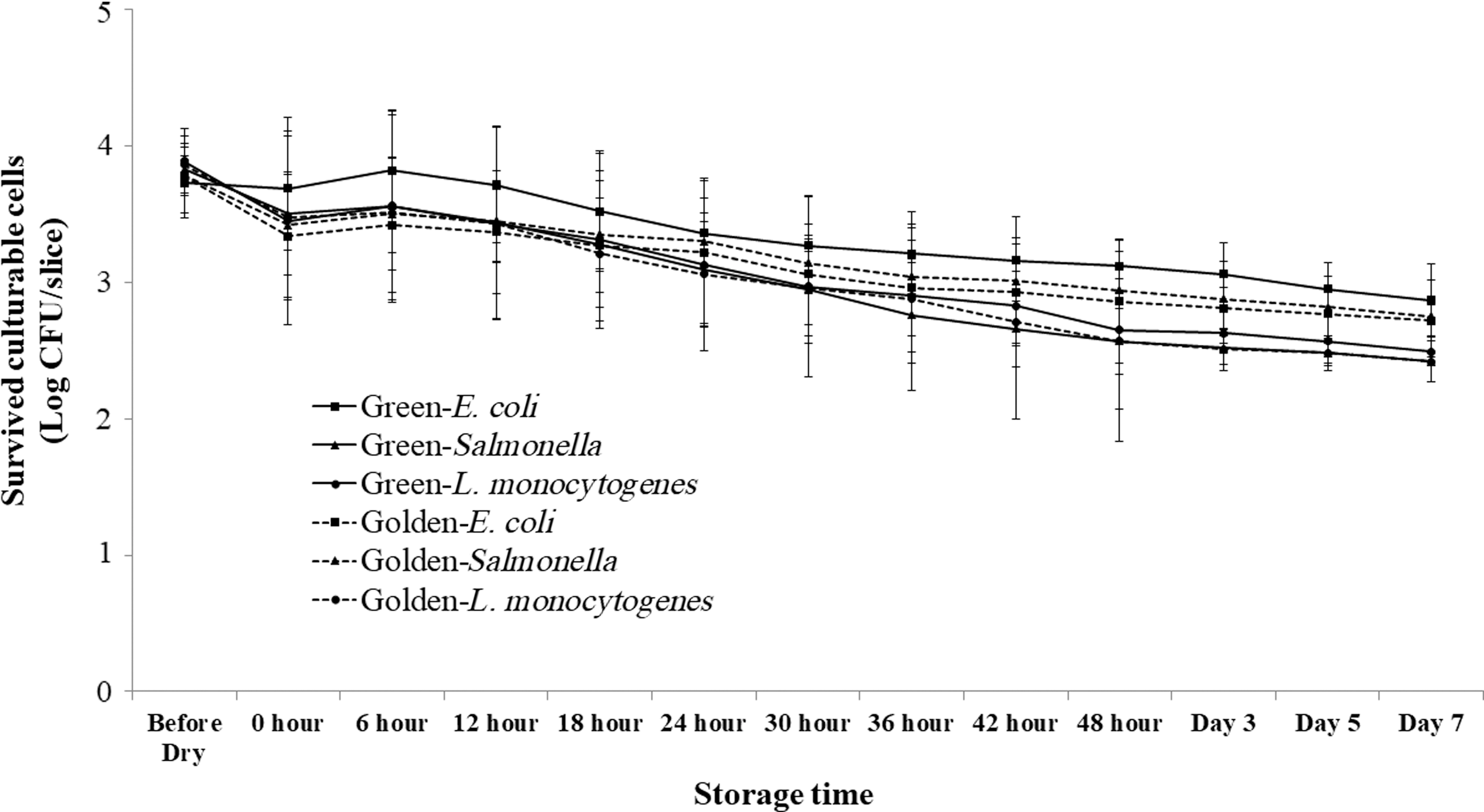
Survival of three pathogens on sliced kiwifruit at 4°C (low inoculated level). Limit of enumeration: 1.70 Log CFU/slice. CFU, colony-forming unit.
Discussion
This study was the first study that evaluated the survival of E. coli O157:H7, Salmonella spp., and L. monocytogenes on whole and sliced kiwifruit. These three pathogens survived for up to 30 days on whole kiwifruit when stored at the room temperature, regardless of the inoculation levels and the kiwifruit variety. The survival of pathogens on fruit surfaces had been shown by other studies. Perez-Rodriguez et al. (2014) found that Salmonella can survive on whole apples for 12 days at room temperature. Collignon and Korsten (2010) showed that E. coli O157:H7, L. monocytogenes, Salmonella Typhimurium, and Staphylococcus aureus could survive on whole peach and plum through the entire simulated commercial export chain. In this study, by using two different kiwifruit varieties, we demonstrated the important role the structure of fruit skin could play during inoculation and survival. The fuzzy and hairy structure of green kiwifruit was a significant factor in supporting the attachment and survival of pathogens.
For sliced kiwifruit, although the pH was low (∼3.5), the tested E. coli O157:H7, Salmonella, and L. monocytogenes cocktails survived for 7 days when stored at 4°C. The sampling stopped at day 7 because fermented smell started to develop after 3 days of storage for golden kiwifruit and 4 days for green kiwifruit. Off-color and off-flavor were significant on day 7. In a challenge study conducted on cut mangoes (pH 4.2) and papayas (pH 5.7) by Strawn and Danyluk (2010), E. coli O157:H7 and Salmonella survived for 28 days when inoculated onto cut mangoes and papayas and stored at 4°C.
The surface of an apple, kiwifruit, plum, or peach is a harsh environment for enteric pathogens to grow, as there are very limited nutrients available for the pathogens. The facts that these pathogens can survive for long periods of time at different storage temperatures on fruit skins highlighted the importance of controlling contamination during fruit production, processing, and preparation. Studies that evaluate the efficiency of novel antimicrobial coating or different modified atmosphere packaging systems will also be beneficial for the control of pathogens on both whole and sliced kiwifruit.
Footnotes
Acknowledgments
This project was supported by the Alabama Agricultural Experiment Station and the Hatch program of the National Institute of Food and Agriculture, U.S. Department of Agriculture.
Disclosure Statement
No competing financial interests exist.