
Abstract
Campylobacter is the leading bacterial cause of human enteritis in developed countries. Human campylobacteriosis is commonly associated with the consumption of undercooked, contaminated chicken, a natural host of Campylobacter. Thus, the control of Campylobacter colonization in poultry at the farm level would reduce the risk of human exposure to this pathogen. Vaccination is an attractive intervention measure to mitigate Campylobacter in poultry. Our recent studies have demonstrated that the outer-membrane proteins CmeC (an essential component of CmeABC multidrug efflux pump) and CfrA (ferric enterobactin receptor) are feasible candidates for immune intervention against Campylobacter. By targeting these two promising vaccine candidates, live attenuated Salmonella-vectored vaccines were developed and evaluated in this study. Briefly, the cfrA and cmeC genes were cloned into expression vector pYA3493 and transferred into Salmonella enterica serovar Typhimurium χ8914, the USDA licensed live attenuated vaccine strain. The oral live Salmonella vaccines producing CfrA or CmeC (truncated or full length) were successfully constructed by using delicate molecular manipulation despite the challenge due to the potential toxic effect of the cloned gene product in the Escherichia coli host. Expression and membrane localization of the target protein in the vaccines were confirmed by immunoblotting. The efficacies of the two live vaccines that produce full-length CfrA or CmeC were evaluated by using broiler chickens. However, oral vaccination of chickens failed to trigger significant systemic and intestinal mucosal immune responses and, consequently, did not confer protection against Campylobacter jejuni colonization chickens. The vaccination regimens of the constructed live Salmonella-vectored vaccine need to be optimized in future studies.
Introduction
Thermophilic
It has been observed that the elevated levels of Campylobacter-specific antibodies are correlated with reduced colonization levels of Campylobacter (Sahin et al., 2003; Cawthraw and Newell, 2010). Therefore, vaccination of poultry against Campylobacter is regarded as an effective intervention strategy to protect food safety (Jagusztyn-Krynicka et al., 2009; Lin, 2009). A successful chicken vaccine should prevent colonization or cause a strong reduction of Campylobacter numbers in chickens (>2 log units) (Rosenquist et al., 2003). However, there is still no vaccine available to date to control Campylobacter infections in poultry; vaccinations of chickens against C. jejuni have had only partial success (Jagusztyn-Krynicka et al., 2009; Lin, 2009), primarily due to a lack of understanding of pathogenesis mechanisms, the antigenic complexity of this organism, and ineffective vaccination regimen (Lin, 2009).
In the past years, we have been actively involved in elucidation of immunogenic and protective antigens in C. jejuni, a primary and critical step toward the design of protective poultry vaccines. Specifically, we have identified and characterized two surface-exposed proteins, CfrA and CmeC, which play an essential role in C. jejuni colonization in the chicken intestine. CfrA is a surface-exposed “gatekeeper” that is essential for C. jejuni colonization by mediating ferric enterobactin high-affinity iron acquisition (Zeng et al., 2009). CmeC is an essential outer-membrane protein component of CmeABC multidrug efflux that plays a critical role in multidrug resistance and C. jejuni colonization (Lin et al., 2002, 2003, 2005; Martinez and Lin, 2006; Zeng et al., 2010). The following findings from our previous studies showed that both CmeC and CfrA have significant advantages compared with other immunogenic/protective antigens identified in C. jejuni (e.g., flagellin, capsule polysaccharide): (1) CfrA and CmeC-specific antibodies greatly inhibited the function of corresponding targets and significantly reduced the growth of C. jejuni (Zeng et al., 2009, 2010); (2) both CfrA and CmeC are prevalent and highly conserved in diverse C. jejuni strains with sequence identity of 89–98% for CfrA and 97.3–100% for CmeC (Lin et al., 2002; Zeng et al., 2009, 2010); (3) CfrA and CmeC are highly induced and produced in the intestinal tract (Lin et al., 2002, 2003; Zeng et al., 2009, 2010); (4) both CfrA and CmeC are immunogenic in poultry and elicit a specific antibody response during C. jejuni infection in poultry (Lin et al., 2002, 2003; Zeng et al., 2009); and (5) inhibition of CmeABC efflux pump by a pump inhibitor increased susceptibility of C. jejuni to multiple antimicrobials and reduced in vivo colonization of C. jejuni in chickens (Martinez and Lin, 2006). Clearly, these comprehensive molecular, immunogenic, functional studies have provided compelling evidence that CmeC and CfrA are promising candidates for developing an effective vaccine against C. jejuni in poultry.
Attenuated Salmonella-based vaccines are an attractive strategy to develop inexpensive and practical oral vaccines for chickens to prevent Campylobacter infections (Curtiss and Hassan, 1996). In this study, by targeting CfrA and CmeC, we aimed at constructing oral Salmonella-vectored vaccines by using Salmonella enterica serovar Typhimurium χ8914, the USDA licensed live attenuated vaccine strain. We also evaluated the immunogenicity and protective efficacy of the live Salmonella-vectored vaccines in broilers.
Materials and Methods
Bacterial strains, plasmids, and growth conditions
The bacterial strains and plasmids used in this study, and their sources, are listed in Table 1. C. jejuni NCTC 11168 (JL241) was used for amplification of cfrA and cmeC genes. JL241 was routinely grown in Mueller-Hinton (MH) broth (BD Difco, Sparks, MD) or on MH agar plates under microaerobic conditions (5% O2, 10% CO2, 85% N2) at 42°C. If needed, MH agar was supplemented with Campylobacter Growth and Preston Campylobacter Supplements (Oxoid, Bashingstoke, Hampshire, England). Escherichia coli χ6097 and Salmonella Typhimurium UK-1 χ8914 were grown in Luria-Bertani (LB) broth (BD Difco) with shaking (250 rpm) or on LB agar plates containing 50 μg/mL diaminopimelic (DAP) acid at 37°C overnight. When necessary, LB media were supplemented with 50 μg/mL of tetracycline or 50 μg/mL of ampicillin.
Bacterial Plasmids and Strains Used in This Study
Polymerase chain reaction
Primers used in this study and the expected sizes of the products are listed in Tables 2 and 3. Each PCR was performed with a 50-μL mixture containing 200 μM deoxynucleoside triphosphates, 200 nM of each primer, 50 ng of JL241 template DNA, 2.5 mM MgSO4, and 5 U PfuUltra II high-fidelity DNA Polymerase (Stratagene, San Diego, CA). The temperature-cycling parameters are typically as follows: 95°C for 5 min for denaturation, 32 cycles of 1 min at 94°C, 1 min at 58°C, 90 s at 72°C, and a final extension step of 45 s at 72°C, though cycling conditions varied according to annealing temperatures of primers and estimated product sizes. PCR products were further purified for cloning procedures or sequencing analysis.
Primers Used in Construction of Full-Length CfrA and CmeC Salmonella-Vectored Vaccines
The restriction site is shown as bold letters.
Primers Used in Construction of Truncated CfrA and CmeC Salmonella-Vectored Vaccines
The restriction site is shown as bold letters.
Sequence analysis of plasmid constructs
Plasmid DNA was extracted from host cells by using Qiagen QIAprep Spin Miniprep Kit (Qiagen, Hilden, Germany). Primer pairs of pYA3493_F/pYA3493_R (Table 2) were used to sequence pYA3493 derivatives. Sanger Capillary Sequencing was performed in the Molecular Biology Resource Facility at the University of Tennessee (Knoxville, TN). For recombinant plasmids, proper insertion of cfrA or cmeC was confirmed by comparing the sequences to those from parent plasmids and the genome of C. jejuni NCTC 11168.
Construction of Salmonella-vectored vaccines
The plasmid pYA3493 (Zekarias et al., 2008; kindly provided by Dr. Roy Curtiss III, University of Florida) was used for preparation of desired recombinant plasmids that were then transferred to the E. coli host and attenuated Salmonella Typhimurium χ8914 successively (Kang and Curtiss, 2003; Zekarias et al., 2008) for live vaccine construction. The pYA3493 is an expression plasmid containing the Ptrc promoter upstream of the β-lactamase signal peptide, and the asdA gene for natural selection. The primer pairs of pYA3493_CfrA_F/pYA3493_CfrA_R and pYA3493_CmeC_F/pYA3493_CmeC_R (Table 2) were designed to amplify full-length cfrA and cmeC genes from NCTC 11168 genomic DNA. Specific PCR product was digested with EcoRI and SalI for directional cloning of the fragment into the pYA3493 that has been digested with EcoRI and SalI. The ligation mixtures were introduced into competent E. coli χ6097, the intermediate host, via electroporation at 4–5 ms and 2.5 kV; preparation of the competent cells is detailed in the next section. Transformants were selected on LB agar plates. The empty vector, pYA3493 was electroporated into E. coli χ6097 to create strain JL1070. After positive identification through plasmid extraction and gel electrophoresis, pYA3493 was then transferred into Salmonella Typhimurium χ8914 to create strain JL1059 (Table 1).
Failure to obtain desired recombinant plasmids bearing full-length cfrA or cmeC genes in the intermediate E. coli host despite extensive efforts led to the design of alternative primer pairs, which included; (1) pYA3493_CfrA_F/pYA3493_CfrA_R (EcoRI) and pYA3493_CmeC_F/pYA3493_CmeC_R (EcoRI) that contain a single restriction enzyme site (EcoRI) for bidirectional cloning (Table 2); (2) pYA3493_CfrA_F/CfrA_B1_R, pYA3493_CfrA_F/CfrA_B7_R, CfrA_B3_F/CfrA_B14_R, and CfrA_B14_F/CfrA_C_R (Table 3) for amplifying different truncated cfrA fragments. The EcoRI and PstI were attached to the 5′ end of the primers for directional cloning; and (3) pYA3493_CmeC_F/CmeC_TM2_R, CmeC_TM2_F/CmeC_TM3_R, and CmeC_TM3_F/CmeC_C_R (Table 3) for amplifying different truncated cmeC fragments. The cloning and transformation procedure are the same as described earlier. The primer pairs pYA3493_CfrA_F/CfrA_B1_R and CmeC_TM2_F/CmeC_TM3_R (Table 3) led to successful construction of pYA3493_tCfrA and pYA3493_tCmeC (Table 1), respectively, for production of desired truncated proteins. The pYA3493_tCfrA and pYA3493_tCmeC were confirmed by sequencing, extracted from the corresponding E. coli host (JL1060 and JL1061, Table 1), and finally electroporated into competent Salmonella Typhimurium χ8914. The Salmonella strains harboring pYA3493_tCfrA and pYA3493_tCmeC are referred to as JL1062 and JL1063 (Table 1). The Salmonella transformants were selected on LB plates. The plasmids from Salmonella transformants were further extracted for sequencing analysis. In addition, production of the specific inserted protein (CfrA or CmeC) in both E. coli (the intermediate host) and Salmonella (the final live vaccine host) was confirmed by immunoblotting by using specific antibodies.
To overcome any potentially lethal effect of full-length CfrA or CmeC on the intermediate E. coli host, the plasmid pBR232 (lacI q) (Zekarias et al., 2008) was transferred to E. coli χ6097 to create a new E. coli host strain JL1080 (Table 1), which can suppress the promoter activity of transformed pYA3493. In addition, primer pairs were redesigned for directional cloning (Table 2, pYA3493_CfrA_F/pYA3493_CfrA_R for cfrA gene and pYA3493_CmeC_F/pYA3493_CmeC_R for cmeC gene). The EcoRI and PstI digested PCR fragment was ligated into the pYA3493 that had been digested with the same enzymes. Ligation mixture was electroporated into JL1080. Transformants were screened and confirmed as described earlier. The recombinant plasmids that carried full-length cfrA or cmeC gene within E. coli JL1109 and JL1110 were extracted and transferred into Salmonella host strain, creating JL1104 and JL1105; the plasmids were validated by sequencing, and production of target gene products was verified by immunoblotting.
The competent cells used for molecular cloning were prepared by following published protocol (Gonzales et al., 2013) with slight modifications. Briefly, a single colony of the E. coli χ6097 or Salmonella Typhimurium χ8914 was grown in LB broth containing DAP to OD600 value of 0.35 to 0.4. The cells were washed with ice-cold water followed by ice-cold 10% glycerol. The cells were suspended in ice-cold GYT media (10% glycerol, 0.125% yeast extract, 0.25% tryptone) to a final concentration of 2–3 × 10¹⁰ cells/mL. The competent cells were aliquoted and stored at −80°C until use.
Preparation of membrane fraction
To measure antibody responses against Salmonella Typhimurium χ8914, the membranes were isolated from a culture of JL1059 containing empty vector pYA3493 (Table 1). One liter of overnight culture (37°C, 250 rpm, in LB broth) was subjected to centrifugation at 2500 × g for 30 min at 4°C, and it was then washed with PBS. Pellets were resuspended in 20-mL ddH2O and sonicated in an ice bath by using Sonic Dismembrator (Model 100; Fisher Scientific, Hampton, NH) three times with 30 s followed by a 1-min rest period between each sonication. Sonicated culture was then centrifuged at 5000 × g, and the supernatant was further centrifuged at 30,000 × g for 60 min. The pellet, representing the membrane fraction was resuspended in 1 mL ddH2O and stored at −20°C.
The live Salmonella vaccines expressing CfrA or CmeC (truncated or full length) were also subjected to membrane fractionation to evaluate whether the cloned foreign proteins were localized in the membrane. Membrane samples were evaluated by immunoblotting analysis.
Sodium dodecyl sulfate-polyacrylamide gel electrophoresis and Western blot analysis
Sodium dodecyl sulfate-polyacrylamide gel electrophoresis (SDS-PAGE) and immunoblotting were performed as previously described with slight modifications (Lin et al., 2002; Zeng et al., 2009). Typically, 5 mL of overnight cultures of E. coli or Salmonella were centrifuged and resuspended in 50 μL of 1 × PBS and 50 μL of 2 × SDS-PAGE sample buffer. Five to 15 μL of whole-cell lysate suspension or positive control protein (purified CfrA or CmeC) (Zeng et al., 2009, 2010) were loaded in each lane and separated by SDS-PAGE with a 12% (w/v) polyacrylamide gel at 80 V for 40 min followed by 160 V for 40 min. After SDS-PAGE, proteins in gels were then electrophoretically transferred to nitrocellulose membranes (Bio-Rad, Hercules, CA) at 90 V for 1 h. The membranes were incubated with blocking buffer (5% Nestle skim milk powder in PBS) overnight at 4°C, followed by a 1-h incubation at 25°C with primary antibodies (1:2000 diluted rabbit anti-CfrA sera or 1:1000 diluted rabbit anti-CmeC sera in blocking buffer). After incubation, the membranes were washed three times with PBS containing 0.05% Tween 20 and incubated with secondary antibody (goat anti-rabbit immunoglobulin G-horseradish peroxidase [KPL, Gaithersburg, MD], diluted 1:2000 for CfrA and 1:1000 for CmeC) for 1 h at 25°C. After incubation, the membranes were washed as described earlier. The membranes were then developed with the 4CN Membrane Peroxidase Substrate System (KPL).
Chicken vaccination and challenge trial
Inoculum preparation
Two vaccine strains, JL1104 (Live-CfrA) and JL1105 (Live-CmeC) (Table 1), were evaluated in this vaccination trial. The major reason for choosing JL1104 and JL1105 strains for the chicken trial was because these strains expressed full length of target proteins, which potentially can trigger diverse antibodies specifically targeting different protective epitopes. Three to five colonies of each live vaccine strain as well as the vector control JL1059 (Table 1) were randomly picked from the LB agar plate and inoculated into 50 mL LB broth overnight; static growth was observed at 37°C. Subsequently, 10 mL of the overnight culture was inoculated into 90 mL LB broth and grown statically for about 6 h to reach an OD600 value between 0.8 and 1.0. Bacteria cultures were then centrifuged and resuspended in sterile PBS to a concentration of ∼5 × 10⁹ colony-forming unit (CFU)/mL. Chickens were inoculated orally with 200 μL of PBS control or the cell suspension, which contained 1 × 10⁹ CFU of live vaccine.
Chicken immunization and sample collection
The chicken trial was approved by the Institutional Animal Care and Use Committee at The University of Tennessee (IACUC No. 2099-0415). One-day-old Cornish x Rock (commercial broiler) chicks (n = 80) were obtained from Hubbard Hatchery (Pikeville, TN) and allocated into 4 treatment groups (Table 4; 20 birds per group). On arrival, cloacal swabs from five randomly selected birds from each group were collected for determining the presence of Salmonella spp. and C. jejuni in the intestine. Specifically, cloacal swabs were placed in 2 mL of PBS containing 1% of gelatin. To examine Salmonella contamination, 100 μL of the suspension was spread on MacConkey (BD Difco) plates and incubated overnight at 37°C. The next day, colonies matching the expected phenotype (nonlactose fermenting, gray colonies) of Salmonella on MacConkey plates were re-streaked on XLT-4 agar plates (XLT Agar base [BD Difco] and 2.4 mL [per 500 mL] Tergitol [Sigma]) and were incubated for 24–48 h for identifying H2S-producing colonies. To isolate Campylobacter, 100 μL of the suspension was spread on MH agar plates that contain Campylobacter-specific selective supplements, followed by incubation at 42°C under microaerobic conditions for 48 h.
Evaluation of the Live Salmonella-Vectored Vaccines in Chickens
All chickens were managed in a sanitized wire-floor cage and provided with water and antibiotic-free feed ad libitum. As shown in Table 4, at 7 days of age, chickens were orally immunized with 200 μL of Live_CfrA (group 3) or Live_CmeC (group 4). Two control groups received either PBS (group 1) or the vector control (group 2). At 28 days of age, all birds were orally challenged with ∼2 × 10³ CFU/bird of C. jejuni NCTC 11168. After challenge, cloacal swabs were collected every 2–3 days for 10 days. Swabs were placed in 1 mL MH broth, and 100 μL was plated on MH agar containing a selective supplement for enumeration of C. jejuni.
Blood samples were collected via the wing vein of 10 randomly selected chickens from each group at 7, 18, 28, and 38 days of age to evaluate systemic IgG and IgA antibodies. Intestinal lavage was taken from each euthanized chick (5 birds per group) on 18, 28, and 38 days, and it was diluted 1:4 in lavage extraction buffer (PBS containing 0.05% Tween 20, 0.05 g/mL of EDTA) and complete mini protease inhibitor (Roche, Mannheim, Germany), which were used to determine specific mucosal IgA and IgG antibodies. The five birds from each group used for collecting intestinal lavage were randomly selected from the group of 10 birds for blood sample collection.
Spleen, liver, and cecum were also taken to evaluate the presence of inoculated Salmonella live vaccine after oral vaccination. Briefly, at 18, 28, and 38 days, spleen, liver, and cecum were asceptically collected (5 birds per group), diluted 1:9 in PBS containing 1% of gelatin, and homogenized by using the Stomacher-80 and filter stomacher bags (Fisher). Tissue homogenates (100 μL) were spread on MacConkey plates. Nonlactose fermenting colonies were then isolated on XLT-4 and re-isolated on MacConkey. Pink colonies containing H2S production on XLT-4 were selected, and the identities of the selected colonies were examined by plasmid profile as well as PCR using cfrA or cmeC-specific primers.
Enzyme-linked immunosorbent assay
CfrA-, CmeC-, and Salmonella Typhimurium χ8914 membrane-specific antibodies in serum and intestinal lavage samples were measured by indirect enzyme-linked immunosorbent assay (ELISA) as previously described with modifications (Zeng et al., 2010). Briefly, microtiter plates (Nunc-Immuno Plate; Thermo Fisher Scientific) were coated with 100 μL of highly purified rCmeC, rCfrA, or Salmonella Typhimurium χ8914 membrane protein (as obtained through sonication and ultracentrifugation as described next) (30 ng/well) in coating buffer (1 M ammonium acetate and ammonium carbonate, pH 8.2) overnight at room temperature. Plates were washed three times with washing solution (0.05% Tween 20, 1 × PBS) and blocked with blocking buffer (1 × PBS, 1% BSA, 0.1% Tween 20) for 1 h at 37°C. Chicken serum and intestinal lavage samples were diluted 1:100 and 1:4 in blocking buffer, respectively, and 100 μL was added, in duplicate, to wells, followed by 1-h incubation. Plates were washed five times with washing solution. Bound IgG and IgA antibodies were detected by addition of 100 μL/well anti-chicken IgG (KPL) and IgA (Bethyl, Montgomery, TX) HRP conjugates diluted 1:2000 in blocking buffer. After 1-h incubation, the plates were washed three times. Plates were developed by using the ABTS Peroxidase Substrate Kit (KPL), and the reaction was stopped after 10 min by using 100 μL stopping solution (1 × PBS, 1% SDS). Absorbance was measured at OD405 nm.
Statistical analysis
Differences in serum and intestinal lavage OD405 nm readings among treatment groups were analyzed by least-square analysis of covariance with the data at day 7 (day of vaccination) as the covariant; main effects were day of sample collection and treatment. Comparison of OD405 nm readings within treatment groups across time was tested by ANOVA. Levels of significance for p value were 5% (0.05). All statistical analyses were performed by using SAS software (v9.03; SAS Institute, Inc., Cary, NC).
Results
Development of Salmonella-vectored vaccines producing truncated proteins
The full-length cfrA and cmeC fragments were first amplified and used for directional cloning into the pYA3493 plasmid. However, despite success in PCR amplification, extensive efforts on molecular cloning did not lead to identification of the E. coli transformants containing the desired recombinant plasmids; all selected transformants were false positives as reflected by the equivalent size of the extracted plasmids to the parental pYA3493 vector (data not shown). This finding suggested that overexpression of the full-length cfrA or cmeC gene was lethal to the E. coli host. To test this hypothesis, the cloning process was repeated by using bidirectional cloning with a single restriction enzyme. As expected, of all selected transformants containing inserted fragments, the cloned gene (either cfrA or cmeC) displayed in the reverse, undesired orientation in the vector by PCR analysis (data not shown).
Subsequently, as an alternative approach, we obtained two E. coli constructs JL1060 and JL1061 containing recombinant plasmid pYA3493_tCfrA and pYA3493_tCmeC only bearing the truncated cfrA or cmeC gene with correct orientation, respectively (Table 1). The two plasmids were extracted from E. coli hosts and then transformed into the Salmonella host vaccine strain, creating live vaccines JL1062 and JL1063 (Table 1) that are expected to produce truncated CfrA and CmeC, respectively. Immunoblotting using whole-cell lysate demonstrated that the truncated CfrA (Fig. 1A) and CmeC (Fig. 1B) proteins were produced in their corresponding E. coli and Salmonella hosts with approximate molecular masses of 15.7 and 19.8 kDa, respectively.

Expression of truncated CfrA and CmeC in the Escherichia coli and Salmonella enterica serovar Typhimurium constructs.
Development of Salmonella-vectored vaccines producing full-length proteins
Production of full-length target protein has advantages for triggering strong and specific immune responses in vivo. Despite the speculated toxic effect of full-length CfrA or CmeC on the E. coli host strain as described earlier, it was possible that full-length CfrA or CmeC protein would not be toxic to Salmonella, the intended vaccine vector. Therefore, to construct Salmonella live vaccines expressing full-length CfrA or CmeC, we first transferred the pBR232 plasmid into the E. coli χ6097 host, creating a new cloning host JL1080 (Table 1). In this new host, due to the presence of the lacIq repressor system in pBR232 plasmid, the Ptrc promoter and downstream target antigen gene cloned in pYA3493 were repressed. Consequently, the pYA3493-derived recombinant plasmids bearing full-length cfrA or cmeC gene were successfully obtained by using this new cloning system. These recombinant plasmids were then extracted from E. coli (strains JL1109 and JL1110, respectively; Table 1) and successfully transferred into the Salmonella live vaccine host, creating a new Salmonella-vectored vaccine Live-CfrA (JL1104) and Live-CmeC (JL1105) (Table 1). Immunoblotting using specific antibodies confirmed that both the full-length CfrA (76 kDa; Fig. 2A) and CmeC (53 kDa; Fig. 2B) were produced in Salmonella. However, numerous bands of lower molecular weight appeared for both CfrA and CmeC within the Salmonella host and were suggestive of degradation of these proteins.
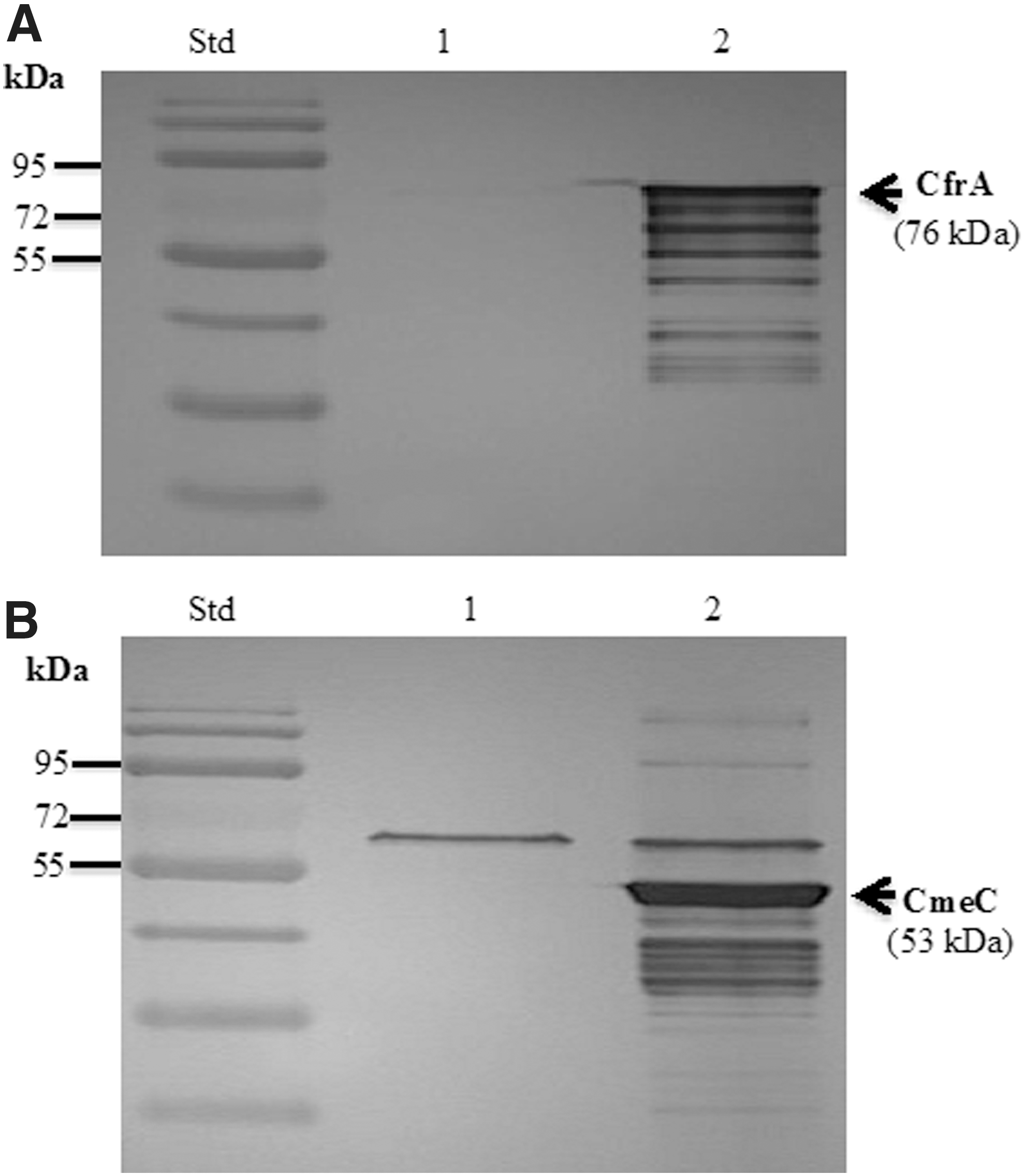
Expression of full-length CfrA and CmeC in the Salmonella Typhimurium vaccine
Membrane localization
Full-length and truncated CfrA proteins (76 and 15.7 kDa, respectively) were expressed and located in Salmonella Typhimurium membrane as demonstrated by immunoblotting (Fig. 3A). Similarly, full-length and truncated CmeC was also expressed (53 and 19.8 kDa, respectively) and localized in the Salmonella Typhimurium membrane (Fig. 3B).
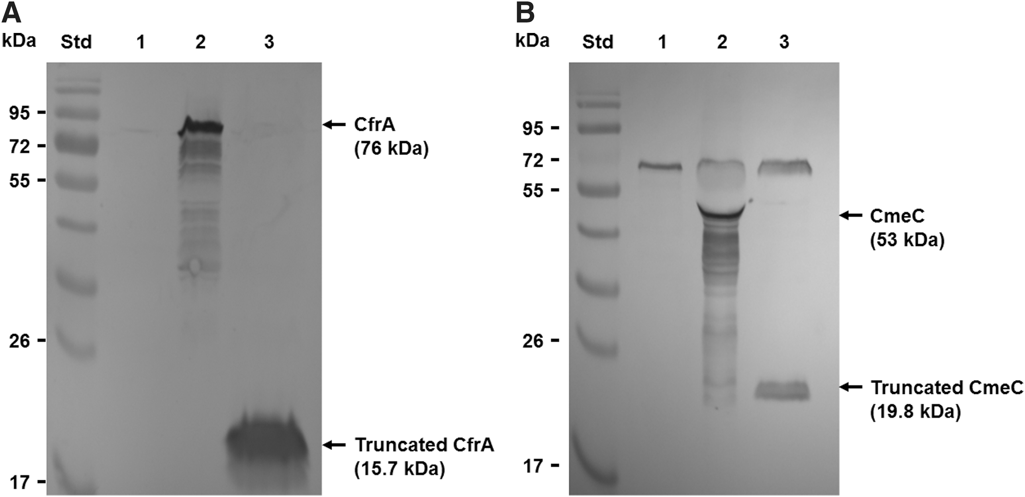
Confirmation of membrane localization of CfrA and CmeC in Salmonella-vectored vaccines by using immunoblotting analysis.
Evaluation of Salmonella-vectored vaccines in commercial broiler chickens
The chicks were free of Campylobacter and Salmonella on arrival, as reflected by initial cloacal swab tests. After oral vaccination with Salmonella-vectored vaccines, a low percentage of the birds in each group were positive for Salmonella Typhimurium vaccine strains in tissues at different time points (Table 5). The levels of control Salmonella Typhimurium (Group 2, Table 5) in cecum and spleen at 11 days postimmunization (dpi) were lower than those of Live-CfrA strain (Group 3, Table 5). By 21 dpi, the inoculated Live-CfrA Salmonella vaccine strain could be still isolated from cecum, spleen, and liver samples with different levels (Group 3, Table 5). The Live-CmeC vaccine strain (Group 4, Table 5) was only detected in the spleen at a relatively low level by 21 dpi. No Salmonella Typhimurium bacteria were recovered from the cecum, spleen, or liver samples at 31 dpi (Table 5).
Recovery of Salmonella Typhimurium Vaccine Strains from Euthanized Chickens at Different Time Points
Cecum, spleen, and liver samples were aseptically collected (five birds per group per time point) and subjected to Salmonella identification (detailed in the Materials and Methods section). The information in the parenthesis following a specific number indicates the Salmonella level in an individual Salmonella-positive bird (CFU/g).
CFU, colony-forming unit; dpi, days postimmunization.
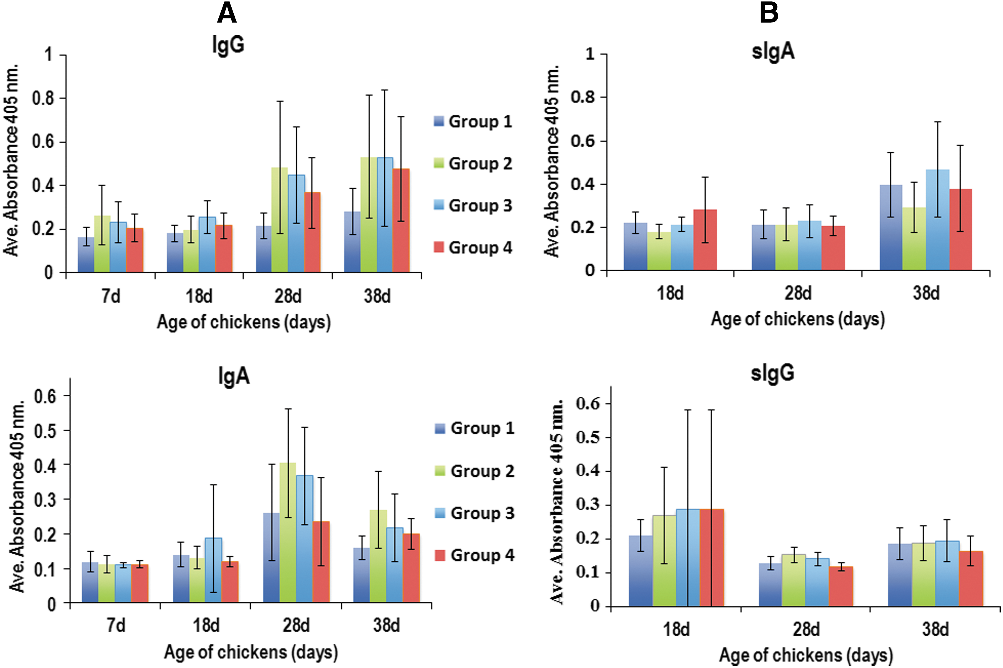
Oral vaccination of chickens with Live-pYA3493 (empty vector), Live-CfrA, and Live-CmeC produced elevated, but not significant, antibody responses directed against Salmonella membrane proteins with respect to systemic IgG and IgA levels (Fig. 4A) and mucosal IgA and IgG levels (Fig. 4B). In terms of CfrA- and CmeC-specific antibody responses, there was no significant enhancement of systemic IgG or IgA or local mucosal IgG or IgA in intestinal lavage samples at different time points postimmunization (Figs. 5 and 6). All groups displayed relatively higher CfrA- or CmeC-systemic IgG before immunization (Figs. 5 and 6), likely due to the presence of maternal antibody (Sahin et al., 2003). Consistent with the patterns of weak systemic and mucosal immune responses observed (Figs. 4 –6), challenge of chickens with NCTC 11168 at age 28 days indicated no difference in the levels of C. jejuni colonization at different time points among the four groups (data not shown).
Host immune responses directed against Salmonella Typhimurium on vaccination of broilers with live Salmonella-vectored vaccines.

Host immune responses directed against CfrA on vaccination of broilers with live Salmonella-vectored vaccines.

Host immune responses directed against CmeC on vaccination of broilers with live Salmonella-vectored vaccines.
Discussion
Campylobacter can quickly infect an entire commercial poultry flock and establish itself at high quantities within the ceca of broilers until time of slaughter (Sahin et al., 2002). Horizontal transmission from numerous potential environmental sources is the major route of Campylobacter introduction into commercial flocks (Sahin et al., 2002). Intervention strategies at the poultry farm level to reduce Campylobacter colonization within poultry create a challenge, but they are necessary to reduce its presence at human consumption (Lin, 2009).
The short life span of the commercial broiler, slaughter age of 6–7 weeks, creates a challenge for induction of an immune response against Campylobacter and a subsequent reduction in colonization. Since Campylobacter primarily colonize in the intestine of poultry with the highest load in ceca, generation of a strong mucosal immune response is highly desired to effectively control Campylobacter colonization. However, mucosal vaccines face several challenges such as dilution and entrapment by mucosal secretions, degradation by proteases, and exclusion by the epithelial barriers (Neutra and Kozlowski, 2006). As observed in our previous CmeC subunit vaccination trial (Zeng et al., 2010), protein-based subunit vaccines stimulated weak mucosal immune responses when administered orally even if a mucosal adjuvant was used. Choice of an optimal vaccine adjuvant is essential to “alert” the host's immune system (Neutra and Kozlowski, 2006). Another important requirement in chicken vaccination that needs to be addressed is the ease of mass administration and cost-effectiveness (de Zoete et al., 2007). Oral administration routes are the most suitable and frequently explored for developing Campylobacter vaccine used in poultry (Lin, 2009). To address this requirement and the challenges faced by mucosal vaccines, in this study we constructed and evaluated oral vaccination of the vaccines targeting CfrA and CmeC, created from a USDA licensed live attenuated Salmonella Typhimurium strain.
The use of live attenuated mucosal vaccine vectors can generate substantial innate immune responses that bolster adaptive immune responses (Neutra and Kozlowski, 2006). The most commonly used bacterial vectors include attenuated Salmonella spp. to carry antigens against viral, bacterial, and parasitic pathogens (Zekarias et al., 2008; Jenikova et al., 2011; Jazayeri et al., 2012). Salmonella is an ideal organism to invade and colonize effector lymphoid tissues and induce T cell response (Kong et al., 2013). In our study, we chose live, attenuated Salmonella Typhimurium χ8914 as a vector to carry our immunogenic CfrA and CmeC proteins. Attenuation in this strain was achieved with deletion in pabA and pabB genes, which encode for 4-amino-4-deoxychorismate synthase required for production of folic acid in Salmonella; which the organism is unable to obtain from the environment (Wang et al., 2011). This strain carries a deletion in the asdA gene, creating a deficiency in DAP acid, a key component of Salmonella peptidoglycan. Deficiency in DAP causes lysis of the cell and since DAP is not found within mammalian tissues, the need for DAP is dire. The expression plasmid pYA3493, used to clone CfrA and CmeC in this study, carries the asdA gene, allowing for retention of the plasmid to be an absolute requirement for the Salmonella vaccine vector survival (Curtiss et al., 1989; Galan et al., 1990). The advantage to this complementation system is the absence of drug-resistant gene markers while achieving selective pressure for retention of the plasmid within the vaccine cells in vivo. Ma et al. (2011) used a recombinant Salmonella choleraesuis vaccine strain carrying the pYA3493 vector expressing p36, p46, p65, and p97R1-Nrdf genes of Mycosplasma hyopneumonaie. These recombinant S. choleraesuis vaccines, when orally administered, produced significantly higher Mycoplasma-specific antibodies as compared with the group intramuscularly injected with the p36, p46, and p65-expressing strains. Another study (Kang and Curtiss, 2003) demonstrated that the type II secretion system within pYA3493 is important for secretion of the antigenic protein into the periplasm and is mounting a higher immune response as compared with other expression plasmids. In addition, the Salmonella Typhimurium χ8914 host strain used in this study is USDA licensed, allowing for a vaccine with proven systemic and mucosal immune enhancement in regards to Campylobacter to be approved in an expedited timeframe.
However, in this study, it was disappointing that both systemic and local mucosal immune responses were weak on oral vaccination of the validated Salmonella-vectored vaccines. Our initial construction of recombinant expression vector demonstrated that overexpression of intact CfrA and CmeC was likely toxic to the E. coli host and required the lacIq repression system from within plasmid pBR232 for cloning. Although the Salmonella live vaccines produced full-length CfrA or CmeC normally in broth medium (Fig. 2), we cannot rule out the possibility that production of CfrA or CmeC can exert a metabolic burden and even detrimental effect on the Salmonella host in vivo. In particular, the CfrA and CmeC, which can form channels in membrane, seemed to be folded normally, reflected by their membrane localization demonstrated in this study (Fig. 6). Consequently, production of potentially toxic heterologous proteins, such as CfrA or CmeC, can lead to hyperattenuation, modified or poorly expressed antigenic proteins, and reduction in viability and colonizing ability, leading to poor immunogenicity overall as previously described (Galan et al., 1990). Using the same pYA3493 expression plasmid and Salmonella χ8914 host strain combination, a previous study (Zekarias et al., 2008) developed and evaluated Salmonella Typhimurium vectored vaccines to control Clostridium perfringens. After immunization of chickens with these vaccines, IgG and IgA antibody titers were low; however, on intranasal booster administration with a purified antigen protein, serum IgG and bile IgA titers increased (Zekarias et al., 2008). Thus, booster administration with rCfrA or rCmeC may be needed to induce specific antibody response in future studies. Finally, it is surprising that vaccination of chickens with both Salmonella control and vaccine strains (Group 2–4, Fig. 4A) only triggered subtle specific systemic immune responses directed against Salmonella cells when compared with PBS control (Group 1, Fig. 4A). In addition, no Salmonella-specific mucosal immune responses were induced between the Salmonella groups and PBS control group (Fig. 4B). Notably, the expression plasmid pYA3493 used in this study carries the asdA gene, which ensures that the live Salmonella vaccine cells in the host must possess the expression vector for producing immunogens. However, if the expression plasmid is lost during Salmonella growth in vivo, the Salmonella vaccine cells would be dead due to the lack of AsdA-mediated production of a DAP in host tissues. Given that it is not feasible to determine the stability of the pYA3493 in the constructed live Salmonella vaccine strain, a higher dosage of live vaccine inoculum to trigger the desired level of immune response may be needed in future studies.
The attenuated Salmonella carrier used in this study has been further modified to relieve the metabolic burden experienced by overexpression of antigenic proteins. Appropriate strains have been created to incorporate the araC PBAD arabinose-promoter, which regulates lacI transcription levels when the strain is grown in the presence of arabinose (Wang et al., 2010). Under the araC PBAD promoter, Ptrc is repressed in vitro in the presence of arabinose, allowing for rapid growth. Once in vivo transcription from the araC PBAD promoter is halted due to the lack of arabinose within the environment, the Ptrc is gradually repressed until it and downstream antigens are constitutively expressed (Wang et al., 2010). These recombinant strains are promising for the induction of immune responses in regards to antigens that are particularly toxic to the vaccine vector strain. However, a study conducted by Kulkarni et al. (2010) using a Salmonella Typhimurium strain, including PBAD, and harboring the pYA3493 plasmid expressing either the alpha toxin or hypothetical protein of C. perfringens showed low levels of colonization of the recombinant Salmonella Typhimurium strain within peripheral tissues, marked by ∼30% vaccine strain recovery from chickens; the lack of effective colonization likely led to a decreased immune response (Kulkarni et al., 2010). In addition, different nutritional attenuation strategies in Salmonella vaccine vectors have been produced and tested to enhance immunogenicity (Wang et al., 2013).
Despite availability of promising Salmonella vector systems as well as the attractive vaccine candidates (e.g., CfrA and CmeC in this study), development of effective live Salmonella-vectored vaccines for Campylobacter control in poultry is still challenging. In future studies, several issues should be addressed, which include inoculum dosage, timing of oral administration to avoid the effect of maternal antibodies, choices of booster immunizations, and chicken breed.
Footnotes
Acknowledgments
The authors would like to thank Dr. Roy Curtiss III (University of Florida) for providing the key plasmids (pYA3493 and pBR232) and strains (E. coli K12 χ6097 and Salmonella Typhimurium χ8914) for Campylobacter vaccine development described in this work. They also thank Barbara Gillespie for editing and proofreading of this article. This work was supported by the Agriculture and Food Research Initiative (AFRI) Competitive Grants No. 2012-68003-19679.
Disclosure Statement
No competing financial interests exist.