




Abstract
This study investigated occurrence and antimicrobial resistance profiles of Campylobacter spp. isolates in beef cattle on five cow–calf operations in South Africa. A total of 537 fecal samples from adult beef cattle (n = 435) and rectal swabs from calves (n = 102) were screened for Campylobacter jejuni, Campylobacter coli, and Campylobacter upsaliensis by culture and polymerase chain reaction. Furthermore, 86 Campylobacter spp. isolates including 46 C. jejuni, 24 C. coli, and 16 C. upsaliensis were tested for antimicrobial resistance against a panel of 9 antimicrobials. Overall, Campylobacter spp. was detected in 29.7% of cattle. Among the 158 Campylobacter spp.-positive cattle, 61.8% carried C. jejuni, 25% carried C. coli, and 10% carried C. upsaliensis. Five animals (3.1%) had mixed infections: three cows carried C. jejuni and C. coli concurrently, one cow had both C. jejuni and C. upsaliensis, and one cow harbored C. coli and C. upsaliensis. Antimicrobial resistance profiling among 86 Campylobacter spp. isolates revealed that 52.3% of the isolates were resistant to one or more antimicrobials. Antimicrobial resistance was observed in 46.7% of C. jejuni isolates, 35.6% of C. coli, and 17.8% of C. upsaliensis. Thirty-six percent of isolates were resistant to clindamycin, 19.7% to nalidixic acid, 18.6% to tetracycline, and 17.4% to erythromycin. Lower resistance rates were recorded for azithromycin (8.1%), florfenicol (3.4%), gentamicin (4.8%), and telithromycin and ciprofloxacin (5.8%). Multidrug resistance (MDR) was observed in 32.5% of isolates. Significantly higher levels of MDR were detected among C. jejuni (36.9%) and C. coli (33.3%) isolates in comparison to C. upsaliensis (18.7%). Two main multiresistance patterns were detected: nalidixic acid/clindamycin (17.8%) and tetracycline/clindamycin (14.2%). To the best of our knowledge, this is the first study which has shown that beef cattle on cow–calf operations in South Africa constitute an important reservoir and a potential source of clinically relevant and antimicrobial resistant Campylobacter spp. strains.
Introduction
Campylobacter spp. is the leading cause of foodborne bacterial gastroenteritis globally (World Health Organization, 2012; EFSA, 2018). Campylobacter jejuni and Campylobacter coli are the most common species of public health or clinical importance (Man, 2011). In addition, Campylobacter upsaliensis has also emerged in humans but to a lesser extent (Lynch et al., 2011; Couturier et al., 2012).
Cattle are an important reservoir and cattle products are a potential source of Campylobacter spp. for humans (Stanley and Jones, 2003; Inglis et al., 2004; Thépault et al., 2018). Human foodborne Campylobacter spp. has been associated with transmission from cattle to consumption of food or water contaminated with animal feces and/or contact with infected animals (Fernández and Hitschfeld, 2009; Wieczorek et al., 2013; El-Zamkan and Hameed, 2016). In Africa, a few reports have detected Campylobacter spp. in raw cow milk and other dairy products (El-Zamkan and Hameed, 2016; Kashoma et al., 2016).
Campylobacter gastroenteritis is usually self-limiting in humans while severe cases and other extraintestinal complications including bacteremia and septic arthritis may need treatment with antimicrobials. However, the misuse and abuse of antimicrobials in clinical medicine and animal husbandry exert selective pressure on pathogenic bacteria including Campylobacter spp. Antimicrobial selective pressure facilitates survival and emergence of antimicrobial resistant Campylobacter spp. (Chang et al., 2015). Consequently, antimicrobial resistant Campylobacter spp. strains have emerged worldwide and this can lead to treatment failure in humans and animals. Particularly, resistance to macrolides (erythromycin) and fluoroquinolones (ciprofloxacin), which are considered antimicrobials of choice in the treatment of human Campylobacteriosis, has emerged among animal and human Campylobacter spp. isolates (González-Hein et al., 2013; Webb et al., 2018).
Although Campylobacter is a recognized zoonotic foodborne pathogen in industrialized countries, in developing countries including South Africa, current studies on the prevalence and antimicrobial resistance patterns of Campylobacter spp. from cattle remain scanty (Platts-Mills and Kosek, 2014). This study investigated occurrence and antimicrobial resistance profiles of C. jejuni, C. coli, and C. upsaliensis in beef cattle on five cow–calf operations in South Africa.
Materials and Methods
Sample source
This study was conducted on five cow–calf operations in the Gauteng and North West provinces of South Africa. The cow–calf operations supply calves to feedlots and are routinely serviced by the Onderstepoort Veterinary Animal Hospital. Only cow–calf operations consisting of more than 20 cows/heifers on which animals were maintained on grazing pasture throughout the year were considered for the study.
Sample collection
A total of 537 fresh fecal samples including 453 from adult cows and 102 from calves were collected. Fresh rectal fecal samples were obtained from adult cows and heifers during routine pregnancy diagnosis checks using a new plastic examination glove for each animal. Rectal swabs were used to collect fecal samples from calves. Fecal samples were placed in sterile specimen bottles, transported on ice to the laboratory, stored at 4°C, and processed in the next 24 h. Each herd was visited once. Cattle herds were designated using alphabetical letters: farm A (n = 65), farm B (n = 102 calves), farm C (n = 76), farm D (n = 181), and farm E (n = 113) from Gauteng and North West provinces in South Africa. Farms A, B, and C were located in Gauteng and farms C and D in North West province.
Campylobacter spp. isolation and identification
Campylobacter spp. were cultured, isolated, and identified according to Karama et al. (2019). Briefly, fecal swabs were spread on Campy CVA agar (Becton Dickinson and Company) plates and incubated in a microaerophilic atmosphere at 37°C for 48–72 h. Bacterial colony sweeps were harvested from Campy CVA agar plates showing growth. DNA was extracted from colony sweeps by the boiling method (Karama et al., 2019). Multiplex polymerase chain reaction (PCR) was used to screen DNA for C. jejuni, C. coli (Forbes and Horne, 2009), and C. upsaliensis (Klena et al., 2004). PCR primers are described in Table 1. C. jejuni ATCC 33560, C. coli ATCC 33559, and an in-house C. upsaliensis dog isolate (Karama et al., 2019) were used as positive controls in PCRs. Campy CVA agar plates that were positive for C. jejuni, C. coli, and/or C. upsaliensis on initial PCR screening were streaked on horse blood agar plates and incubated at 37°C for 48–72 h to obtain single colonies. At least, three suspect Campylobacter spp. colonies were obtained from each horse blood agar plate and multiplied separately on horse blood agar for purification. Once again, DNA was extracted from purified single colony bacterial sweeps by the boiling method and screened for C. jejuni, C. coli (Forbes and Horne, 2009), and C. upsaliensis (Klena et al., 2004) by multiplex PCR. Confirmed C. jejuni, C. coli, and C. upsaliensis isolates were stored at −80°C until further processing.
Oligonucleotide Primers Used in This Study
Antimicrobial susceptibility testing by the broth microdilution method
A total of 86 Campylobacter spp. isolates (1 isolate per animal) including 46 C. jejuni, 24 C. coli, and 16 C. upsaliensis were tested for resistance against a panel of 9 antimicrobials including ciprofloxacin, nalidixic acid, azithromycin, erythromycin, tetracycline, florfenicol, telithromycin, clindamycin, and gentamicin by the broth microdilution method (CLSI, 2015). Sensititre™ Campylobacter minimum inhibitory concentration (MIC) plates (Sensititre; TREK Diagnostic Systems Ltd.) were used for antimicrobial resistance testing according to the manufacturer's instructions. C. jejuni ATCC 33560 and C. coli ATCC 33559 were used as control strains. MICs were read visually using a VetMIC-Reading Mirror (Statens Serum Institute). MIC breakpoints for azithromycin, ciprofloxacin, erythromycin, gentamicin, tetracycline, and nalidixic acid were evaluated according to the Clinical and Laboratory Standards Institute (CLSI, 2015) interpretive criteria. However, florfenicol, telithromycin, and clindamycin MIC breakpoints were interpreted using the National Antimicrobial Resistance Monitoring System (NARMS, 2006) interpretive criteria for enteric bacteria. Initially, each isolate was assigned to the susceptible (S), intermediate, or resistant category. However, in the final interpretation of antimicrobial susceptibility results, intermediate readings were assigned to the resistant (R) category. Multidrug resistance (MDR) was defined as resistance to two or more classes of antimicrobials.
Statistical analysis
Data were summarized and described using percentages in Microsoft Excel spreadsheets. The Fisher's exact test was used to determine statistical differences between the prevalence rates of Campylobacter spp. in the two provinces surveyed. The chi-square test was used to determine whether there were statistical differences among the prevalence rates of C. jejuni, C. coli, and C. upsaliensis. A p value ≤0.05 was considered statistically significant. The sample size for this study was calculated based on convenient sampling approach. Fecal samples were collected from all cattle that were screened for pregnancy diagnosis. However, to adjust for the clustering effect (intra-cluster) in the cattle herds surveyed, the 95% confidence interval was calculated by taking into account the cluster size and assuming an intra-class correlation coefficient of 0.1 using the formulas by Dohoo et al. (2003). Statistical analysis was performed using R software (R Foundation for Statistical Computing, 2017, Vienna, Austria;
Results
Occurrence of Campylobacter spp. in beef cattle
Of the 537 cattle fecal samples tested, PCR revealed that 29.4% (158/537; 95% confidence interval: 16.23–42.57%) of cattle carried Campylobacter spp., of which 62.6% (99/158) were identified as C. jejuni subsp. jejuni, 25.3% (40/158) as C. coli, and 10.1% (16/158) as C. upsaliensis (Fig. 1). Mixed infections were observed in 3.1% (5/158) of cattle, including three cows that carried both C. jejuni and C. coli, one cow was C. jejuni and C. upsaliensis positive, and one cow had C. coli and C. upsaliensis (Fig. 1). The difference between the prevalence of C. jejuni, C. coli, and C. upsaliensis was statistically significant (p < 2.2 × 10⁻¹⁶). Furthermore, significant differences were detected between the prevalence of C. jejuni–C. coli (p = 4.92 × 10⁻¹¹), C. jejuni–C. upsaliensis (p < 2.2 × 10⁻¹⁶), and that of C. coli–C. upsaliensis (p = 7.031 × 10⁻⁴).
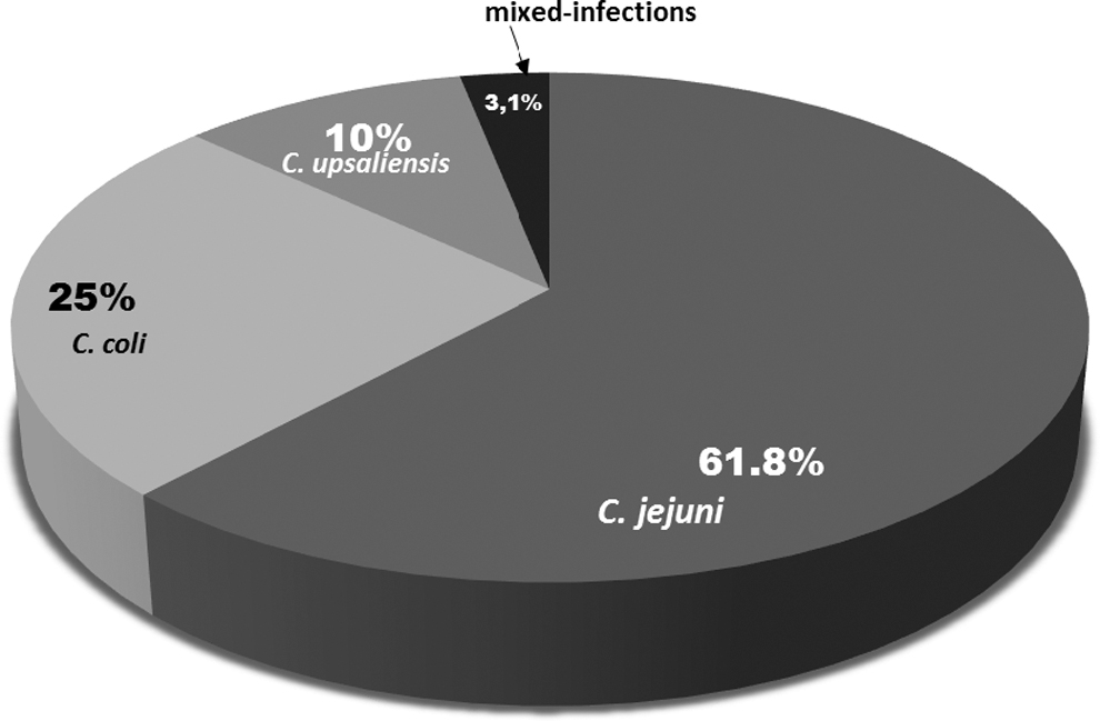
Distribution of different Campylobacter spp. in cattle.
Distribution of Campylobacter spp. in cattle and calves on different farms
Of the 435 adult cows, 33.8% (147/435) harbored Campylobacter spp. Among the 147 Campylobacter spp.-positive animals, 61.2% (90/147) carried C. jejuni, 27.2% (40/147) C. coli, and 9.5% (14/147) C. upsaliensis. Of the 102 calves tested, 10.8% (11/102) carried Campylobacter spp., including 81.8% (9/11) which had C. jejuni and 18.1% (2/11) which carried C. upsaliensis. C. coli was not detected in calves. The overall prevalence of Campylobacter spp. on farm A was 32.3% (21/65). On farms B and C, Campylobacter spp. was detected in 10.8% (11/102) of calves and 53.9% (41/76) of cattle. On farms D and E, 53.9% (41/76) and 19.5% (22/113) of cattle were contaminated with Campylobacter spp., respectively. The distribution of different species of Campylobacter by farm is depicted in Table 2.
Prevalence of Campylobacter spp. in Cattle on Cow–Calf Operations in Gauteng and North West Provinces
Antimicrobial resistance profiles of Campylobacter isolates
Broth microdilution was carried out to determine the antimicrobial resistance profiles of 86 confirmed Campylobacter isolates including 46 C. jejuni subsp. jejuni, 24 C. coli, and 16 C. upsaliensis. Overall, 52.3% (45/86) of the tested Campylobacter spp. isolates were resistant to one or more antimicrobials (Table 3). Among the 45 antimicrobial resistant isolates, 46.7% (21/45) were C. jejuni, 35.6% (16/45) as C. coli, and 17.8% (8/45) as C. upsaliensis.
Antimicrobial Resistance in Campylobacter jejuni, Campylobacter coli, and Campylobacter upsaliensis
Of the 46 C. jejuni isolates, 32.6% (15/46) were resistant to clindamycin, 19.5% (9/46) to nalidixic acid, 17.3% (8/46) to tetracycline, and 15.2% (7/46) to erythromycin. Three C. jejuni isolates (6.5%) were resistant to azithromycin and ciprofloxacin, whereas two C. jejuni isolates (4.3%) showed resistance to telithromycin. Two C. jejuni isolates were each resistant to gentamicin and florfenicol, respectively (Table 3). Among the 24 C. coli antimicrobial resistant isolates, which were resistant to one or more antimicrobial classes, 50% (12/24) were resistant to clindamycin, 25% (6/24) to nalidixic acid and tetracycline, 20.8% (5/24) to erythromycin, and 12.5% (3/24) to azithromycin and gentamicin. Resistance to ciprofloxacin, florfenicol, and telithromycin was low among the C. coli isolates (two isolates for each antimicrobial) (Table 3). Among the 16 antimicrobial resistant C. upsaliensis isolates tested, 25% (4/16) were resistant to clindamycin, 18.7% (3/16) to erythromycin, and 12.5% (2/16) to tetracycline and nalidixic acid. Lower resistance levels were observed against azithromycin and telithromycin (one isolate per antimicrobial). None of the C. upsaliensis isolates was resistant to ciprofloxacin, gentamicin, and florfenicol (Table 3).
MDR (resistance to two or more antimicrobials classes) was recorded in 32.5% (28/86) of isolates and was more frequent among C. jejuni, which showed multiresistance to erythromycin, tetracycline, nalidixic acid, and clindamycin mainly. Of the 46 C. jejuni isolates tested, 36.9% (17/46) were multiresistant (resistant to 2 or more antimicrobials classes): 19.5% (9/46) were resistant to 3 antimicrobials, 13.0% (6/46) were resistant to 4 antimicrobials. One C. jejuni isolate, 2.1% (1/46) was resistant to five antimicrobials and another isolate showed resistance to six antimicrobials including azithromycin, erythromycin, tetracycline, nalidixic acid, telithromycin, and clindamycin. Among C. coli isolates, 33.3% (8/24) were multiresistant, 12.5% (5/24) were resistant to two antimicrobials, one was resistant to four antimicrobials, and two isolates (8.3%) were resistant to all nine antimicrobials tested. C. coli isolates (50%; 4/8) were mainly multiresistant against clindamycin+tetracycline+nalidixic acid. Among C. upsaliensis isolates, only three isolates (3/16; 18.7%) displayed MDR to three antimicrobial agents each.
Discussion
Campylobacter spp. occurrence rates in healthy cattle are variable and can range from 5.3% to 78.5% in different countries (Rahimi and Tajbakhsh, 2008; Ramonaitė et al., 2013; Wieczorek et al., 2013; Smith et al., 2018). Overall, the prevalence of Campylobacter spp. in the beef fecal samples tested in this study was 29.7%, consistent with rates that have been reported in beef cattle in Malaysia (33%) (Premarathne et al., 2017), Chile (35.9%) (Fernández and Hitschfeld, 2009), and Finland (31.1%) (Hakkinen et al., 2007). However, much lower rates of Campylobacter spp. have been reported in Ghana (13.2%) (Karikari et al., 2017), Cambodia (5.3%) (Osbjer et al., 2016), and Iran (5.3%) (Rahimi and Tajbakhsh, 2008).
C. jejuni was the most frequent (62.6%) species in beef cattle, followed by C. coli (25.3%) and C. upsaliensis (10.1%). The predominance of C. jejuni over C. coli and C. upsaliensis in cattle is in agreement with previous reports from South Africa (Uaboi-Egbenni et al., 2012), United States (Cha et al., 2017), Canada (Webb et al., 2018), France (Thépault et al., 2018), and Finland (Hakkinen, 2007). However, some reports from the United States (Sanad et al., 2012; Smith et al., 2018) and Ghana (Karikari et al., 2017) showed that C. coli was the most frequent species in cattle samples.
C. upsaliensis was detected in 10.1% of beef cattle. The occurrence of C. upsaliensis in 10.1% of beef cattle was of particular interest, as this emerging and clinically important Campylobacter spp. is not commonly detected in cattle but mainly found in dogs (Acke, 2018). Similar studies that have searched for C. upsaliensis in Ghana and Lithuania did not detect C. upsaliensis in cattle (Ramonaitė et al., 2013; Karikari et al., 2017). In addition to mild gastroenteritis, C. upsaliensis has been incriminated in bloody diarrhea and extraintestinal infections including bacteremia in debilitated and immunocompromised patients, spontaneous abortion, hemolytic uremic syndrome, and Guillain–Barre syndrome in humans (Bourke et al., 1998; Couturier et al., 2012). The occurrence of C. upsaliensis in the cattle may point toward the presence of a dog reservoir, which may be acting as a source of Campylobacter spp. for cattle on the farms, which were surveyed. However, further investigations will be needed to evaluate to what extent cattle constitute a reservoir of less common but clinically relevant Campylobacter spp. such as C. upsaliensis.
The prevalence of Campylobacter spp. in calves was low (6.9%), in contrast to studies that have reported higher rates of Campylobacter spp. in calves in France (99.4%) and Lithuania (86.5%) (Ramonaitė et al., 2013; Thépault et al., 2018). The low rate of Campylobacter spp. in calves in this study is in agreement with similar reports from Austria (14.9%) and Algeria (14.0%) (Klein et al., 2013; Guessoum et al., 2016). However, it should be noted that calves were tested on one farm only, and the number of samples tested was not representative to conclusively explain differences between the proportions of Campylobacter spp. found in calves in this study and the aforementioned reports from other countries. The expectation is that calves should have higher Campylobacter spp. rates, as they are more susceptible to Campylobacter spp. colonization because of a weak immune system (Klein et al., 2013). However, the low prevalence of Campylobacter spp. in calves in this study shows that Campylobacter spp. carriage levels in calves may not only be dependent on a weak immune system but also on other factors that still remain to be identified.
Antimicrobial resistance profiling revealed that 52.3% of Campylobacter isolates were resistant to one or more antimicrobial agents. Low rates of antimicrobial resistance were detected in this study in comparison to Campylobacter spp. resistance levels that have been reported in beef cattle in the United States (83.7%), France (64.6%), and Poland (65.4%) (Chatre et al., 2010; Wieczorek et al., 2013; Cha et al., 2017). The low antimicrobial resistance rates observed in this study may be ascribed to the fact that the cattle herds which were surveyed were fed on grazing pasture all year round with no exposure to antimicrobials that exert selective pressure on bacteria and facilitate the survival of resistant Campylobacter spp.
Among the nine antimicrobials tested, the highest resistance rate was recorded for clindamycin (36%). Resistance to clindamycin was mostly observed among C. coli (50%), followed by C. jejuni (32.6%) and C. upsaliensis (25%). High resistance to clindamycin was in contrast to reports from Canada and the United States, which have documented much lower proportions of Campylobacter spp. isolates that were resistant to clindamycin (Cha et al., 2017; Tang et al., 2017; Webb et al., 2018).
Lower resistance levels were recorded for nalidixic acid (19.7%), tetracycline (18.6%), and erythromycin (17.4%) in C. coli isolates mainly, consistent with previous reports from the United States, Malaysia, Iran, and Japan (Rahimi and Tajbakhsh, 2008; Haruna et al., 2013; Cha et al., 2017; Premarathne et al., 2017; Tang et al., 2017). However, much higher resistance rates to nalidixic acid (70.4%), tetracycline (51.4%), and erythromycin (up to 97%) have been previously recorded in France, Poland, and Ghana, respectively, mainly among C. coli isolates (Chatre et al., 2010; Wieczorek et al., 2013; Karikari et al., 2017). Resistance to nalidixic acid, tetracycline, and erythromycin is intriguing as these antimicrobials are supposed to be minimally used on cow–calf operations and for therapeutic purposes only.
Very low resistance rates were recorded for azithromycin (8.1%), florfenicol (3.4%), gentamicin (4.8%), and telithromycin and ciprofloxacin (5.8%). Resistance to ciprofloxacin in this study was comparable to that reported in beef cattle isolates in Canada (Webb et al., 2018). However, higher Campylobacter resistance rates to ciprofloxacin have been reported previously in C. jejuni (33.3%) and C. coli (56.3%), which were recovered from dairy cattle in South Africa (Uaboi-Egbenni, 2012).
Resistance against erythromycin and ciprofloxacin is of clinical significance because these antimicrobials are used to treat Campylobacter-associated gastroenteritis. A number of surveys have shown that for C. jejuni and C. coli, there is generally agreement between nalidixic acid and ciprofloxacin, and similarly between erythromycin and azithromycin resistance rates (Englen et al., 2007; Chatre et al., 2010; Rahimi et al., 2013; Wieczorek et al., 2013). However, in this study, no agreement was observed between nalidixic acid and ciprofloxacin, or erythromycin and azithromycin antimicrobial resistance rates, consistent with a few of studies, which have reported similar results (Gormley et al., 2010; Kashoma et al., 2015, 2016). Furthermore, Webb et al. (2018) observed that upon arrival at cow feeding operations, there was no agreement between ciprofloxacin and nalidixic acid resistance rates among C. jejuni cattle isolates. However, ciprofloxacin and nalidixic acid resistance rates become similar or marginally different only as animals are progressively fed rations supplemented with antimicrobial growth promoters. The absence of selective pressure due to lack of antimicrobial promoters in the diet of the cattle population studied may have precluded the parallel development of resistance against ciprofloxacin and nalidixic acid and perhaps erythromycin and azithromycin among the Campylobacter spp. isolates tested.
This study revealed that 35.2% (28/86) of cattle Campylobacter isolates were multiresistant. Similar proportions of multiresistant isolates were previously observed among Campylobacter spp. isolates, which were recovered from cattle fecal samples (Noormohamed and Fakhr, 2014; Cha et al., 2017; Premarathne et al., 2017; Tang et al., 2017). MDR to nalidixic acid+clindamycin (17.8%) and tetracycline+clindamycin (14.3%) was the most common resistance profile. Multidrug was mostly (60.7%) observed among C. jejuni isolates in contrast to other studies that have recorded much higher levels in C. coli isolates from cattle (Wieczorek et al., 2013; Okunlade et al., 2015).
Conclusions
To the best of our knowledge, this is the first study that has investigated the occurrence of Campylobacter spp. of public health significance in beef cattle in South Africa. The cattle surveyed represent an important reservoir and a potential source of clinically relevant and antimicrobial resistant Campylobacter spp. for humans. Data from this study will be useful for understanding the epidemiology of Campylobacter and formulating policies aimed at mitigating human zoonotic foodborne campylobacteriosis.
Footnotes
Disclosure Statement
No competing financial interests exist.
Funding Information
Funding from the National Research Foundation (NRF) of South Africa-Research Technology Fund (RTF14012762427) and the Gauteng Department of Agriculture and Rural Development (GDARD Grants-2013-2015) is gratefully acknowledged.