
Abstract
Staphylococcus aureus is a Gram-positive bacterium that causes intramammary infections and bulk tank milk (BTM) contamination in dairy operations around the world in spite of on-farm application of preventive measures. The study was conducted on a 30-cow dairy farm in the Ñuble Region of Chile. For BTM culture and somatic cell count (SCC) analysis, three consecutive BTM samples were collected. Samples for bacterial culture (n = 16) were collected from macroscopic adherence on previously washed, sanitized, and dry milking equipment surfaces in direct contact with milk during milking or cooling. A total of 48 S. aureus isolates from BTM, milking equipment, and cows' quarters with intramammary infections were analyzed by pulsed-field gel electrophoresis (PFGE). Selected milking equipment pieces were removed for biofilm visualization using scanning electron microscopy (SEM). S. aureus was isolated from all three BTM samples; the average SCC for the three BTM samples was 1,429,333 cells/mL. Fourteen of the 16 samples of milking equipment (87.5%) were culture positive for S. aureus. Biofilms were visualized by SEM in all four removed milking equipment pieces. Microorganisms observed by SEM in those biofilms were mainly coccus-shaped bacteria, and microbiological culture of these biofilms yielded viable S. aureus isolates in all samples. All pulsotypes observed among S. aureus isolates from BTM were indistinguishable from those in milking equipment surfaces. All PFGE pulsotypes observed among S. aureus isolates from biofilms on rubber liners were indistinguishable from isolates from intramammary infections in cows. Our findings suggest that milking equipment films may act as source of S. aureus contamination for BTM and cows during milking, thus compromising the microbiological quality of milk used for manufacturing dairy products.
Introduction
S taphylococcus aureus is a Gram-positive bacterium that causes intramammary infections (IMIs) in dairy herds around the world. S. aureus shedding in milk of infected cows may contribute to increased bacterial count in herd's bulk tank milk (BTM). High bacterial counts in raw milk may not only be a reason for reduction in the payment for BTM to producers, but they also have been linked to defects in manufactured dairy products and reduced conversion efficiency (Barbano et al., 2006; Murphy et al., 2016). From the public health perspective, consumption of raw milk contaminated with S. aureus may pose a threat to human health, especially if enterotoxin-producing or methicillin-resistant S. aureus strains are present in this milk (Korpysa-Dzirba and Osek, 2019; Abdel-Hameid et al., 2019; Omwenga et al., 2019), in milk commercialized directly to consumers (Ding et al., 2016), or in dairy products such as cheese (Valík et al., 2018; Samelis et al., 2019).
Milk from S. aureus-infected cows may also be a continuous on-farm source of S. aureus contamination for the milking machine and milking equipment surfaces. If standard cleaning and sanitation protocols of milking equipment are not properly conducted, milk residues may not be completely removed after milking. This situation can create ideal conditions for bacterial proliferation between milkings (Murphy and Boor, 2000) and may trigger the subsequent colonization of surfaces and eventual formation of biofilms (Latorre et al., 2010; Latorre and Munoz, 2015). The presence of S. aureus strains in dairies and their ability to form biofilms have been previously reported (Lee et al., 2014; Thiran et al., 2018; Unlu et al., 2018), and S. aureus can be part of the various microbial communities present in biofilms formed on milking equipment surfaces (Latorre et al., 2016). These biofilms may create an additional source of bacteria for BTM as milk gets in contact with milking equipment, milk lines, and bulk tank surfaces during milking and cooling, respectively, as well as a result of dispersion of cells from the biofilms (Alonso and Kabuki, 2019). Furthermore, if S. aureus-containing biofilms become established on pieces of milking equipment that are in close contact with teats and teat ends during milking, these parts of the equipment may act as a source for S. aureus IMI in cows.
This study describes sources of S. aureus contamination of BTM on a commercial dairy farm in Chile and the potential role of S. aureus-containing biofilms in the epidemiology of IMIs.
Materials and Methods
Study farm
The study was conducted in June 2015 on a 30-cow dairy farm in the Ñuble Region of Chile. The study farm had 21 lactating cows at the moment of the farm visits. The cows were milked twice a day and had an average daily milk production of 10.5 L/d. Milking was done using three milking units that diverted milk, using long milk tubes, directly to three milk cans. Once each milk can was full, the milk was immediately transferred to a milk tank for cooling and storage. The milk was picked up every other day in a milk truck to be transported to a dairy processor where it was pasteurized for further processing. Routine milking equipment cleaning consisted in a rinse after milking using cold water (∼16°C), manual wash using a solution of alkaline detergent prepared in lukewarm water (∼35°C), and a final soak in an acid solution (∼31°C). Rubber liners were replaced every 6–7 months of use (equivalent to 2520–2940 milkings), while other pieces of the milking equipment were only replaced when broken.
Microbiological analysis
Three consecutive BTM samples were aseptically collected following National Mastitis Council guidelines (1999). Each BTM sample corresponded to milk of a single milking. For evaluating the somatic cell count (SCC), BTM samples were aseptically collected in 50-mL vials with bronopol preservative, and for bacteriological culture purposes, sterile 100-mL vials with no preservative were used (Jayarao et al., 2004). All samples were immediately transported to the laboratory in coolers with ice packs and stored at 4°C until analysis within 24 h of collection. BTM samples were shipped to an ISO-certified milk laboratory (INIA Carillanca, Temuco, Chile) for SCC analysis using automated flow cytometry (Foss®).
For S. aureus testing, milk samples were cultured in Baird–Parker agar (Oxoid, Basingstoke, United Kingdom), following the protocol described by Jayarao et al. (2004). Putative S. aureus colonies were confirmed by PCR (Riffon et al., 2001).
Milking equipment surfaces in contact with milk during milking or cooling were inspected to assess the presence of macroscopic adherence or films (i.e., visible films or adherence attached to surfaces, persisting after cleaning and sanitation procedures). All surface inspections were conducted using an ∼10.000 lx flashlight and after the regular cleaning and sanitizing protocol of milking equipment was performed (Latorre et al., 2010) and the surfaces were dry (Elmoslemany et al., 2009). Swabs or sponges were collected as described by Latorre et al. (2009) from surfaces having macroscopic adherences, including milking units (i.e., liners, collector, collector valves, and short milk tubes), long milk tubes, milk cans, bulk tank outlet, inner surface of the tank, and agitator blades. Swab samples from surfaces selected for removal for electron microscopy analysis (Microscopy assessment of milking equipment surfaces section) were collected on a second farm visit, right before removal of the parts.
All surface samples were enriched with brain-heart infusion broth for 48 h at 37°C (Latorre et al., 2010). After enrichment, 10 μL was streaked onto CHROMagar™ S. aureus (CHROMagar, France) and incubated for up to 48 h at 37°C. Putative S. aureus colonies (i.e., pink to mauve colonies) were identified and transferred to Baird–Parker agar for purification and subsequent confirmation by PCR according to Riffon et al. (2001). Per sample, depending on the culture density and number of isolated colonies available on each CHROMagar S. aureus plate or Baird–Parker agar, up to six S. aureus colonies were purified and then stored at −80°C for further PCR and pulsed-field gel electrophoresis (PFGE) analysis.
No individual cow milk samples were collected in this study. Nevertheless, S. aureus isolates from individual quarter milk samples were available from two other studies conducted on this farm in August 2015 and September 2017. These isolates were also included for comparison purposes in the PFGE analysis of this study.
Microscopy assessment of milking equipment surfaces
Milking equipment pieces whose surfaces had visible macroscopic adherences and could be detached from the equipment were removed and replaced. Biofilm visualization on the surfaces was conducted using scanning electron microscopy (SEM).
The pieces of milking equipment selected for removal and subsequent microscopy analysis were aseptically transported and cut as described by Latorre et al. (2010) using sterile scissors. Preparation of samples for SEM was done according Latorre et al. (2010), with minor modifications. After staining, samples were put in 2.5% glutaraldehyde solution prepared in 1 × PBS (pH 7.0–7.5). After fixation, samples were washed three times with 1 × PBS at room temperature and then gradually dehydrated using a graded series of 30%, 50%, 70%, 80%, 90%, and 100% ethanol (15 min each). Dehydration of samples was completed with an additional soak in 100% ethanol.
The samples were then critical point dried, attached to a specimen support, and coated with gold. Biofilm formation was confirmed when both bacteria attached to a surface and an exopolymeric matrix were visualized by SEM.
PFGE typing
Forty-eight S. aureus isolates were analyzed by PFGE. Thirty-seven of these isolates were randomly selected among all isolates from BTM and surface S. aureus-positive samples (n = 9 and n = 28, respectively) (Table 1). Up to three isolates per sample were analyzed by PFGE, thus covering at least 50% of the available isolates in the case of more populated plates. Complementarily, 11 S. aureus isolates obtained from milk of quarters with S. aureus IMI were also included in the analysis for comparison purposes, as described earlier (individual quarter samples from other studies; one isolate per sample).
Summary of the Type and Number of Samples Analyzed on the Study Farm for Detection of Staphylococcus aureus, Number of S. aureus Isolates Recovered and Analyzed by Pulsed-Field Gel Electrophoresis, and Presence of Biofilms in Removed Pieces of Milking Equipment
Pulsotypes observed through visual inspection by two independent observers.
(+) Biofilm = visualization of bacteria attached to a surface and the exopolymeric matrix.
N/A, not assessed; PFGE, pulsed-field gel electrophoresis.
Molecular typing was conducted according the CDC PulseNet protocol (
For analyses of band patterns from SmaI-digested DNA, visual inspection by two independent observers was done. The criteria described by Tenover et al. (1995) were used to characterize genetic relatedness (i.e., indistinguishable, closely related, possibly related, or unrelated strains). To build a dendrogram for clustering of fingerprint data, automated cluster analysis using BioNumerics 7.6 was done for the 48 S. aureus isolates by using the Dice coefficient (tolerance of 1.5%) and unweighted-pair group method with arithmetic averages, with a similarity score value of 100% (Latorre et al., 2009, 2011).
Results
The SCCs for the three BTM samples were 1,756,000, 1,312,000, and 1,220,000 cells/mL, respectively, and S. aureus was isolated from all three BTM samples.
Fourteen of 16 milking equipment surface samples (87.5%) were culture positive for S. aureus (Table 1). Biofilms were visualized in all four surface types analyzed by SEM (inner surface of liners, internal side of one collector valve, short milk tubes, and one of the long milk hoses) (Figs. 1 and 2). The microorganisms observed by SEM in the biofilms were mainly coccus-shaped bacteria (Figs. 1 and 2) and, reciprocally, viable S. aureus strains were isolated from cultures of biofilms from all these samples.

Scanning electron microscopy image of a biofilm on the surface of a rubber liner.
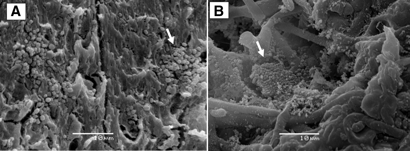
Scanning electron microscopy image of a biofilm on the surface of the long milk hose
Visual inspection of PFGE of SmaI-digested DNA of 48 S. aureus isolates showed 8 pulsotypes (pulsotypes 1–1.7), all of which were identified as closely related S. aureus strains (Fig. 3). Pulsotype 1.4 was the most frequently observed among the isolates that were analyzed (n = 18; 37.5% of isolates; Fig. 3). This pulsotype 1.4 was present in all S. aureus-positive sample sources, except for the milk collector valve (Table 1). In addition, pulsotype 1.4 was also observed on three IMI isolates (two milk samples collected in 2015 and one milk sample collected in 2017). S. aureus pulsotype 1 was the second type that was more frequently observed (n = 12; 25% of isolates), being found in BTM, rubber liners, hose connection of a milk can, and individual quarter samples from three different cows (in one IMI milk sample collected in 2015 and two IMI milk samples collected in 2017) (Fig. 3). The pulsotypes observed among S. aureus isolates from BTM samples were indistinguishable from those observed in visible films on both milking equipment and milk tank surfaces (Fig. 3). Furthermore, pulsotypes observed among S. aureus isolates from biofilms on rubber liners (i.e., pulsotypes 1, 1.1, and 1.4) were indistinguishable from those observed in IMI milk from individual cows, including milk samples collected in 2017. The only exception to these findings of indistinguishable biofilm-IMI milk pulsotypes was S. aureus pulsotype 1.3, which was only observed in milk from individual cows, but not in rubber liners, although it was present in BTM and milking equipment samples (Fig. 3).
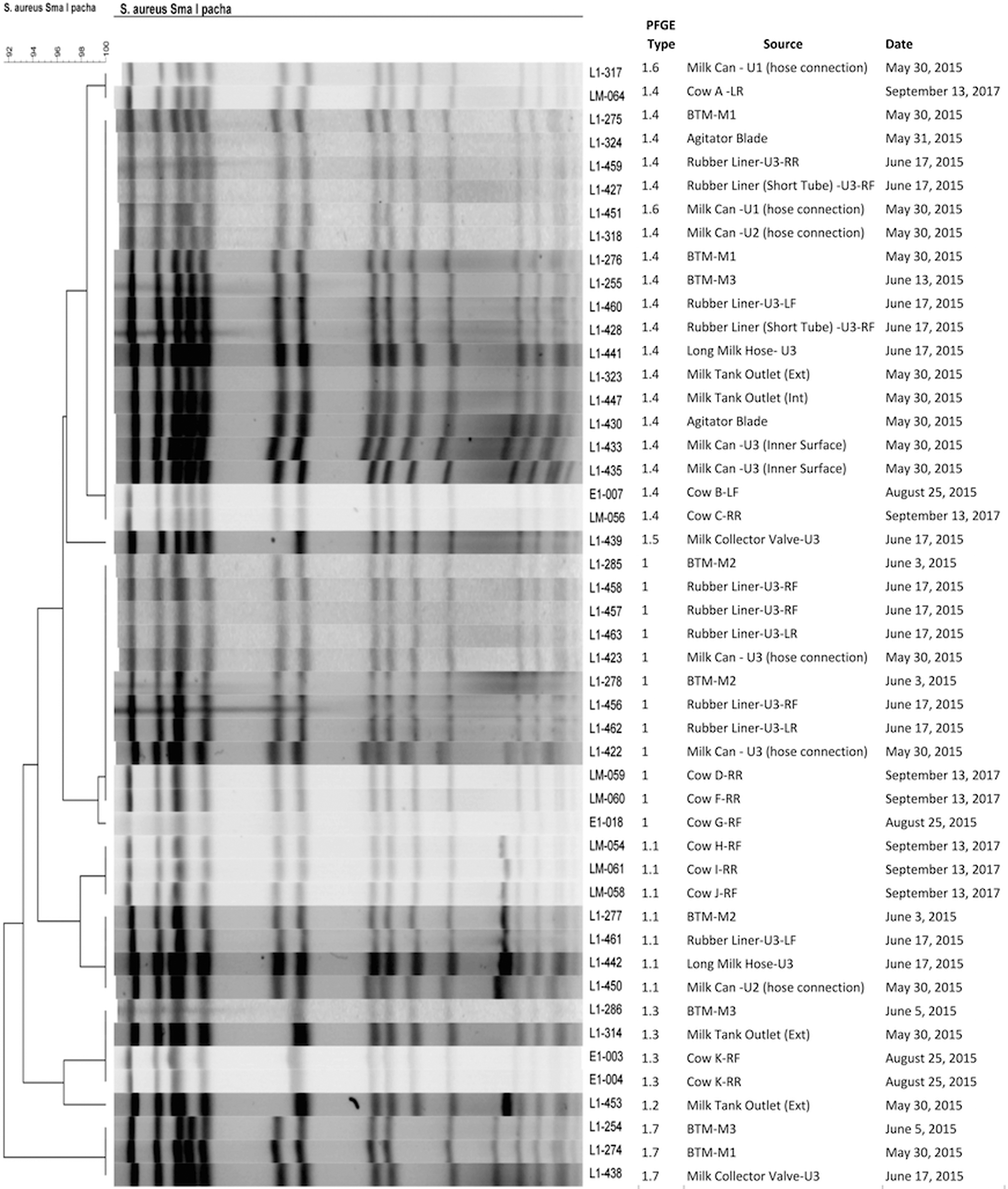
Dendrogram of Staphylococcus aureus isolates obtained from BTM, milking equipment, and individual cows on the study farm. The dendrogram is based on the analysis of SmaI-digested DNA profiles using a cutoff of 100%. PFGE types assigned by visual inspection by two independent observers of the SmaI-digested DNA profiles are shown at the right of the dendrogram after the identification number of the isolate, followed by the source of the sample, and sampling date. U = milking unit position in the parlor; LF, LR, RF, and RR = left front, left rear, right front, and right rear udder quarters, respectively; BTM = bulk tank milk; M1, M2, or M3 = consecutive milk sample 1, 2, or 3, respectively; Ext = external area of milk tank outlet (truck hose connection); Int = internal area of milk tank outlet (milk tank portion); PFGE, pulsed-field gel electrophoresis.
Discussion
Management issues observed in the study farm, such as high SCC and low daily milk production, constitute part of the real-life diversity of dairy production systems not only in Latin America but also around the globe (Edmondson, 2017; Vissio et al., 2018).
We report the presence of biofilms in milking equipment on a poorly managed dairy farm, but the role of milking equipment biofilms as a source of BTM contamination has also been documented in a well-managed dairy farm in the United States (Latorre et al., 2009, 2010), and milking equipment as an alternative source of pathogens for cows and/or BTM has been reported for other pathogens as well (Munoz et al., 2007; Giacometti et al., 2015). Therefore, although this study was conducted on a single dairy farm under a rather suboptimal productive management system and with poor milk quality and production parameters, the findings observed here could also be related to other parts of the world. Furthermore, exploring the role of reservoirs (other than cows) inside dairy premises can contribute to explain why eradication of S. aureus in dairies is difficult and commonly unsuccessful even when S. aureus-positive cows are being culled or sold at a high rate. Moreover, even when the role of dairy cows in the persistence of S. aureus on dairy farms has been more frequently documented or studied (Rainard et al., 2017; Leuenberger et al., 2019), the milking equipment as a plausible source of S. aureus contamination of milk intended for human consumption should not be ruled out.
In this study, almost all samples of visible films on milking equipment were S. aureus positive. All samples were collected from the milking equipment once the equipment was dry after cleaning and sanitation protocols were performed. No milk or milk residues from the previous milking were present at the moment of sampling; therefore, the presence of S. aureus in milking equipment due to residual milk from subclinically infected cows is unlikely. Biofilms that persisted after cleaning and sanitization protocols were extensively present in all the samples removed for microscopy analysis. The presence of biofilms in milking equipment may contribute to contamination of BTM due to dislodging of areas of the biofilm on surfaces during milking (Donlan, 2002; Hall-Stoodley et al., 2004). In addition, multiplication of bacteria (including S. aureus) within the biofilm between milkings may have also contributed to BTM contamination with these bacteria.
Even though we could not confirm by SEM that the cocci visualized on the biofilm were indeed S. aureus strains, this microorganism was isolated in the form of viable bacteria from all the biofilms on surfaces, and the size of the cocci observed by SEM matched with the size described for Staphylococci (0.5–1.5 μm. Seo and Bohach, 2013). Furthermore, in a separate study done using isolates from this farm, 92% and 100% of S. aureus isolates recovered from BTM and equipment surfaces were icaA and icaD positive, respectively (unpublished data), which play an important role in biofilm formation. The ability of S. aureus to form biofilms has been previously reported in studies conducted on dairy isolates (Fox et al., 2005; Thiran et al., 2018) as well as in human clinical settings (Fernandes and Dias, 2013; Ledwoch et al., 2018). The presence of S. aureus-containing biofilms in milking equipment could not only negatively affect the microbiological quality of BTM but also serve as a vehicle and reservoir for transmission of S. aureus to cow's mammary glands.
As not all S. aureus isolates recovered from milking equipment were analyzed by PFGE and not many IMI isolates from cows were available, the complete genomic heterogeneity of S. aureus from these sources could not be assessed (Döpfer et al., 2008). However, it is noteworthy that the same S. aureus pulsotypes were observed among isolates obtained from rubber liners and individual cows, even for isolates in milk samples that were collected 2 years after this study. This finding might be explained by the milking equipment serving as an alternative source of S. aureus to susceptible uninfected cows during milking, other than the shedding of infected quarters.
The close contact of teat ends with contaminated milking surfaces provides the chance for entrance of pathogens into the mammary glands (Zadoks et al., 2002; Munoz et al., 2007). The high SCC and the presence of S. aureus in all three BTM samples collected suggest the presence of S. aureus intramammary infection in cows (Murphy and Boor, 2000; Jayarao et al., 2004), which may have also contributed to the presence of S. aureus and increased the SCC in BTM.
On this farm, indistinguishable S. aureus pulsotypes and closely related pulsotypes were observed in films on the milking equipment pieces throughout the direction of the milking flow. In addition, all pulsotypes (except one) that were observed on milking equipment surfaces overlapped and were also observed in BTM. This finding supports the fact that the presence of S. aureus in BTM had an important contribution of S. aureus from the surface biofilms of the milking equipment.
Although in our study it is not possible to determine if subclinical IMIs of cows were caused by exposure to contaminated milking surfaces or if the S. aureus-containing biofilm in milking equipment was caused by S. aureus shedding from infected cows, the potential role of milking equipment as a reservoir of mastitis pathogens should not be disregarded. In addition, the role of milking equipment as a potentially major contributor to BTM bacterial counts should be considered during troubleshooting on specific, high, colony-forming unit counts.
Good microbiological parameters of raw milk are crucial for manufacturing high-quality dairy products. Human exposure to S. aureus due to unpasteurized milk or dairy products contaminated with S. aureus could pose a threat to public health, especially if enterotoxin-producing or antimicrobial-resistant strains are present (Abdel-Hameid et al., 2019; Omwenga et al., 2019). To protect the health of consumers, assurance quality systems in the dairy industry are crucial, along with prevention of milk contamination starting in the early stages of the milk production–consumption chain, that is, the farm and farm's animals. The latter may be achieved by implementation of good management practices on farms, including strict protocols for cleaning, sanitation, and maintenance of milking equipment.
Conclusions
This study demonstrates that biofilms on milking equipment surfaces can play a role as a source of S. aureus for both BTM contamination and dairy cows' mammary exposure. S. aureus biofilm reservoirs in milking equipment may play a role in the understanding of the epidemiology of S. aureus IMIs in many herds around the world.
Footnotes
Acknowledgments
The authors thank the participating producer who kindly allowed them to conduct this study on his dairy farm. The authors acknowledge the valuable technical assistance of Mr. Luis Aranguiz-Pimentel during farm sampling of milking equipment pieces for SEM analysis.
Disclosure Statement
No competing financial interests exist.
Funding Information
Financial support was provided by the Chilean Commission for Scientific and Technological Research, FONDECYT Project No. 11130343.