
Abstract
Microorganisms detected on dairy factory surfaces after disinfection can cause product contamination, leading to economic losses and health hazards for consumers. In this study, the presence of Staphylococcus spp. and Coliform in a total of 450 samples taken from food-contact and non-contact surfaces (stainless steel, plastic, cloth, and tiles surfaces), raw milk and final product (white cheese, kashar cheese, butter, yogurt, and cream) samples in five dairy factories was investigated by conventional techniques. The isolates obtained were identified by matrix-assisted laser desorption/ionization-time of flight mass spectrometry. In this study, a total of 54 Staphylococci (16.7% S. aureus and 81.5% coagulase-negative Staphylococci [CNS]) and 44 coliform isolates were identified at the species level. The most common CNS isolated by samples was S. epidermidis followed by S. saprophyticus, S. capitis, S. succinus S. carnosus. S. xylosus, S. sciuri, S. equorum, S. warneri, and S. hominis. Eighteen of the coliform isolates (41%) were identified as E. coli; 13 (29.5%) as E. cloacae; 3 (6.9%) as E. kobei, C. freundii, and K. oxytoca; 2 (4.5%) as K. pneumoniae; 1 (2.3%) as E. ludwigii and C. farmeri. The contamination rate of non-food contact surfaces (71.3%) was found to be higher than food contact surfaces (10.4%), and contaminated surfaces were found to be effective in product contamination. Study results show that some bacterial species obtained from raw milk, surfaces, and final products are factory specific and surface-associated bacteria are prominent in the product microbial profile.
Introduction
In industrial milk processing plants, the high nutritional content of milk and dairy products supports microbial growth, and milk processing lines offer a wide range of microenvironments in which pathogenic and saprophytic microorganisms can multiply. Accordingly, problems arise regarding maintaining sanitation conditions during production, processing, storage, distribution, and marketing (Quijada et al., 2018; Sharma and Anand, 2002).
It is stated that microorganisms in production lines in food processing facilities may pose a risk to food quality and safety by causing cross contamination. In order to eliminate this risk, microbiological threats should be routinely checked by sampling the production surfaces (Ismaïl et al., 2013; Stellato et al., 2015; Xu et al., 2023). In surface contamination studies conducted in food businesses, sampling areas are generally classified as food-contact and non-food-contact surfaces. According to the U.S. FDA (2018), a food contact surface is defined as a surface of equipment or apparatus that food normally comes into contact with, or a surface of equipment or apparatus from which food may flow, drip, or splash. Non-food contact surfaces are surfaces such as walls, floors, grates, and drains near food processing areas, and equipment surfaces such as forklifts and wheelbarrows used in food processing areas. Locker rooms, cafeterias, and break rooms outside the food processing area, as well as outdoor floors and sewers, are classified as remote non-food contact surfaces (Jones et al., 2020; US. FDA, 2018).
Studies analyzing the presence of microorganisms before and after sanitation procedures applied in food processing processes have revealed that many organisms persist after sanitation procedures (Jones et al., 2020; Schön et al., 2016; Xu et al., 2023). Microorganisms remaining on surfaces after sanitation stand out as an important factor in final product contamination (Querido et al., 2019). In this study, it was aimed to determine priorities for eliminating microorganisms remaining on surfaces by monitoring processing lines in five different dairy factories. For this purpose: (i) the aim was to detect Staphylococci and coliform contamination in samples taken after sanitation from food-contact and non-food-contact surfaces, as well as in raw material (raw milk) and final product (white cheese, kashar cheese, yogurt, butter, and cream) samples, and (ii) to identify contamination sources of species isolated from samples at each factory.
Material and Methods
Sampling procedures
During the sampling period, five dairy factories in Niğde and Konya provinces, producing white cheese, kashar cheese, yogurt, butter, and cream, were visited twice between August 2019 and December 2020. A total of 450 samples (20 raw milk, 100 dairy products, and 330 surface swab samples) were collected after the routine sanitation procedure and transported to the laboratory in a cool box.
Swab samples were taken from equipments, containers, and pipes surfaces, and tools by ISO 18593 (2018). The surfaces from which swab samples will be taken are classified as food contact surfaces and non-food contact surfaces located near food contact surfaces. (ISO 18593, 2018; US. FDA, 2018). Accordingly, all pipes carrying raw materials and products to processing areas, inner and outer surfaces of tanks, plastic molds, knives, scoops, presses, and cloths, gaskets used in processing were classified as food contact surfaces (n = 250). Wastewater pipes, channel drainage grates, and wall tiles were classified as surfaces that do non-food contact surfaces (n = 200) (Table 1). For sampling of raw milk and dairy products, 10 mL of milk samples and 25 g of dairy products samples were collected, homogenized in 0.1% peptone water and serial dilutions were prepared.
Types of Samples Collected from Dairy Factories
Microbiological analyses
Coliform bacteria isolation
Violet Red Bile Lactose (VRBL, Merck, 101406) Agar was used for isolation of coliforms. Medium was incubated at 30°C for 24 h. Purplish red colonies with a zone of at least 0.5 mm in diameter were evaluated as coliforms. Atypical colonies were inoculated in Brilliant Green and Lactose Broth. After 24 h of incubation at 37°C, gas formers in the Durham tube were considered coliform (ISO 4832, 2006).
Staphylococcus spp. isolation
The method of ISO 6888-1:2018 was applied in the isolation of Staphylococci using Mannitol Salt Phenol Red Agar (MSA, Merck 1.05404) and Baird-Parker Medium (BA, OxoidTM) containing Egg-Yolk Tellurit (BPA, Merck 1.03785.0001. Gray-black colonies with a halo formed after 24 h of incubation at 37°C on BPA were considered as presumptive colonies of coagulase-positive Staphylococci. Selected isolates were also inoculated on MSA to determine the mannitol fermentation. Yellow colonies with a yellow zone were mannitol positive. Gram staining, oxidase, catalase, and coagulase tests were also performed on colonies (ISO 6888-1:1999/Amd 2:2018).
Identification by matrix assisted laser desorption/Ionization-Time of flight mass spectrometry
According to the results of biochemical tests, the strains accepted as coliform and Staphyloccus were re-inoculated into VRBL, MSA, BA, and Blood Agar (Merck 110886) with the aim of reducing single colony by streak plate method. The colonies were examined by stereo microscope and a total 130 randomly selected strains with different colony morphology. Matrix-assisted laser desorption/ionization-time of flight mass spectrometry (MALDI-TOF MS) Bruker Microflex LT model FlexControl 3.0 software (Bruker Daltonics, ABD) was used for measurements and data processing. Experimentally obtained protein spectra were compared with MALDI Bruker’s Biotyper-specific database and results were obtained with numerical scores between 0 and 2510. The software recommends an acceptable score cut-off of ≥2.0 for identification at the species level, 1.70–1.99 at the genus level, and <1.69 at the unreliable genus level. Measurements were performed in linear positive mode (m/z 800–1800 Da). Measurements were carried out in the linear positive ion mode, in the mass range of 2000–20.000 Da, with the appropriate method optimized for microorganism identification in the system.
PCR amplification of some virulence and enterotoxin genes genes
In this study, the existence of classical enterotoxin genes (sea, seb, sec, sed, see) in Staphylococci and six major virulence genes (fliC, rfbE, stx1, stx2, eaeA, hlyA) in E. coli isolates were examined by multiplex PCR, following the methods described by previous studies (Karadal et al., 2024; Karadal et al., 2013).
Statistical analyses
Samples and five different factories were compared in terms of Staphylococcus spp. and coliform positivity rates using Pearson’s chi-square analysis using the Jamovi package program. Statistical significance level was determined as p < 0.05.
Results
In our study, 130 isolates, including 60 coliforms and 70 Staphylococci, were recovered from the analyzed samples using conventional methods. MALDI-TOF MS identified 98 (75.4%) of the 130 isolates at the species level, including 54 Staphylococci and 44 coliforms, while 13 (10%) were recognized at the genus level (Staphylococcus, Macrococcus, Micrococcus, Enterobacter, and Klebsiella). However, 14.6% (19/130) of the isolates were identified as non-staphylococcal and non-coliform species. MALDI-TOF MS analysis results obtained for 98 isolates are detailed in Figure 1. Briefly, of the isolates identified as Staphylococcus spp. by MALDI-TOF MS, 16.7% (9/54) were identified as coagulase-positive (S. aureus) and 81.5% (44/54) as coagulase-negative Staphylococci (CNS). The distribution of CNSs species was identified as 40.9% (18/44) S. epidermidis; 22.7% (10/44) as Staphylococcus saprophyticus; 9.1% (4/44) as Staphylococcus warneri; Staphylococcus succinus, Staphylococcus sciuri, Staphylococcus xylosus each at 6.8% (3/44) and Staphylococcus capitis, Staphylococcus hominis, Staphylococcus equorum and Staphylococcus carnosus each at 2.8% (1/44). In addition, of the 44 coliform isolates, 18 were E. coli (41%), 13 were Enterobacter cloacae (29.5%), 3 each were Enterobacter kobei, Citrobacter freundii, and Klebsiella oxytoca (6.9%), and 2 were Klebsiella pneumoniae (4.5%), 1 each were Enterobacter ludwigii and Citrobacter farmeri (2.3%) (Fig. 1).
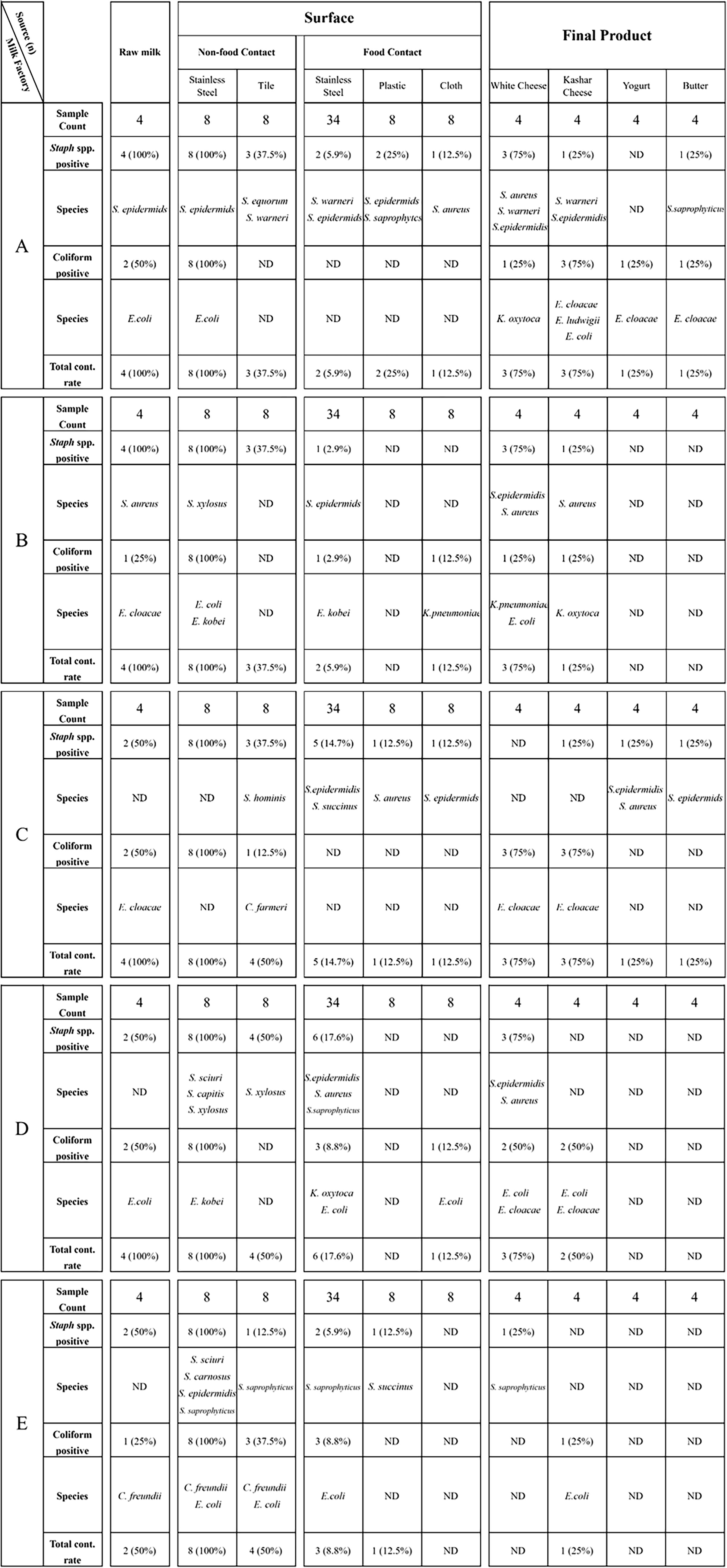
Staphylococcus and coliform contamination rates and distribution of species in raw milk, surface, and final product samples.
Among the analyzed samples, 100% (40/40) of non-food contact stainless steel wastewater pipes and drainage grates were contaminated with Staphylococci and coliforms; 42.5% (17/40) of the non-food contact tile surfaces were found to be contaminated. Contamination on these surfaces was determined as 32.5% Staphylococcus (13/40), 10% coliform bacteria (4/40), and 5% both (2/40). Moreover, 10.4% (26/250) of food contact surfaces were contaminated with these bacteria (staphylococci: 25/250, coliforms: 10/250); contamination was observed on stainless steel surfaces (18/170), plastic surfaces (4/40), and fabric surfaces (4/40) (Fig. 1).
Staphylococcal and coliform contamination was detected in 70% (14/20) and 40% (8/20) of raw milk samples, respectively; in final products (cheese, kashar cheese, yogurt, and butter), from factories the contamination rates were 16% (16/100) and 19% (19/100), respectively (Fig. 1). It was determined that there was no contamination in the cream samples. None of the E. coli and Staphylococcus isolates contained tested virulence genes.
There was a significant difference between the samples in terms of Staphylococcus spp. and coliform positivity rates, while no difference was observed between the factories where the samples were taken (p > 0.05) (Fig. 2).
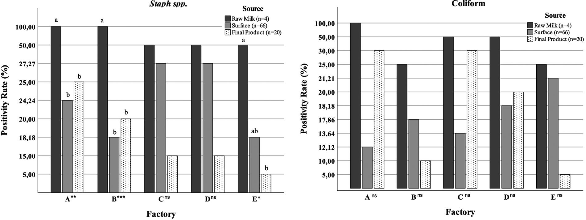
Staphylococcus spp. and coliform positive sample rate (%) according to sample sources *:p < 0,05; **:p < 0,01; ***:p < 0,001; ns: Not significant; a,b,c: Differences between sample sources in the same factory.
Discussion
In the study, Staphylococci and coliform species were isolated from raw material, food contact, and non-food contact surfaces, and final product samples were taken from five different dairy factories. When the overall contamination rate of Staphylococci and coliforms was examined, a significant difference was found between the raw milk, surface, and final product samples (Fig. 2). All raw milk samples analyzed in this study (70%) were contaminated with staphylococci and 40% with coliform species. Staphylococcal and coliform contamination in raw milk samples used in milk processing facilities has also been reported in previous studies (Gajewska and Chajęcka-Wierzchowska, 2020; Irkin, 2010; Schlegelová et al., 2010; Sharma and Anand, 2002; Temelli et al., 2006). The microbial quality of raw milk, which is the raw material of the dairy industry, is the most important factor determining the quality of dairy products. It is stated that microbial contamination in raw milk can originate from animal udder, milking equipment, personnel, air, or soil, and the microbial profile may change after transfer from the farm to the factory (Falardeau et al., 2019; Kable et al., 2016; Sharma and Anand, 2002). In this study, E. cloacae, which was found in raw milk at factory C, was also found in kashar cheese and white cheese, but could not be isolated from any of the surface samples (Fig. 1). This situation is thought to be caused by inadequate pasteurization, contamination with raw milk after pasteurization, or workers.
In the study, the total contamination rate of non-food contact surfaces (71.3%) was higher than that of food contact surfaces (10.4%), with all (100%) samples from wastewater pipes and grates and 42.5% from tile surfaces being contaminated (Fig. 1). Unlike this study, Dittmann et al. (2017) reported S. aureus contamination rates of 4.2% on non-food contact surfaces in 2 of 5 dairy farms producing cheese and 8.1% on food contact surfaces in 4 of them. In this study, some bacterial species from non-food contact surfaces were also found in final dairy products: S. warneri was detected on wall tiles and in kashar and white cheese, while E. coli was found in kashar cheese and its production unit’s drainage channel in factory A. In factory E, the drainage channel grate and wall tiles in the white cheese production unit, as well as the white cheese, were contaminated with S. saprophyticus. Non-food contact surfaces are considered as high or low nutrient areas depending on their potential to contain food residues that will enable microorganisms to grow. Floor drains and wastewater pipes are classified as “high nutrient” areas due to their rich product residue content. It is stated that contamination levels are high because they are exposed to a wide variety of microorganisms and that these surfaces are one of the spaces where biofilm formation can occur (Schön et al., 2016; Xu et al., 2023). Schön et al. (2016) stated that they isolated C. freundii and K. pneumonia from the floor drainage of the cheese production facility, and similar to the results obtained in this study, product-associated bacteria were dominant in the floor drainage microbial communities. The nutrient levels of walls and ground surfaces are classified as “unknown” (Xu et al., 2023). In this study, C. freundii was isolated from raw milk and kashar cheese production unit wall and drainage channel grade in factory E, and E. coli was isolated from kashar cheese production unit tile wall and kashar cheese. Similar to this study, Temelli et al. (2006), Irkın (2010) and Quijada et al., (2018) also reported that they isolated coliform and staphylococcal species on tile materials such as floors and walls in cheese production facilities. It is stated that less focus on the sanitation of wall and floor surfaces compared with food contact surfaces increases the contamination rate. It is thought that routine cleaning measures are insufficient to eliminate microorganisms on non-food contact surfaces that can contaminate the final product through cross contamination resulting from sanitation practices such as diffusion, condensation, airflow, and high pressure washing. Cleaning and sanitation are reported to control bacterial contamination in food processing facilities, but also contribute to high temperatures and humidity in the environment, serving as a potential source of cross-contamination and selective pressure for microbial communities (Quijada et al., 2018; Schlegelová et al. 2010; Temelli et al., 2006; Xu et al., 2023).
In this study, it was seen that some bacterial species isolated from food contact surfaces were also isolated from products (Fig. 1). S. warneri isolated from the separator inlet pipe and kashar cheese and white cheese samples, and S epidemidis isolated from white cheese, kashar cheese, white cheese vat, and kashar cheese ripening box in factory A. K. pneumoniae was isolated from white cheese press cloth and white cheese in factory B. In factory D, E. coli was isolated from raw milk, cream pasteurizer and white cheesecloth, white cheese and kashar cheese; S epidermidis was obtained from white cheese press and white cheese. It has also been reported in previous studies that cheese vats and cheesecloth can be a source of contamination for cheese (Griffin et al., 2020; Kamimura et al., 2020; Temelli et al., 2006).
In this study, it was determined that some bacterial species that could not be isolated from raw materials and surface samples were found in the products: E. cloacae was found in white cheese and kashar cheese samples obtain from D factory, but it was not found in raw milk and surface samples. Similarly, Sharma and Anand (2002) reported that Staphylococcus and Citrobacter species, which are not found in raw milk, appeared at the pasteurizer inlet. It is thought that E. cloacae may have contaminated the final product with rennet or brine, which were not sampled in this study but were mentioned as a source of contamination in previous studies (Irkın, 2010; Kousta et al., 2010; Temelli et al., 2006). It was obtained from the outer surface of the E. kobei yoghurt production tank and the yoghurt production unit drainage grate in factory B, but was not found in the yoghurt. It is thought that bacteria transmitted to the outer surface of the yoghurt tank due to air or personnel do not reach the final product.
It was noted that 81.5% (44/54) of the staphylococcal strains from dairy factories were CNS, while 16.7% were S. aureus. S. epidermidis was the most abundant species with 40.9%, followed by S. saprophyticus at 22.7% and S. warneri at 9.1%. Also, S. succinus, S. sciuri, S. xylosus, S. capitis, S. hominis, S. equorum, and S. carnosus were isolated in this study. Similarly, previous studies conducted in dairy farms also reported that CNS were isolated at higher rates (Cherif-Antar et al., 2016; Gündoğan and Ataol, 2013; Zand et al., 2021). Schlegelová et al. (2010) have also reported that S. epidermidis was prevalent on milk processing surfaces, followed by S. warneri, S. saprophyticus, and S. xylosus.
Among the coliform species identified in the study, 41% were E. coli and 29.5% were E. cloacae. E. kobei, C. freundii, K. oxytoca, K. pneumoniae, E. ludwigii, and C. farmeri were also identified, but in lower frequencies. Similarly, Cherif-Antar et al. (2016) detected E. coli, K. pneumoniae, and Enterobacter spp. at rates of 33%, 23%, and 9%, respectively, among the Gram-negative microorganisms isolated from the milk processing plant. Stellato et al. (2015) reported that Enterobacteriaceae species were isolated from cheese vats, curd distribution tanks, curd benches, curd knives, ladles, hands, cheese stretchers, cleavers, and cheese boxes, and Escherichia species from curd knives, cheese stretchers, and boxes. It is stated that microorganisms carried by personnel, air and water also contribute to this microbiota, and depending on hygiene practices, the relationship between dairy products and the environment has the potential to affect dairy production processes and the quality of final products (Irkın, 2010; Schlegelová et al., 2010; Stellato et al., 2015; Temelli et al., 2006). Therefore, the hygienic design of equipment used in processing facilities, comprehensive sanitation practices including drying, and personnel trained in food safety management can be effective in preventing the proliferation of these microorganisms (Quijada et al., 2018; Schlegelová et al., 2010; Temelli et al., 2006; Xu et al., 2023).
In conclusion, our results suggests that the microbial quality of dairy products is influenced by processing practices, with surface contamination playing an important role in the cross-contamination of final products. Moreover, the isolates recovered from this study might cause foodborne illnesses or premature spoilage of the final products. Therefore, our data highlighted that routine microbial monitoring, with samples taken from all processes and final products starting from raw materials, is essential for understanding contamination problems in dairy factories and for developing an appropriate sanitation program to prevent microbial risks.
Footnotes
Authors’ Contributions
F.K.: Constructing hypothesis for research, planning methodology, literature revıew, laboratory analysis, interpretation of results, and writing the article. N.E.O.: Planning methodology, literature review, interpretation of results, writing the article, and reviewing the article before submission. C.B.: Laboratory analysis, interpretation of results. Y.U.Y.: Interpretation of results, and reviewing the article before submission. H.H.: Planning methodology, and literature review. Z.G.: Interpretation of results, and reviewing the article before submission. S.A.: Interpretation of results, and reviewing the article before submission.
Disclosure Statement
The authors declare no conflict of interest.
Funding Information
This research was supported by Niğde Ömer Halisdemir University Scientific Research Council, Niğde, Türkiye (Project No. SSB 2017/04-BAGEP).