
Abstract
The beneficial effects of probiotics on alcohol-induced liver injury have been studied, but the mechanisms by which Enterococcus regulates liver function are still under investigation. In this study, we examined Enterococcus faecium (Efm) and E. faecium-derived extracellular vesicles (EfmEVs) to provide a protective effect against ethanol-induced liver injury in rats. We evaluated the impact of EfmEVs on liver histological lesions, antioxidative function, alanine aminotransferase (ALT), aspartate aminotransferase (AST) activities, and serum ALT, AST, blood alcohol concentration. The results demonstrated that pretreatment with Efm significantly ameliorated ethanol-induced liver injury. Efm pretreatment mitigated the decline in ethanol-induced liver antioxidant indicators (malondialdehyde, superoxide dismutase, and glutathione peroxidase. Additionally, Efm pretreatment significantly reduced ethanol-induced ALT activities in the liver and serum, potentially by lowering blood ethanol concentration. Further, functional studies on three bioactive components (inactivated Efm, EfmEVs, and EVs-free supernatants) from the bacterial culture revealed that EVs were primarily responsible for the liver-protective effect. Moreover, EVs secretion contributed to the overall liver-protective effect of Efm. In summary, EfmEVs mediated the protective effect of Efm against ethanol-induced liver injury, potentially by improving antioxidative function and lowering blood ethanol concentration. These findings suggest that EfmEVs could serve as a potential antioxidative strategy to alleviate alcohol-induced acute liver injury.
Introduction
According to the World Health Organization (WHO), alcohol consumption is the third leading risk factor for global disease and disability, causing 3 million deaths yearly, or 4% of all deaths (Sanyal, 2011; WHO, 2024). The liver, which metabolizes most consumed alcohol, is particularly susceptible to oxidative stress and inflammation from alcohol metabolism, leading to alcohol-induced liver disease (ALD) (Sundar and Saraswathi, 2023). Oxidative stress plays a key role in acute alcoholic liver injury, contributing to the progression of ALD. Currently, treatment for alcoholic liver injury is inadequate, with no effective clinical drugs available (Teschke, 2018).
As we know, bilateral communication between the liver and the intestine occurs through the portal vein, bile ducts, and blood circulation, which is known as the enterohepatic axis. Chronic alcohol intake often disrupts the intestinal barrier, increasing permeability that allows the translocation of gut-derived pathogen-associated molecular patterns or even bacteria into the bloodstream, which can then be captured by the liver (Seitz et al., 2018). This process can trigger liver inflammation and exacerbate ALD. Recently, probiotics have gained attention for their positive effects on intestinal health, including promoting intestinal development and mucosal immunity, reducing oxidative stress, alleviating diarrhea, and supporting barrier function, which may help mitigate alcohol-induced liver damage (Lee et al., 2024; Yang et al., 2025). Among these probiotics, Enterococcus faecium (Efm) has drawn attention for its antioxidant activity, immunomodulatory, antitumor, and hepatoprotective properties. It shows particular promise in reducing hepatic inflammation by modulating the intestinal microbiota and limiting lipopolysaccharide (LPS) production. Probiotics, including Efm, can reduce LPS levels by competitively excluding Gram-negative bacteria, secreting antimicrobial peptides, and enhancing gut barrier function (Zhang et al., 2021; Zheng et al., 2016). However, its exact mechanism in preventing alcohol-induced liver injury (ALI) remains unclear.
Extracellular vesicles (EVs) are nanometer-scale lipid bilayer vesicles released by cells across all domains of life, including eukaryotes and bacteria. They contain a variety of biomolecules, including proteins, lipids, and nucleic acids, which contribute to their biological activities (Brown et al., 2015; Luo et al., 2024c; Macia et al., 2019). Due to their stability, low toxicity, and ability to carry therapeutic agents, EVs have significant potential in treating liver and gut disorders (Fábrega et al., 2017; Kang et al., 2013). Recent research has highlighted the beneficial effects of probiotic-derived EVs in improving ALD in animal models (Cheng et al., 2024; Dao et al., 2016; Gu et al., 2021). Efm, known for its antioxidant, immunomodulatory, antitumor, and hepatoprotective properties, emerges as a potential candidate (Costantini et al., 2021; Luo et al., 2024b). However, the specific liver-protective mechanisms of EfmEVs, which carry bioactive molecules that may mediate these effects, in the context of ALI remain underexplored.
In this study, we investigated the effects of Efm and EVs on liver histological lesions, antioxidative capacity, and the activities of alanine aminotransferase (ALT) and aspartate aminotransferase (AST) in both serum and liver tissues in rats. Therefore, this study aims to determine whether EfmEVs have a preventive effect on ethanol-induced liver injury in rats, thereby confirming its potential as a promising new probiotic candidate for preventing or improving ALD.
Materials and Methods
Identification and culture of Efm
Efm was received from Animal Nutrition Laboratory of Foshan University laboratory. Bacteria culture was performed as previously described (Gu et al., 2021). Bacteria were cultured in MRS medium at 37°C, shaking at 200 rpm for 14–16 h to reach an OD600 of 0.4–0.6.
Inactivation of Efm
Inactivation of bacteria was performed as previously described (Luo et al., 2024b). Bacteria were harvested by centrifugation (12,000 rpm, 10 min, 4°C), washed in phosphate buffered saline (PBS), and inactivated by incubation at 85°C for 30 min. Agar plating confirmed complete inactivation.
Isolation and characterization of EfmEVs
EfmEVs were isolated from culture supernatants by differential ultracentrifugation: 2000 × g (10 min, 4°C), 10,000 × g (30 min, 4°C), and 120,000 × g (90 min, 4°C). The pellet was washed with PBS, resuspended in sterile PBS, and stored at −80°C. EfmEVs were characterized by dynamic light scattering (DLS; ZETASIZER Nano-ZS, Malvern Instruments, UK) and transmission electron microscopy (JEM-2000EX, Jeol, Japan). Protein concentrations were measured using a Bicinchoninic Acid assay (BCA) Protein Assay Kit (Beyotime Biotechnology, China) (Choi et al., 2020; Luo et al., 2024b).
Stimulation of EfmEVs secretion
Stimulation of EfmEVs secretion was performed as previously described (Luo et al., 2024b). Linezolid (LZD), a synthetic antibiotic, inhibits bacterial protein synthesis by binding to rRNA on ribosomal subunits. To assess LZD’s effect on EV secretion, Efm (1 × 10⁵ CFU [colony-forming unit]/mL) was cultured in MRS medium containing LZD (0.2 µg/mL; MIC 2 µg/mL) or DMSO (control) at 37°C overnight with shaking (200 rpm). Conditioned media were collected for EfmEV isolation as described. EfmEV protein concentrations were quantified using a BCA Protein Assay Kit, and particle size distribution was analyzed by DLS. Total protein content was assessed by SDS-PAGE (30 µg protein per lane) with Coomassie Brilliant Blue staining.
Animals
Sprague Dawley (SD) rats (specified pathogen-free, female, 190–230 g, 8 weeks of age) were obtained from the Guangdong Medical Laboratory Animal Center (Foshan, Guangdong, China). Animals were housed at 22 ± 2°C in 12 h light/dark cycles and domesticated for a week before the experiment. A standard diet was fed, and water was provided ad libitum.
Experimental design
To explore the protective effect of Efm on ethanol-induced liver injury, 24 female SD rats were randomly divided into three groups (n = 8): normal control group (PBS), ethanol model group (EtOH), and Efm group (Efm). The PBS and EtOH groups were orally administered with 5 mL/kg body weight (BW) of PBS, while the Efm groups received 2 × 10⁹ CFU/mL of Efm (volume 5 mL/kg BW) (Luo et al., 2024b) every other day for a total of three times. The animals were fasted for 12 h and allowed water access before the last treatment. The animals in EtOH and Efm groups were orally administered with a single dose of absolute ethanol (5 mL/kg BW) to induce acute liver injury at 2 h after the last treatment (Chang et al., 2013); (Luo et al., 2024b), while the PBS group received 5 mL/kg BW of PBS.
In our experiment, each 1 mL overnight culture contained about 2 × 10⁹ CFU Efm and 20 µg EfmEVs. Therefore, in the subsequent tests, 1 mL extracellular vesicle-free supernatants (EVFS) was comparable to 2 × 10⁹ CFU-inactive Efm and 20 µg EfmEVs. To determine which fraction of the bacterial cultures is mostly responsible for the liver-protective effect, we studied the protective effect of different active components of bacterial cultures against liver injury. Twenty-four female SD rats were randomly divided into three groups (n = 8): inactivated Efm group (iaEfm), EfmEVs group (EVs), and EVFS group. The iaEfm, EVs, and EVFS groups were orally administered with 2 × 10⁹ CFU/mL of iaEfm (volume 5 mL/kg BW), 20 µg/mL of EfmEVs (volume 5 mL/kg BW), or 5 mL/kg BW of EVFS, respectively, every other day for a total of three times. Fasting and ethanol induction were performed as described above.
To further investigate whether EV secretion is beneficial for the gastroprotective effect of Efm, 24 female SD rats were randomly divided into three groups (n = 8): LZD group, Efm group, and Efm + LZD-cotreated group (Efm-LZD). The LZD, Efm, and Efm-LZD groups were orally administered with 0.2 µg/mL of LZD (volume 5 mL/kg BW), 2 × 10⁹ CFU/mL of Efm (volume 5 mL/kg BW), and Efm mixed with LZD (2 × 10⁹ CFU/mL of Efm, 0.2 µg/mL of LZD, and volume 5 mL/kg BW) every other day for a total of three times. Fasting and ethanol induction were performed as described above.
Biochemical indicators detection
The rats’ blood was collected and allowed to coagulate naturally at room temperature for 20–30 min, and then centrifuged at 4°C for 20 min (3000 rpm) to obtain rat serum. An appropriate amount of liver tissue was grinded with saline, and then centrifuged at 4°C for 10 min (1000 g) to obtain the supernatant extract. The levels of malondialdehyde (MDA) and the activities of superoxide dismutase (SOD) and glutathione peroxidase (GSH-Px) in liver tissue, as well as the activities of AST and ALT in both liver tissue and serum and blood alcohol concentration (BAC), were measured using commercial kits (Jiancheng Bioengineering Institute, Nanjing, China) according to the manufacturer’s instructions. These biomarkers were selected as indicators of oxidative stress, liver damage, and alcohol metabolism, which are crucial for assessing the hepatoprotective effects in the context of ALI.
Histopathological assessment of liver injury
The liver was rinsed with sterile PBS and fixed in 4% paraformaldehyde (vol/vol) for 48 h. Tissues were then dehydrated and embedded in paraffin. Afterward, the fixed tissues were dehydrated with gradient ethanol (70%, 80%, 90%, 95%, and 100%), and then the samples were transparent twice in xylene and embedded in paraffin. Full-thickness sections (5 µm) were prepared and then stained by hematoxylin and eosin (H&E) to evaluate liver histological damage as described previously. Sections were photographed using a Nikon Eclipse E200 microscope (Nikon, Tokyo, Japan).
Statistical analysis
All statistical analyses were performed using SPSS 26.0 software (IBM Corporation, Armonk, NY, United States). Data were presented as means ± standard error of the mean. Differences were tested by one-way analysis of variance. p Values of <0.05 were considered statistically significant.
Results
Oral administration of Efm alleviated ethanol-induced liver injury
Efm pretreatment mitigates histopathological damage in liver tissues of rats with acute ALI
The schematic diagram of the experimental procedures is shown in Figure 1a. Light microscope observations showed that hepatic lobules were intact in the PBS group, with normal hepatocyte structure and morphology; the liver cells were arranged radially, closely packed with clear hepatocyte outlines, and no necrosis (Fig. 1b). In the EtOH group, the arrangement of hepatic cells was disordered, and hepatocytes were significantly swollen. There were diffuse fat vacuoles of different sizes in the cytoplasm and extensive infiltration of inflammatory cells. Compared with the EtOH group, the Efm treatment group showed significantly alleviated histopathological changes of liver injury. The inflammatory cell infiltration and necrosis in the liver were decreased, with only slight swelling of some hepatocytes. Most of the liver cell structures were tight, and few areas of inflammatory infiltration were observed.

Pretreatment with Efm attenuated ethanol-induced liver injury in rats.
Efm pretreatment enhanced antioxidant capacity in alcohol-induced acute liver injury in rats
To test the effects of Efm against oxidative stress induced by ethanol, the levels of MDA, and the activities of SOD and GSH-Px in the liver tissues were measured. As shown in Figure 1c–e, ethanol exposure significantly increased MDA levels compared with the PBS group (p < 0.01). Efm pretreatment effectively reduced MDA levels (p < 0.05), indicating an improvement in the antioxidant capacity of the liver.
Efm pretreatment exhibited hepatoprotective effects in rats with alcohol-induced acute liver injury
To assess liver injury, AST and ALT activities in liver tissue and serum were measured. As shown in Figure 1f and g, ethanol exposure slightly elevated AST activity in liver tissue compared with the PBS group. While Efm pretreatment reduced AST activity indicating a potential protective effect. Similarly, ALT activity in the EtOH group increased, but Efm pretreatment significantly reduced (p < 0.01), suggesting that Efm alleviates ethanol-induced liver injury.
In serum (Fig. 1h and i), ethanol exposure elevated both AST and ALT activities compared with the PBS group. Interestingly, Efm pretreatment further increased AST activity but significantly reduced ALT activity (p < 0.01), This discrepancy suggests that Efm pretreatment may partially mitigate liver injury that may involve distinct regulatory mechanisms for AST and ALT.
In serum, as shown in Figure 1j, BAC contents were significantly higher in the EtOH group compared with the PBS group (p < 0.01). Efm treatment significantly reduced BAC levels (p < 0.01).
EfmEVs pretreatment alleviates ethanol-induced acute liver injury in rats
EfmEVs pretreatment reduces histopathological damage in liver tissues of rats with acute ALI
To determine which fraction of the bacterial culture is primarily responsible for the liver-protective effect, we studied the effects of three different active components: iaEfm, EfmEVs, and EVFS, and conducted histopathological analysis of liver tissue. The schematic diagram of the experimental procedures is shown in Figure 2a.
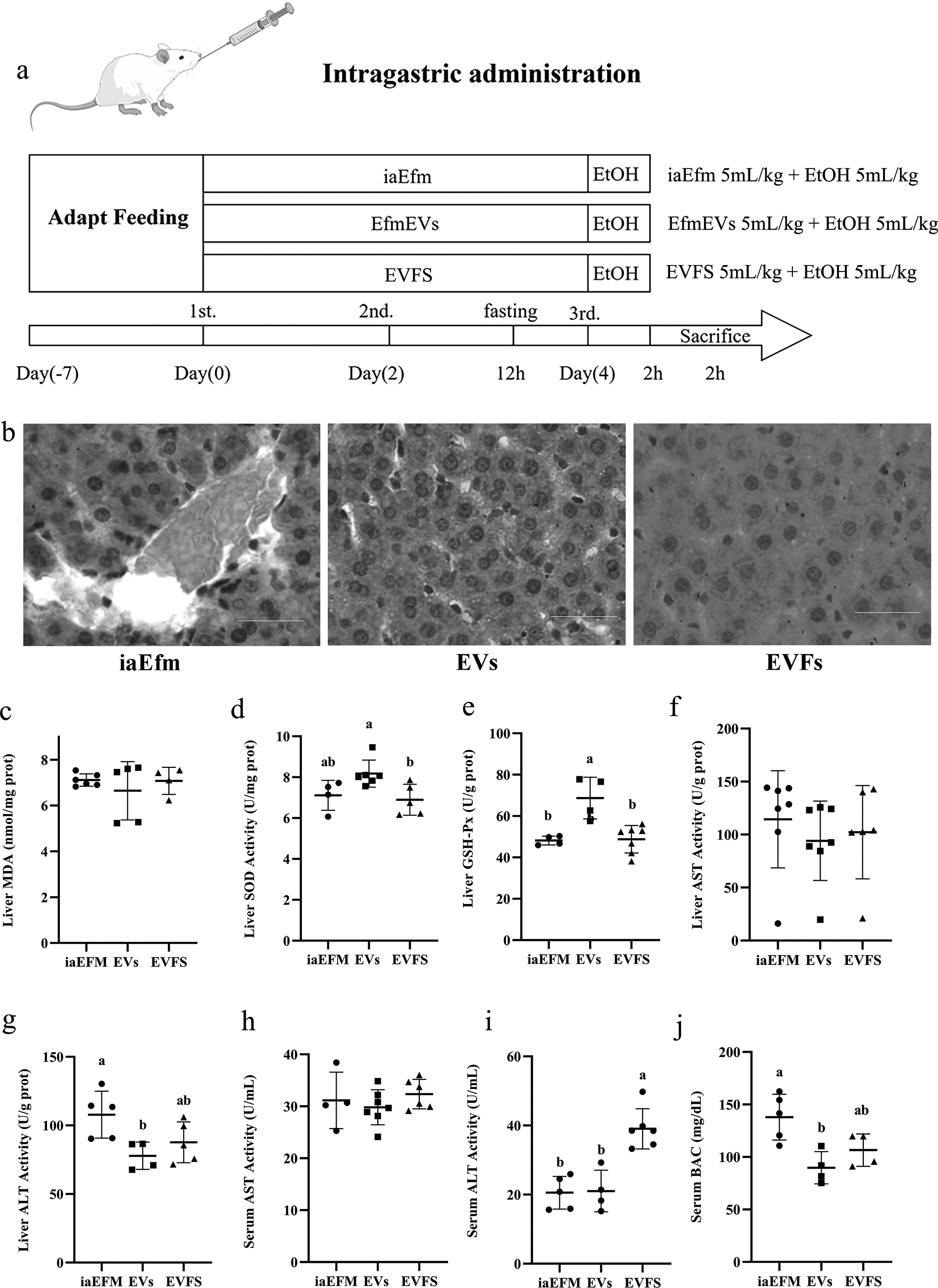
Protective effect of different active components of bacterial culture against liver injury in rats.
As shown in Figure 2b, in the iaEfm group, the basic architecture of liver cells was disrupted, displaying apparent steatosis, and mild bleeding was observed in the portal vein and surrounding areas. In the EfmEVs group, liver cells exhibited clear hepatocyte outlines and nuclei, with no necrosis observed. Minor intercellular gaps were present, which were likely due to mild fatty degeneration. EVFS treatment group showed significantly alleviated histopathological changes of liver injury, with decreased inflammatory cell infiltration and necrosis in the rat’s liver.
EfmEVs pretreatment enhanced antioxidant capacity in alcohol-induced acute liver injury in rats
To evaluate the antioxidant effects of EfmEVs, MDA levels and the activities of SOD and GSH-Px in liver tissues were measured. As shown in Figure 2c–e, MDA levels were lowest in the EVs group, while SOD and GSH-Px activities were highest in the EVs group. These results indicate that EfmEVs significantly reduced oxidative stress by decreasing MDA levels and enhancing the activities of SOD and GSH-Px in the liver.
EfmEVs pretreatment exhibited hepatoprotective effects in rats with alcohol-induced acute liver injury
We further assessed liver AST and ALT activities. In liver tissue (Fig. 2f and g), ALT and AST activities were lowest in the EVs group and highest in the iaEfm group. Similarly, in serum (Fig. 2h and i), the EVs group exhibited the lowest AST activities, significantly lower than the iaEFM group and EVFS group. For ALT activity in serum, the EVFS group showed the highest levels, while the EVs group and iaEFM group exhibited similar levels, with a slight but nonsignificant increase in the EVs group. Overall, EfmEVs significantly reduced the activities of AST and ALT.
In serum, EfmEVs pretreatment displayed the lowest BAC contents among the three groups. (p < 0.01; Fig. 2j). The results showed that EfmEVs pretreatment could thereby effectively reduce BAC.
Efm and LZD coadministration improves hepatoprotection through increased EVs secretion
Increased EVs secretion by Efm pretreatment reduces histopathological damage in liver tissues of rats with acute ALI
To explore the role of EVs in Efm’s hepatoprotective effect, Efm was coincubated with LZD, and their intragastric coadministration was assessed in rats. The schematic diagram of the experimental procedures is shown in Figure 3a. Significant differences were observed among the groups, as shown in Figure 3b. In the LZD group, most hepatocytes were deformed, accompanied by inflammatory cell infiltration, disorganized cell arrangement, and mild-to-moderate hemorrhage in the veins. In contrast, the Efm group showed notable improvements compared with the LZD group, with only disorganized hepatocyte arrangement observed. Among these groups, the Efm-LZD group demonstrated superior hepatoprotective effects, surpassing those of the other two groups. In the EfmEVs group, hepatocytes were tightly arranged with clear cell outlines and nuclei, showing no necrosis and only slight fatty degeneration.
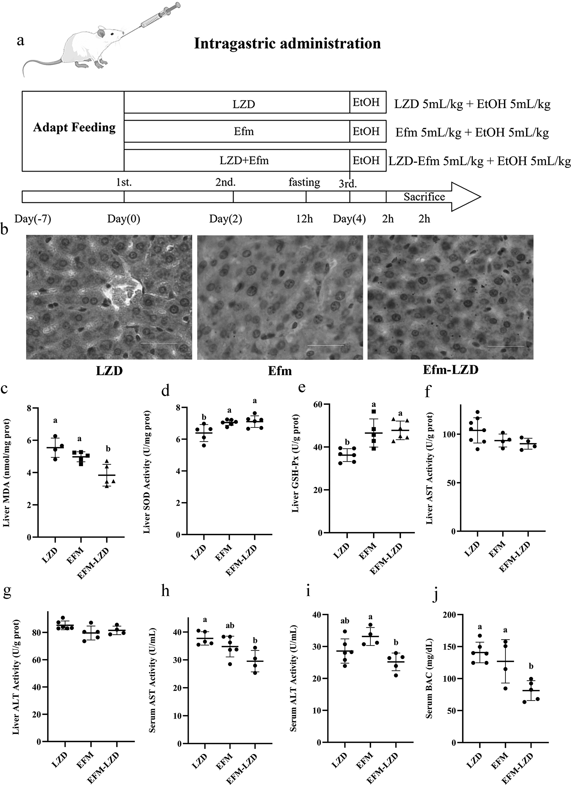
The protective effect of Efm was improved by increasing EVs secretion in rats.
EVs secretion enhanced antioxidant capacity in alcohol-induced acute liver injury rats
We further assessed liver antioxidant activities. As shown in Figure 3c–e, Efm and LZD coadministration displayed the lowest MDA and highest GSH-Px activities in the liver among the three groups. Efm and LZD coadministration significantly increased liver SOD and GSH-Px activities compared with the LZD group (p < 0.05 or p < 0.01). The findings suggest that enhanced hepatic antioxidant activity plays a critical role in alleviating ethanol-induced liver injury, potentially mediated by EVs.
EVs secretion exhibited hepatoprotective effects in rats with alcohol-induced acute liver injury
We further assessed the activities of AST and ALT in the liver and serum. There was no significant difference in AST and ALT among the three groups in the liver (Fig. 3f and g). In the serum, AST and ALT in the Efm-LZD treatment group were significantly decreased (p < 0.05; Fig. 3h and i). Overall, EVs significantly reduced the activities of AST and ALT in serum. Considered together, these results suggested that the Efm-LZD combination effectively mitigates ALI, as reflected by the reduction in serum biomarkers.
In serum, compared with the Efm group, the BAC in the LZD group was significantly higher, while Efm-LZD treatment significantly decreased it (p < 0.01; Fig. 3j). The results showed that Efm and LZD coadministration pretreatment could thereby effectively reduce BAC.
Discussion
ALD is a life-threatening condition and a significant global health issue (WHO, 2024). Growing evidence links intestinal microflora imbalance with liver injury, where acute ethanol exposure disrupts this balance, damages mucosal integrity, increases intestinal permeability, and facilitates endotoxin translocation to the liver (Kirpich et al., 2008; Malaguarnera et al, 2014). ALD triggers inflammation and oxidative stress, and recent treatments focus on anti-inflammatory, antioxidant, and microbiota-regulation strategies (Liu et al., 2021). Probiotics are advantageous in this context due to their low toxicity, minimal side effects, and reduced risk of drug resistance (Plaza-Diaz et al., 2019). Alterations in the gut microbiome are associated with metabolic, inflammatory, and infectious diseases (Lynch and Pedersen, 2016), underscoring the importance of probiotics in protecting the liver (Hong et al., 2019).
Many studies have demonstrated the liver-protective effects of probiotics. For instance, Bacteroides thetaiotaomicron has been shown to improve hepatic steatosis, while Bifidobacterium longum and Bifidobacterium adolescentis reduce ALI and enhance gut health (Li et al., 2019; Sangineto et al., 2022). However, further clinical studies are needed to validate their efficacy in ALD.
In this study, we investigated the therapeutic potential of EfmEVs against ethanol-induced liver injury in rats, while also evaluating the effects of Efm itself. H&E staining results showed that ethanol (5 mL/kg BW) orally administered resulted in severe liver damage and Efm pretreatment markedly attenuated liver injury.
Oxidative stress plays a significant role in the pathogenesis of ethanol-induced liver injury. MDA is a byproduct of lipid peroxidation, typically reflecting the extent of oxidative damage to cellular membrane lipids. SOD and GSH-Px are pivotal enzymes in the antioxidant defense system (Frijhoff et al., 2015). In this study, alcohol induced a reduction in serum GSH-Px activities (Fig. 2e) and an increase in liver and serum MDA levels (Fig. 2c), which was consistent with previous research reports (Wu et al., 2021). Efm pretreatment effectively restored GSH-Px activity and normalized MDA levels.
AST and ALT are hepatic-specific enzymes that reflect the degree of acute liver damage and are frequently elevated after excessive alcohol intake (Ding et al., 2021). Liver cell damage disrupts membrane integrity, releasing these enzymes into the bloodstream and leading to increased serum AST and ALT levels, both of which play essential roles in metabolism (Kobayashi et al., 2011).
Interestingly, while Efm pretreatment reduced ALT activity in both the liver and serum, it unexpectedly increased serum AST levels despite lowering liver AST. This discrepancy suggests that although Efm may exert a protective effect, it does not fully prevent liver injury. Since AST elevation is generally associated with liver damage (Malnick et al., 2022), the rise in serum AST in the Efm group complicates its hepatoprotective role, potentially indicating residual liver injury. In contrast, EfmEVs treatment consistently reduced both AST and ALT levels in the liver and serum, suggesting a more comprehensive protective effect (Fig. 1f–i). These findings highlight the complexity of AST and ALT dynamics in liver injury and recovery, warranting further investigation to clarify their distinct roles.
EfmEVs treatment significantly reduced serum BAC contents (Fig. 2j), indicating its protective role against ALI. This effect may be attributed to restoring gut barrier integrity, which reduces ethanol absorption and systemic toxicity, as previously reported (Luo et al., 2024a). In the study of inflammatory bowel disease, damage to the intestinal mucosa disrupts tight junction proteins, leading to increased intestinal permeability to small molecular organic compounds, such as ethanol, which allows ethanol to more easily enter the bloodstream and be absorbed (Rocco et al., 2014).
Consistent with previous studies, exogenous EVs show potential in liver protection. For example, probiotic-derived nanoparticles inhibit ALD by modulating miRNA and farnesoid X receptor (FXR) pathways (Kim et al., 2023), while plant-derived vesicles alleviate alcohol-induced liver damage (Jiang et al., 2025). The protective effects of EfmEVs are likely due to bioactive components, such as proteins, lipids, and nucleic acids, which modulate immune responses, reduce oxidative stress, and enhance gut barrier integrity. However, the precise mechanisms by which EfmEVs modulate liver inflammation and oxidative stress remain unclear (Fig. 4).
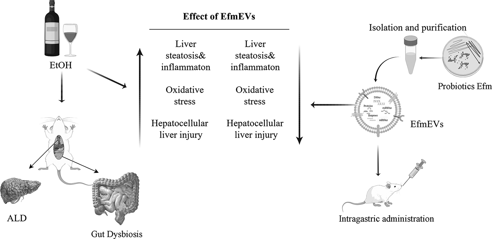
Summary of the protective effects of EfmEVs against ALI. The diagram illustrates the mechanisms by which EfmEVs alleviate ethanol-induced liver steatosis, inflammation, oxidative stress, and hepatocellular injury. Upward arrows indicate the exacerbation of these parameters by alcohol exposure, while downward arrows represent the attenuation achieved by EfmEVs pretreatment. This figure provides a comprehensive overview of the potential of EfmEVs as a probiotic candidate for preventing ALD. ALD, alcohol-induced liver disease; ALI, alcohol-induced liver injury; EfmEV, E. faecium-derived extracellular vesicles.
Future studies should explore whether EfmEVs regulate hepatoprotective pathways via miRNA modulation, TLR signaling, or gut-liver axis interactions. These findings highlight the potential of EfmEVs as a novel therapeutic strategy for ALD. However, future research is required to elucidate their molecular mechanisms, optimize dosage, and validate their clinical efficacy and long-term safety in human trials.
Conclusions
In summary, our study demonstrated that EfmEVs exhibit significant protective effects against alcohol-induced acute liver injury in rats. These findings suggest that EfmEVs could serve as a novel therapeutic approach for managing ALI. Future research should focus on identifying the active molecules within EfmEVs and elucidating their mechanisms of action, as well as exploring their potential in long-term models of ALD to develop effective therapeutic strategies.
Authors’ Contributions
Q.Q. contributed to the study conceptualization, data curation, formal analysis, validation, and visualization. Methodology, investigation, and writing—original draft were performed by Y.Z., X.Z., and W.H. Funding acquisition was performed by Q.Q. H.Z. contributed to the project administration. Writing—reviewing and editing were performed by Q.Q. and X.F. All authors have read and agreed to the published version.
Footnotes
Author Disclosure Statement
No competing financial interests exist.
Funding Information
This research was funded by the National Natural Science Foundation of China (Grant No. 31902228), Key Construction Discipline Research Ability Improvement Project of Guangdong Province (Grant No. 2022ZDJS040), and Special Fund for Science and Technology Innovation Cultivation of Guangdong University Students (Grant No. pdjh2024b397).
Animal Welfare Statement
All animal care and treatment procedures were approved by the Animal Care and Use Committee of Foshan University, which meets the ethical standards in laboratory animal guidelines for ethical review of animal welfare (The National Standard of the People’s Republic of China GB/T 35892-2018).