




Abstract
Introduction: The aim of this study was to design new potentially antineoplastic agents by combining nitrogen mustard with steroidal skeleton, in an effort to improve specificity and simultaneously to reduce systemic toxicity. The steroidal part is aimed to act as a biological platform enabling the alkylating moiety to approach its site of action by altering its physicochemical properties. Materials and Methods: The compounds tested have, as alkylating agents, either p-N,N-bis(2-chloroethyl)aminophenyl-butyrate or p-N,N-bis(2-chloroethyl)aminophenyl-acetate esterified with a modified steroidal nucleus. The four newly synthesized compounds were compared on a molar basis, regarding their ability to induce sister chromatid exchanges and modify proliferation rate indices in cultured human lymphocytes. Life span of BDF1 mice inoculated with L1210 leukemia was also estimated (antileukemic activity). Results: A compound having p-N,N-bis(2-chloroethyl)aminophenyl-acetate as the alkylator and two ketone groups in the steroidal part demonstrated the highest statistically significant enhancement of sister chromatid exchanges and suppression of proliferation rate indices, and also caused significant antineoplastic activity. The other compounds proved less active. Conclusion: These results suggest that cytogenetic and antileukemic activity of alkylating steroidal esters depends on the configuration of the whole molecule and the appropriate combination of the alkylator with the steroidal molecule.
Introduction
T
Nitrogen mustards are highly reactive compounds with an inherent chemical affinity toward the nucleophilic sites of several biomolecules such as plasma proteins; at the same time, they are rapidly hydrolyzed and in some cases are deactivated by cellular resistance mechanisms (Koutsourea et al., 2008a). These disadvantages result in a significant loss of the active drug before reaching the DNA target and at the same time increase their toxic effects.
The conjugation of nitrogen mustards with steroidal skeletons may increase their lipophilicity and alter their physicochemical properties, resulting in an easier and more quantitative penetration through the cellular and nuclear membranes and consequently improving their antileukemic activity and reducing their toxicity. Especially, steroidal hormones have been reported to influence the growth of many cancers, and the presence of tumor-associated receptors for these hormones offers the opportunity for targeting useful drug-hormone conjugates (Catsoulacos, 1984). The hormones can lead nitrogen mustard directly to the nuclei, where they naturally act as transcription factors through binding to their receptors or possibly through nongenomic action (Wasan and Waxman, 1996). By combining the results of some studies, it has been confirmed that the steroidal part of these molecules not only acts as a simple biological carrier as has been speculated for many years, but also plays a significant role in their mechanism of action (Papageorgiou et al., 2005).
In a previous study, we have shown that minor functional changes in the B steroidal ring (e.g., 7-keto-group) of a steroidal ester (androstan) had considerable effects on the molecule's antileukemic, genotoxic, and cytotoxic activity, leading us to assume that this modification is fundamental to the design of more effective molecules (Fousteris et al., 2007).
Therefore, in this study we extended those observations by synthesizing four new potentially chemotherapeutic compounds (Fig. 1). These compounds contain p-N,N-bis(2-chloroethyl)aminophenyl butyrate (CHL) or p-N,N-bis(2-chloroethyl)aminophenyl acetate (PHE), as an alkylating agent conjugated with a steroidal ester modified in the B and D steroidal rings. These agents were comparatively studied on a molar basis regarding their ability to induce SCEs and disturb cell kinetics (proliferation rate index [PRI]) in normal human lymphocyte cultures. The synthesized compounds were also tested for their antileukemic potency against L1210 leukemia-bearing mice. The potential increase in SCE frequency and the decrease in PRIs caused by genotoxic agents are considered as indicators of cytogenetic damage (genotoxicity and cytostatic activity, respectively); therefore, SCEs appear to have an application in the clinical prediction of tumor sensitivity to potential chemotherapeutics (Mourelatos, 1996). Moreover, the SCE assay has been proposed as having a predictive value as a clinical assay for drugs for which a strong correlation between cell killing and induction of SCEs has been established (Deen et al., 1986).
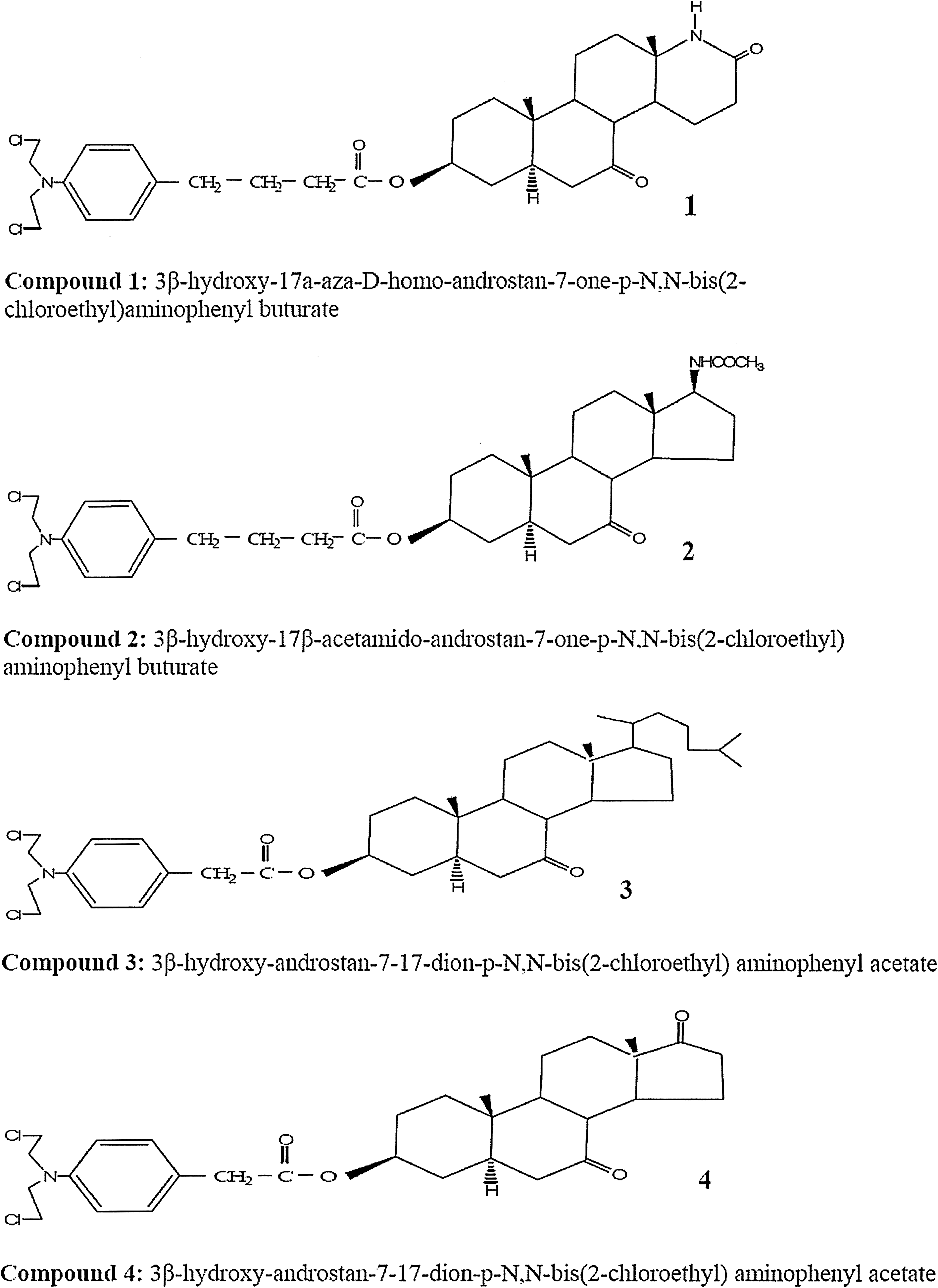
Newly synthesized compounds.
Materials and Methods
Newly synthesized compounds and concentrations tested
All newly synthesized compounds (Fig. 1) have a ketone group in the B-ring of the steroidal nucleus. More specifically, compounds 1 and 2 contain CHL as the alkylating agent in conjunction with a steroidal nucleus having a lactam group (-NHCO-) at the D ring (endocyclically and exocyclically, respectively). Compound 4 has PHE as the alkylating agent in conjunction with a steroidal nucleus having a second ketone at the 17 position of the D-ring, while compound 3 includes a cholestene group. The steroidal esteric derivatives were prepared according to procedures described in the literature (Koutsourea et al., 2008b)
Cell culture
Lymphocyte primary cultures were set up (Karapidaki et al., 2009) by adding 11 drops of heparinized whole blood obtained from three normal subjects to 5 mL of a cell culture medium, Roswell Park Memorial Institute (RPMI) (1640). The individuals who donated their blood (25-35 years old) were healthy, were not taking any medication, and were nonsmokers and no alcohol consumers. All donors were informed about the study and they willingly signed permission for using their blood samples for scientific purposes.
In vitro SCE assay
For SCE demonstration, 5 μg/mL of 5-BromodeoxyUridine and the solutions of the newly synthesized compounds at five concentrations (0.2, 0.4, 0.6, 0.8, and 1.0 μM) were added at the beginning of the culture life. The cultures were incubated for 72 h at 37°C. Metaphases were collected during the last 2 h with 0.3 μg/mL colchicine. Air-dried preparations were stained by the Fluorescence-plus Giemsa procedure (Goto et al., 1978).
Assessment of the PRI
The PRI was calculated according to the formula PRI = (M1 + 2M2 + 3M3+)/N, where M1, M2, and M3+ indicate the number of metaphases corresponding to first, second, and third or subsequent divisions, respectively, and N is the total number of metaphases scored (at least 100) for each culture.
In vivo acute toxicity and anti-tumor experiments
The antileukemic study was performed according to procedures described in the literature (Papageorgiou et al., 2005).
Statistical analysis
For the statistical evaluation of the experimental data, Student's t-test was performed for the SCE frequencies, to determine whether any values deviated significantly from the controls (p < 0.05), whereas the χ2-test was used for PRIs to assess significant deviations compared to controls (p < 0.05).
The in vivo experimental data were statistically evaluated by the Wilcoxon test, considering p-values less than 5% as significant.
Results
Enhancement of SCE frequency and reduction of PRI values in human lymphocyte cell cultures
The cytogenetic effects of the tested compounds are shown in Table 1. The genotoxic and cytostatic effects of the compounds are evaluated by calculating the SCEs and PRIs, respectively, induced in normal human lymphocyte cultures of three normal donors (in vitro). Controls of mean SCEs and of mean PRI values from the three donors are estimated at 8.15 ± 0.51 and 2.40, respectively.
Statistically significant increase in SCEs compared to the control (p < 0.01, by t-test).
Statistically significant decrease in PRIs compared to the control (p < 0.01, by χ2-test).
SCE, sister chromatid exchange; SE, standard error; PRI, proliferation rate index.
All newly synthesized compounds induced statistically significant (p < 0.01) increases in SCEs at almost all concentrations tested. Moreover, compounds 1 and 2 were identified to be effective, on a molar basis, at concentrations higher than 0.6 μM, while compound 4 was effective at 0.4 and 0.6 μM in causing cell division delays. Compound 1 induced a statistically significant increase (p < 0.01) in SCE levels at all concentrations tested; its effect, however, was lower compared to the rest of the compounds studied. Further, statistically significant PRI depression is calculated for compound 1 at 0.6, 0.8, and 1.0 μM (p < 0.01). Compound 2 induced a statistically significant increase of SCE values (p < 0.01). The higher increase appeared at 0.6 μM (SCE = 14.75 ± 0.87). The PRI is 1.87, 1.76, and 1.65 (p < 0.01) at 0.6, 0.8, and 1.0 μM, respectively. Compound 3 induced a statistically significant increase of SCE values (p < 0.01) only at the 0.6 μM concentration, but the decrease of PRI was not statistically significant at any concentration. Compound 4 was found to have the most pronounced genotoxic activity by increasing the frequency of SCEs up to 95.02 ± 3.36 (p < 0.01) as compared to the control (8.15 ± 0.51 SCEs/cell). Statistically significant decreases in PRI values were observed at 0.4 and 0.6 μM.
The four tested compounds showed a different pattern, concerning their genotoxic potential. Compound 1 caused a statistically significant increase in SCEs beginning from the lowest concentration of 0.2 μM and showing a greater increase at 0.8 μM. Compound 2 caused a statistically significant increase in SCEs starting from the lowest concentration of 0.2 μM, but this increase reached a plateau at the concentrations 0.4 and 0.6 μM. Finally, compound 4 displayed a clearly concentration-dependent pattern of toxicity, as SCE frequencies increased at increasing concentrations of this compound.
Newly synthesized compounds increase the life span of mice bearing lymphoid L1210 leukemia
The effective compounds were tested for their antineoplastic activity in vivo against the transplantable lymphoid L1210 leukemia (Table 3).
LD50 values were estimated graphically, where the percentage of deaths due to the toxicity of each does is shown in the ordinate, while the administered doses are indicated on the abscissa on semilogarithmic paper. For chemotherapy testing the highest dose used for a single treatment was LD10. Therefore, the drugs in the survival experiments were compared at equitoxic doses.
Medial survival time ± standard error of the mean.
T/C (%) is the percent increase in median life span of treated animals over the control.
Significant increase (p < 0.01, by the Wilcoxon test) over the control.
In the antileukemic experiments we calculated the therapeutic dose to evaluate the LD10 acute toxicity (D). On lymphoid L1210 leukemia-bearing mice (BDF1) the dose given was D/2 × 3 on days 1, 4, and 7 after tumor transplantation (Table 2). Compounds 2 and 4 showed a statistically significant increase in the life span of BDF1 mice (Table 3). Compound 4 showed the most pronounced increase of the life span up to 145% (Table 3), while compound 2 showed a borderline increase of the life span up to 126%. The order of antineoplastic activity of compounds on lymphoid L1210 leukemia-bearing mice was 4 > 2. Compounds 1 and 3 were inactive.
Discussion
In an effort to extend previous observations on the genotoxic effects of newly synthesized compounds (Papageorgiou et al., 2005; Fousteris et al., 2007), we investigated in this study the cytogenetic and antileukemic effects of four different newly synthesized compounds that contain CHL and PHE as alkylating agents and are also conjugated with steroidal ester modified in the B and D steroidal rings.
The investigation was based on SCE and PRI analysis methods in lymphocyte cultures in vitro and on T/C% calculation in L1210 leukemia-bearing mice. SCEs have been frequently used as highly sensitive indicators of DNA damage and/or subsequent repair (Mourelatos, 1996). Nonrepaired DNA damage expressed as SCEs in normal cells caused by certain chemicals may indicate inability to repair the damage induced by the same chemicals in cancer cells since both cell types have similar DNA repair mechanisms (Karayianni et al., 2003). There are findings indicating that the effectiveness in SCE induction by potential antitumor agents in cancer cells in vitro and in vivo is positively correlated with in vivo tumor response to these agents (Papageorgiou et al., 2005). This suggests that the SCE assay could be used to predict both the sensitivity of human tumor cells to chemotherapeutics and the heterogeneity of drug sensitivity of individual tumors (Mourelatos, 1996). Other studies investigating a relationship between SCE induction and other expression of genotoxicity have also shown a positive relationship between SCE induction and reduced cell survival and alteration in cell cycle kinetics (Deen et al., 1986). According to our results, compounds 1, 2, and 4, compared on a molar basis, have been proved to be cytogenetically active by inducing SCEs and reducing PRI values in normal human lymphocyte cultures. The order of magnitude of the cytogenetic activity of the compounds was 4 > 2 > 1 > 3.
All derivatives have a keto group in the B ring, whereas they differ in the D steroidal ring and in the alkylator congener (Fig. 1). Compound 1 with -NHCO- group endocyclically has been proved to be less effective in the increase of SCEs and the reduction of PRIs as compared to compound 2, which contains an -NHCO- group exocyclically. Compound 4 has been proved more active than compound 3, which was inactive in almost all concentrations tested. Therefore, the insertion of a cholesten group in 17 position of steroidal nuclei has been proved ineffective. The most active compound 4 presented a dose-dependent, statistically significant increase of SCEs as compared to compounds 1, 2, and 3. Compound 4 has been proved to be cytogenetically active, while it seems that the modifications in the steroidal skeleton have an important role in the activity of the compounds (Papageorgiou et al., 2005; Fousteris et al., 2007).
The acute toxicity values of the studied compounds are reported in Table 2. Comparing the toxicity of the alkylating steroidal nucleus in all tested compounds, it is obvious that the toxicity depends not only on alkylating agents but also on the modified steroidal nucleus. The results from antileukemic experiments showed that compound 4 was the most active in increasing the life span of mice, up to 145% in lymphoid L1210 leukemia-bearing mice. Compound 2 presented lower antileukemic activity than compound 4, whereas compounds 1 and 3 were inactive.
Comparing compound 2 with compound 1, we can conclude that the exocyclical insertion of -NHCO- group in the D steroidal ring (compound 2) offers a better cytogenetic and antileukemic activity than the endocyclical insertion of -NHCO- group (compound 1). When comparing compound 4 with compound 2, it seems that compound 4 has been proved more active in cytogenetic and antileukemic efficacy. These results could be attributed apart from the presence of PHE as the alkylator (Karapidaki et al., 2009) to the insertion of a keto group at the B and D rings offering a positive influence on their cytogenetic and antileukemic potency. The presence of the two keto groups in the steroidal rings affects their activity, indicating that they may interact by a different way or by different cellular moieties (Papageorgiou et al., 2005) in comparison to the presence of -NHCO- group, which interacts with similar groups present in the proteins and nucleic acids, destabilizing DNA structure and facilitating DNA-protein or DNA-DNA interstrand cross-links and detectable strand breaks (Fousteris et al., 2002).
In conclusion, the current study showed that newly synthesized compounds 2 and 4 caused a significant increase in the frequency of SCEs and decrease in the corresponding PRI values and a relevant antileukemic activity. Despite that compounds 1 and 3 showed a slight genotoxic activity, they were inactive in antileukemic experiments. Compound 2 showed a significant genotoxic and antileukemic activity, but this activity was much more pronounced for compound 4. The SCE assay in vitro has been established as a highly predictive method for the clinical behavior for drugs for which a correlation between cell killing and induction of SCEs has been established, as it seems applicable for compound 4. It has been reported from earlier studies that both the chemical structure of the nitrogen mustard and the configuration of the steroidal moiety determine the antineoplastic effect of these compounds (Papageorgiou et al., 2005; Fousteris et al., 2007; Karayianni et al., 2003).
Our observations support the previous notion that the cytogenetic and antileukemic specificity of nitrogen mustard's steroidal esters depends on the configuration of the whole molecule and the appropriate combination of nitrogen mustard with a steroidal molecule. Moreover, similar chemical properties of antineoplastic drugs usually result in similar biological responses; at the same time, minor differences in chemical properties may result in pronounced differences in biological responses.
Footnotes
Disclosure Statement
No competing financial interests exist.