




Abstract
Aims: Our study aimed to evaluate the diagnostic and prognostic values of microRNA-21 (miR-21) expression levels in peripheral blood mononuclear cells (PBMCs) for prostate cancer (PCa). Methods: Between February 2010 and June 2014, 75 consecutive patients with localized PCa confirmed by radical prostatectomy and biopsy were enrolled as the case group. Among them, 25 patients were confirmed with recurrence or metastasis (R/M) (PCa with R/M group) and 50 patients without R/M (PCa without R/M group). During the same period, 75 healthy volunteers were enrolled as the control group. Blood was collected from all subjects, and PBMCs were isolated. Relative miR-21 expression levels from the PBMCs were determined by fluorescence real-time quantitative polymerase chain reaction (RT-qPCR). Results: The relative miR-21 expression levels in the preoperative case group was significantly higher than that in the postoperative case group and the control group (both p<0.001). miR-21 expression levels were associated with tumor differentiation, clinical stage, and lymph node metastasis (all p<0.001). Furthermore, miR-21 expression levels in PCa patients with R/M were significantly higher than that in PCa patients without R/M and healthy controls (both p<0.001). Receiver operating characteristic (ROC) curve analysis revealed that the cut-off point of miR-21 for diagnosis of PCa was 0.9 with a sensitivity of 87.5% and a specificity of 85.7%. Conclusions: Our study demonstrates that high miR-21 expression levels in PBMCs were correlated with the presence, recurrence, and metastasis of PCa and that this may be a useful biomarker for screening PCa and monitoring the risk of PCa recurrence and metastasis.
Introduction
I
A great number of genetic and epigenetic alternations are evidenced to be associated with tumorigenesis and the development of numerous cancers (Shinjo et al., 2012; Achyut et al., 2013; Li et al., 2014). Particularly, it is becoming increasingly obvious that microRNAs (miRNAs), an abundant class of small nonprotein coding RNAs regulating gene expression by targeting mRNA for the repression of translation or cleavage, are correlated with the development and progression of PCa (Brase et al., 2011; Martens-Uzunova et al., 2012). Recently, studies have shown overexpressed miR-21 in various experiments designed for detecting miRNAs that are dysregulated in PCa, detectable in the plasma/serum (Li et al., 2009, 2012; Melbo-Jorgensen et al., 2014). Fujita et al. (2008) revealed a correlation between the promoter of miR-21 and androgen receptor, and Ribas et al. (2009) showed a sensitivity of miR-21 to androgen in a positive PCa cell line of androgen receptor. Also, Coppola et al. (2013) have demonstrated a novel role of the overexpression of miR-21 in driving prostate cells toward acquiring a malignant phenotype through suppressing BTG2 levels and promoting the acquisition of luminal markers and epithelial-mesenchymal transition in prostate cells. Many studies have shown the presence of miR-21 in plasma or serum and its potential application as new noninvasive biomarkers of PCa; however, few studies assess the role of miR-21 in peripheral blood mononuclear cells (PBMCs) in PCa patients (Shen et al., 2012; Heitzer et al., 2013; Sita-Lumsden et al., 2013). Therefore, miR-21 in PBMCs was chosen as our research point. In our present study, we compared the expression of miR-21 in PBMCs among PCa patients with recurrence or metastasis (R/M), PCa patients without R/M, and healthy controls to assess the diagnostic and prognostic values of miR-21 expression in PBMCs for PCa.
Materials and Methods
Ethics statement
The study was approved by the Ethics Committee of Xiangya Hospital, Central South University, Changsha, Hunan Province. Written informed consent was provided by each eligible patient or the patient's next of kin and the study conformed to the Declaration of Helsinki (M, 2014).
Subject population
The study was conducted in the Department of Urology of Xiangya Hospital, Central South University, Changsha, Hunan Province. A total of 75 consecutive patients (mean age, 55.4±11.3 years) with localized PCa confirmed by radical prostatectomy and biopsy were enrolled from February 2010 to June 2014 as the case group. Patients who (1) have been treated with surgery, radiotherapy, and chemotherapy or (2) were associated with other malignancies, cardiovascular disease, diabetes, kidney disease, or other chronic diseases that may affect miRNA expression, such as leukemia, pancreatitis, hepatitis B, arthritis, pulmonary obstruction, and osteoporosis, were excluded. Clinical characteristics of all patients, including age, pathological grade, Whitmore-Jewett stage (Oesterling et al., 1993), and lymph node status, were recorded. During the same period, 75 healthy volunteers (mean age, 58.9±9.5 years) were enrolled as the control group. All localized PCa patients with no absolute contraindications to surgery were treated with retropubic radical prostatectomy, after which serum of all patients was collected and follow-up was conducted; at the end of follow-up, serum was collected again from patients and controls. Fifty PCa patients without R/M were defined as the PCa without R/M group and 25 PCa patients with R/M (recurrence, n=18; metastasis, n=17) as the PCa with R/M group. Whole blood samples (5 mL) were collected from all subjects, stored in procoagulant tubes for 2 h, and centrifuged at 4000 rpm for 10 min at 4°C. The supernatant was collected, placed in a 1.5-mL enzyme-free Eppendorf tube, and stored in an ultralow temperature freezer at −80°C.
Extraction of PBMCs
Peripheral venous blood (2 mL) collected in vials containing ethylenediaminetetraacetate was diluted and well-mixed with an equal volume (2 mL) of phosphate-buffered saline (PBS) solution. The diluted blood was carefully layered on the top of Ficoll solution (Ficoll separation solution: diluted blood, 1:2), ensuring that the Ficoll interface was not disturbed. After centrifugation in a swing-out bucket at 2000 rpm at room temperature for 20 min, the mixture separated into three layers, and a milky turbid cell layer was obtained at the Ficoll/blood interface. The cell layer was carefully withdrawn using a Pasteur pipette, transferred into another centrifuge tube, then diluted with PBS (1:5), and centrifuged at 1500 rpm for 5 min. The supernatant was discarded and the PBS wash was repeated once. The isolated PBMCs were resuspended with PBS, and the cell concentration was adjusted to 1×10⁶/mL.
Isolation of total RNA from serum
Isolation of total RNA was conducted by using the standard TRIzol method (Qiagen, Hilden, Germany) according to the kit's instructions (Haimov-Kochman et al., 2006). The isolated total RNA was dissolved in sterile distilled water treated with 20 μL diethylpyrocarbonate and stored at −70°C. In addition, the purity and concentration of RNA samples were determined through measuring UV absorbance at D260 and D280 using a NanoDrop®ND-1000 spectrophotometer (NanoDrop Technologies, Inc., Wilmington, DE). RNA samples with D260/D280 value between 1.8 and 2.1 were obtained for further use.
Real-time quantitative reverse transcriptase-polymerase chain reaction
Reverse transcriptase (RT) was conducted using a One-Step PrimeScript miRNA cDNA Synthesis Kit (Takara Bio, Inc., Shiga, Japan) in accordance with the manufacturer's protocol. TaqMan® MicroRNA Assays (ABI, Forest City, CA) were performed using the stem-loop method. The MiR-21-specific stem-loop primer for RT was (5′-GTCGTATCCAGTGCGTGTCGTGGAGTCGGCAATTGGCACTGGATACGACTCAACATC-3′). The RT primer for U6 was 5′-AACGCTTCACGAATTTGCGT-3′. The reaction system comprised 10 μL miRNA Reaction Buffer Mix, 2 μL 0.1% bovine serum albumin, 2 μL miRNA PrimeScript® RT Enzyme Mix, 1 μL total RNA, and 5 μL RNase-Free dH2O. Reaction conditions were as follows: poly (A) tailing and RT reaction at 37°C for 60 min, enzyme inactivation at 85°C for 5 s, and followed by cDNA stored at −70°C.
Polymerase chain reaction (PCR) amplification was conducted with cDNA synthesized from RT as template. PCR primers for miR-21 were designed by using Premier 6.0 software (Premier Biosoft, Palo Alto, CA): upstream forward primer, 5′-ACACTCCAGCTGGGTAGCTTATCAGACTGATG-3′; downstream reverse primer, 5′-CTCAACTGGTGTCGTGGA-3′. Amplification reaction conditions were as follows: 40 cycles of predenaturation at 95°C for 5 min, denaturation at 95°C for 10 s, annealing at 60°C for 20 s, and a final extension at 72°C for 20 s. Amplification reaction was performed on an ABI7500 real-time PCR system (Applied Biosystems, Darmstadt, Germany). All primers in our study were supplied by the Shanghai Sangon Company (Shanghai, China). The PCR products (10 μL) were electrophoresized on 10% polyacrylamide gels, followed by staining and photographing. Total reaction system of the real-time quantitative PCR (20 μL) included 1.3 μL cDNA template, 1 μL miR-21/U6-specific primer, 10 μL TaqMan Universal PCR master Mix (ABI), and 7.7 μL H2O. All reactions were set up in three wells. The computed tomography (CT) value of each reaction was recorded, and results were analyzed by the relative quantitative method, with 2−ΔΔCt representing fold changes in miR-21 expression in the case group relative to the control group. ΔΔCt=(CTmiR-21−CTU6)case group−(CTmiR-21−CTU6)control group.
Statistical analysis
Data statistical analysis was performed using SPSS20.0 software (SPSS, Inc., Chicago, IL). All measurement data were verified by the normality test using the Shapiro-Wilk test. Multiple comparisons of normally distributed data between multiple groups were conducted, after homogeneity test of variances with variance calibration for heterogeneity, using the one-way ANOVA, least significant difference, and honestly significant difference. Comparison of non-normally distributed data between the two groups was conducted using the Mann-Whitney U test, a nonparametric method for assessing whether two independent samples came from the same distribution. Comparisons of non-normally distributed data between multiple groups were conducted using the Kruskal-Wallis H test. The diagnostic value in PCa was evaluated by using a receiver operating characteristic (ROC) curve, and the area under the ROC curve (ROC-AUC), sensitivity, and specificity were obtained by the Hanley-McNeil nonparametric method. All p-values were two tailed and <0.05 were considered statistically significant.
Results
PCR amplification products of miR-21 and U6
The 10% polyacrylamide gel electrophoresis revealed that the length of PCR amplification products of miR-21 and U6 was 66 and 99 bp, respectively, as seen in Figure 1.
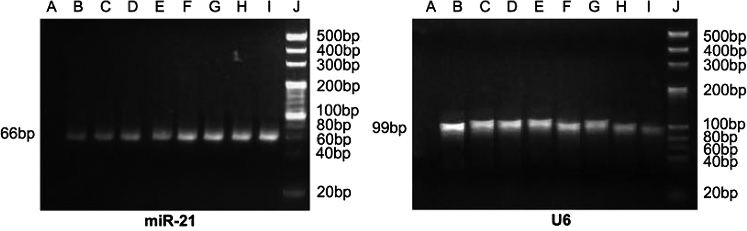
Electrophoretogram for polymerase chain reaction amplification products of microRNA-21 (miR-21; 66 bp) and U6 (99 bp). A, blank control group; B-E, control group; F-I, case group; J, Marker (20 bp).
Expression of miR-21 in PBMCs
The appearance of single peaks for the miR-21 during melting curve analysis indicated that miR-21 was efficiently extracted and specifically amplified (Fig. 2B). The amplification plot of miR-21 showed that the CT value of miR-21 in the preoperative case group was apparently lower than that in the postoperative case group and the control group (Fig. 2A). The 2−ΔΔCt in the control group, preoperative case group, and postoperative case group was 0.53±0.33, 1.85±0.89, and 0.99±0.72, respectively, the differences of which showed statistical significances (F=71.16, p<0.001) (Fig. 3). The relative miR-21 expression in the preoperative case group was apparently higher than that in the postoperative case group and control group (both p<0.001). Compared with the control group, the relative expression of miR-21 in the postoperative case group increased, but showed no statistical significance (p>0.05).

miR-21 and U6 amplification plots
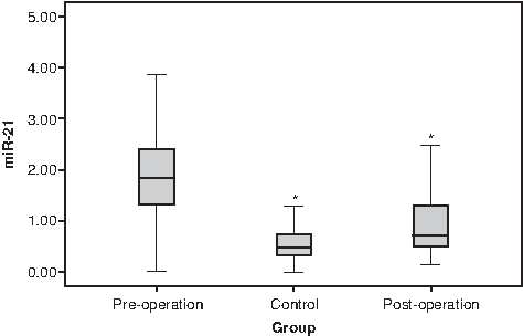
Comparison of the relative expression level of miR-21 in PBMCs between pre- and postoperative samples from prostate cancer (PCa) patients and healthy controls. *Compared with pre-operation group, p<0.05.
Association of miR-21 expression with clinicopathological factors
It was revealed that tumor differentiation, clinical stage, and lymph node metastasis were associated with miR-21 expression in PCa patients (Table 1). MiR-21 expression in PCa patients with poorly/moderately differentiated tumors was higher than that in the patients with well-differentiated tumors (p<0.001). MiR-21 expression in PCa patients with clinical stage A/B was lower than that in the patients with clinical stage C/D (p<0.001). Compared with PCa patients without lymph node metastasis, the expression level of miR-21 in the patients with lymph node metastasis was higher (p<0.001). No association of age with miR-21 expression in PCa was detected (p>0.05).
miR-21, microRNA-21; PBMCs, peripheral blood mononuclear cells.
Association of miR-21 expression with the risk of recurrence and metastasis
During follow-up, the relative miR-21 expression was compared in the control group, PCa without R/M group, and PCa with R/M group, as seen in Figure 4. Relative miR-21 expression in the control group, PCa without R/M group, and PCa with R/M group was 0.53±0.33, 0.61±0.22, and 1.78±0.73, respectively, revealing that relative miR-21 expression in the PCa without R/M group was greatly higher than that in the control group and PCa without R/M group (both p<0.001), and relative miR-21 expression in the PCa without R/M group was higher than that in the control group (p>0.05).

Comparison of the relative expression level of miR-21 in PBMCs between control group, PCa without recurrence or metastasis (R/M) group, and PCa with R/M group. *Compared with PCa with R/M group, p<0.05.
Diagnostic value of miR-21 for PCa
The expression level of miR-21 in the 75 patients confirmed with PCa was 1.85±0.89, ranging from 0.12 to 4.42. In the control group, the miR-21 expression level was 0.53±0.33, ranging from 0.13 to 1.38. The resultant sensitivities and specificities were used to generate the ROC curve, as seen in Figure 5. To generate the ROC curve, the sensitivity was plotted on the ordinate (y-axis) against the specificity plotted on the abscissa (x-axis) for all possible miR-21 relative expression values of the test. The ROC-AUC was 0.833 (95% CI=0.764-0.902). ROC curve analysis revealed that the cutoff point of miR-21 for diagnosis of PCa was 0.9 with a sensitivity of 87.5% and a specificity of 85.7%, which suggested a good diagnostic value of miR-21 (Table 2).
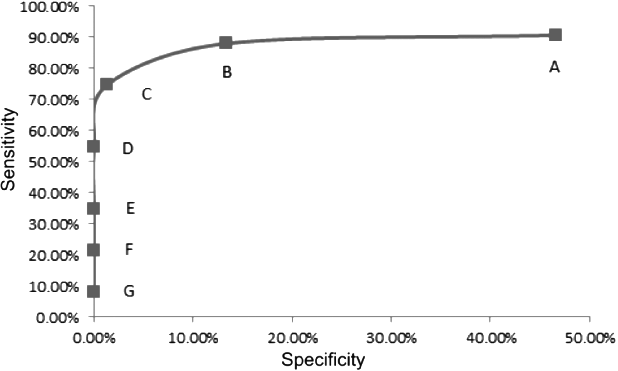
Receiver operating characteristic (ROC) curve analysis in the miR-21 assay for detecting PCa. A-G, diagnostic threshold values; A, 0.9, showed a sensitivity of 87.5% and a specificity of 85.7%, which suggested a good diagnostic value of miR-21.
A (+), positive case group; B (−), negative case group; C (+), positive control group; D (−), negative control group.
Discussion
In fact, several studies have revealed miR-21 expression profile in PCa, and the differential expression of miR-21 relating to clinicopathological factors (Li et al., 2012; Reis et al., 2012). Especially, a study investigating miR-21 in plasma/serum yields a promising field for clinical application (Wang and Zhang, 2012). However, serum/plasma tumor markers are lacking sufficient sensitivity and specificity to improve early cancer detection (Morimura et al., 2011). Thus, the significance of detecting new biomarkers utilizing a less invasive diagnostic assay for PCa should be emphasized. In our present study, we evaluated the miR-21 expression level in PBMCs of PCa patients compared with healthy controls and explored the diagnostic value and prognostic significance of miR-21 in PCa.
The results of our present study showed that miR-21 expression was significantly higher in PBMC samples from PCa patients without treatment of retropubic radical prostatectomy, compared with the patients after surgery and healthy controls, suggesting that miR-21 expression level in PBMCs may be associated with the occurrence and progression of PCa. Our results are in accordance with the findings of previous studies, such as Ribas et al. (2009), which have indicated that elevated expression of miR-21 enhanced PCa tumor growth and may explain the pathogenesis of PCa (Amankwah et al., 2013; Zheng et al., 2014). However, the molecular mechanisms responsible for the altered expression of miR-21 in PBMCs from PCa are still unclear. It was suspected that the potential mechanisms involve multilevel regulatory controls such as apoptosis, proliferation, and invasion-related genes. For instance, miR-21 was recently noted to target a number of well-known tumor suppressor genes, including TPM123 and PDCD4, which are related to the inhibition of tumor invasion (Frankel et al., 2008). In addition, miR-21 may influence PCa cell motility and invasion by regulating MARCKS, which is a protein with roles in cell adhesion, cell spreading, membrane trafficking, and cell motility based on its regulation of the actin cytoskeletal structure (Rumsby et al., 2011).
In addition, our study found miR-21 in PBMC samples was associated with clinicopathology of patients with PCa. MiR-21 expression was upregulated in PBMC samples from PCa patients with poorly/moderately differentiated tumors, clinical-stage A/B tumors, and lymph node metastasis, which indicated that the level of positive miR-21 expression may be a useful indicator of PCa progression. A higher expression level of miR-21 in PBMC samples, also dramatically related to the recurrence and metastasis of PCa, revealed a potential relationship between miR-21 in PBMCs and poor prognosis in PCa. Therefore, our current study suggests that miR-21 represents a potential novel drug target for treating PCa, which may reduce PCa relapse after removing androgenic steroids. For example, Li et al. have indicated that inhibiting miR-21 by antagomir-21 dramatically decreases the tumor formation of DU145 cells in vivo, which is an androgen-independent cell line, supporting the use of miR-21 as a new drug target for PCa treatment (Li et al., 2012). Amankwah et al. (2013) also have documented that positive expression of miR-21 was in relation to poor prognosis and forecast the risk of recurrence in PCa patients after radical prostatectomy. Accordingly, gene therapy utilizing the strategy of miR-21 inhibition may prove useful for PCa therapy.
In the present study, we chose miR-21, reported to present high levels in PCa previously, in PBMC samples as a new candidate miRNA, to assess its diagnostic value in PCa. The ROC curve analysis revealed a high sensitivity and specificity of miR-21 in diagnosing PCa, which suggested a good diagnostic value of miR-21. Our results are consistent with another study, which has demonstrated that representation of the data utilizing an ROC plot displayed obvious separation between the PCa patients and healthy controls, with a good diagnostic value (Morimura et al., 2011).
In conclusion, this study clearly revealed that high miR-21 expression in PBMCs is correlated to the progression, recurrence, and metastasis of PCa, thereby high miR-21 in PBMCs can supply a useful biomarker for screening PCa and monitoring the risk of recurrence and metastasis in PCa after retropubic radical prostatectomy. Although an evaluation of miR-21 expression in PBMCs in larger populations is needed, our results indicate that miR-21 in PBMCs may be a promising candidate as a molecular prognostic biomarker and therapeutic target for PCa.
Footnotes
Acknowledgment
We would like to acknowledge the reviewers for their helpful comments on this article.
Author Disclosure Statement
No competing financial interests exist.