
Abstract
Aims:
Evaluation of nucleic acids in plasma exosomes is a noninvasive method that can be used to detect different types of cancer. The aim of this study was to determine the value of exosomal long noncoding RNAs (lncRNAs) in detecting lung squamous cell carcinoma (LSCC).
Materials and Methods:
A total of 75 LSCC patients and 79 negative control subjects were enrolled in the study. Twenty differentially expressed lncRNAs were evaluated as potential candidates. Exosomes were isolated by ultracentrifugation, and lncRNA levels in exosomes were determined using real-time polymerase chain reaction. Receiver Operating Characteristic (ROC) curves were used to determine specificity and sensitivity.
Results:
Exosomal SOX2-OT was significantly upregulated in LSCC patients and showed the strongest power in detecting LSCC. The area under the ROC curve was 0.815, and the sensitivity and specificity were 76% and 73.17%, respectively. Moreover, exosomal SOX2-OT levels were significantly correlated with tumor size, TNM stage, and lymph node metastasis. Exosomal SOX2-OT levels were significantly decreased in the postoperative plasma of LSCC patients.
Conclusion:
SOX2-OT may serve as a promising noninvasive plasma-based tumor biomarker for LSCC.
Introduction
Lung cancer is a leading cause of cancer death worldwide (Torre et al., 2016). Lung squamous cell carcinoma (LSCC) is a common type of lung cancer that is mostly diagnosed at the advanced stage, with a 5-year survival rate of less than 15% after surgery and anticancer treatments. In routine clinical practice, lung cancer is usually diagnosed by pathological biopsy, which is an invasive procedure that carries risks of pleural reaction, hemothorax, pneumothorax, and infection; in addition, surgery can only be performed if the solid tumor size is large enough. A blood-based test is an ideal noninvasive method to detect and diagnose cancer sooner, as accurate and early detection of lung cancer is critical for effective treatment and reduced mortality. Serum biomarkers, such as carcinoembryonic antigen (CEA) and squamous carcinoma antigen (SCC), can identify many types of cancer, including LSCC; however, a lack of sensitivity and specificity has limited their value. Thus, identification of new LSCC-specific biomarkers with high sensitivity is needed.
Long noncoding RNAs (lncRNAs) are more than 200 bp in length with varied functions, including signaling, guiding ribonucleoprotein complexes, and molecular decoys. lncRNA expression has greater tissue specificity than protein-coding mRNAs, and only 1% of lncRNAs are ubiquitously expressed (Zhang et al., 2016c). In addition, lncRNAs can act either as oncogenes or as antioncogenes; these properties make them potential biomarkers for cancer diagnosis and prognosis.
lncRNA-HEIH expression in serum and exosomes is increased in patients with Hepatitis C virus-related hepatocellular carcinoma (Zhang et al., 2017a). The expression of H19 is significantly increased in breast cancer tissues and plasma compared with healthy controls and, therefore, may serve as a potential biomarker for breast cancer early screening and prognosis monitoring (Zhang et al., 2016b). LINC00857 plays an important role in the regulation of response to platinum-based chemotherapy and may serve as a novel prognostic and predictive biomarker for monitoring cisplatin resistance in patients with muscle-invasive bladder cancer (Dudek et al., 2018).
Exosomes are microvesicles released by cells and are 30-100 nm in diameter (Zabeo et al., 2017). lncRNAs are carried as cargo in exosomes and transferred into the blood. Exosomal RNAs promote pulmonary metastasis by recruiting neutrophils (Liu et al., 2016) and affecting lung fibrosis pathogenesis (Huleihel et al., 2017). Recently, it was demonstrated that circulating nucleic acids, particularly RNAs in exosomes, may be potential biomarkers for lung cancer (Rabinowits et al., 2009; Giallombardo et al., 2016; Wang et al., 2016; Reclusa et al., 2017). According to Qu et al. (2016), exosomal lncARSR serves as a predictor and potential therapeutic target for sunitinib resistance. ZFAS1 may also be protected by exosomes to enhance gastric cancer cell proliferation and migration (Pan et al., 2017). Differentially expressed lncRNAs in LSCC tissue and cell lines have been reported; however, their expression and clinical significance in plasma exosomes are unknown.
In this study, we selected 20 lung cancer-associated lncRNAs from an analysis of five datasets from the gene expression omnibus and evaluated their expression in plasma-derived exosomes (Yang et al., 2014). Real-time polymerase chain reaction (PCR) was used to evaluate expression levels in LSCC patients and negative control (NC) subjects, and Receiver Operating Characteristic (ROC) curves revealed the diagnostic value of SOX2-OT as a novel candidate biomarker.
Materials and Methods
Patients and sample collection
Clinical samples were obtained from the Medical Examination Center of the First Affiliated Hospital of China Medical University from April to December 2016. Protocols were approved by the Clinical Research Ethics Committee of the First Affiliated Hospital of China Medical University according to the tenets of the Declaration of Helsinki, and all participants provided informed consent. LSCC was confirmed by pathological diagnosis. Blood samples were collected in EDTA-K2 tubes before any anticancer treatment, and postoperative samples were collected the seventh day after surgery. A two-step separation was used to separate plasma from whole blood within 4 h after collection. Samples were first centrifuged at 800 g for 10 min at 4°C to spin down blood cells and then supernatants were centrifuged at 12,000 g for 10 min at 4°C to spin down the platelets. Plasma samples were used to isolate exosomes immediately.
Isolation of plasma exosomes
To isolate exosomes, 10 mL plasma was diluted at a ratio of 1:1 in phosphate-buffered saline and filtered using a 0.22 μm filter (Millipore). The collected media was ultracentrifuged at 120,000 g for 2 h at 4°C, and supernatants were discarded.
Transmission electron microscopy
A volume of 20 μL of exosomes was placed on a 400-mesh copper grid and incubated for 3 min at room temperature, followed by incubation with 3% phosphotungstic acid (pH = 6.5) for 5 min. The grid was dried at 37°C for 15 min and was then observed using transmission electron microscopy (TEM) (Hitachi H-7650).
Western blot analysis
Total protein was extracted from exosomes. Protein concentration was measured using a BCA Protein Assay Kit. sodium dodecyl sulfate-polyacrylamide gel electrophoresis (SDS-PAGE) was used to separate lysates, and PVDF membranes were used to transfer the proteins. After incubation with antibodies, the target bands were detected using an E3 imaging system.
Total RNA isolation
Total RNA was isolated from exosomes using the miRNeasy Serum/Plasma Kit (Qiagen) according to the manufacturer's instructions. Briefly, total RNA was isolated from a 250 μL-exosome suspension and eluted in 30 μL RNase-free water. Then, a 2 μL RNA solution was used for quantification with a NanoDrop 1000 spectrophotometer (Thermo Scientific); RNA samples were stored at −80°C.
Quantification of lncRNA by real-time PCR
RNA was reverse transcribed using a PrimeScript RT Reagent Kit (Takara) according to the manufacturer's instructions. PCR was performed using SYBR Premix Ex Taq (Takara) according to the manufacturer's recommended cycling conditions. Reverse transcription reactions were performed using an Eppendorf Mastercycler EP Gradient S (Eppendorf, Germany), while PCRs were performed using a LightCycler® 480 System (Roche Applied Science). The primers of lncRNAs and reference genes are shown in Supplementary Table S1.
Serum CEA and SCC determination
CEA and SCC levels in the serum were measured using a commercial kit (Roche Diagnostics, Mannheim, Germany) according to the manufacturer's instructions using a Roche Cobas E-601 immunoassay analyzer.
Statistical analysis
Relative RNA expressions were calculated using the following formula:
2−ΔCt (ΔCt = Cttarget − Ctreference). Kolmogorov-Smirnov and Shapiro-Wilk normality tests were used to determine characteristics of the data. Mann-Whitney, Wilcoxon, or Kruskal-Wallis tests were used to analyze differences in lncRNA expression. Area under the curve (AUC) and sensitivity and specificity of lncRNAs were determined by ROC analysis. All statistical tests were two sided, and p < 0.05 was considered to be statistically significant. Statistical analysis was performed using SPSS software.
Results
Patient characteristics
Plasma samples were taken from a total of 154 participants (75 LSCC patients and 79 NC subjects). The NC group consisted of 33 healthy subjects, 17 lung adenocarcinoma patients, 12 small cell lung cancer patients, 6 benign tumor patients, and 14 patients with other types of lung tumor (adenosquamous carcinoma or carcinoid). The clinical characteristics of LSCC patients are summarized in Supplementary Table S2.
Characterization of plasma exosomes
Exosomes were isolated and purified from the plasma of LSCC patients and NC subjects and were observed using a TEM. The size distribution was mainly between 30 and 100 nm (Fig. 1B). Western blot analysis of exosomal markers CD63 and CD9 and the endoplasmic reticulum marker calreticulin further confirmed the identity of the exosomes (Fig. 1A).
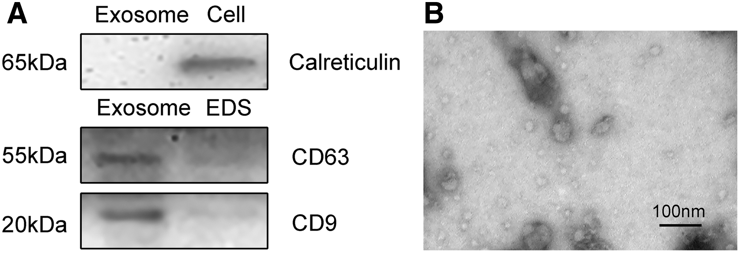
Characteristics of exosomes.
Evaluation of potential endogenous controls of exosomal lncRNAs
To select the optimal internal control for a lncRNA analysis of exosomes, we examined three candidate reference genes (GAPDH, β-actin, and TBP) in 10 exosome samples (5 LSCC patients and 5 NC subjects). GAPDH was the optimal endogenous control calculated by BestKeeper, which is a common algorithm. Raw Ct values are shown in Figure 2.
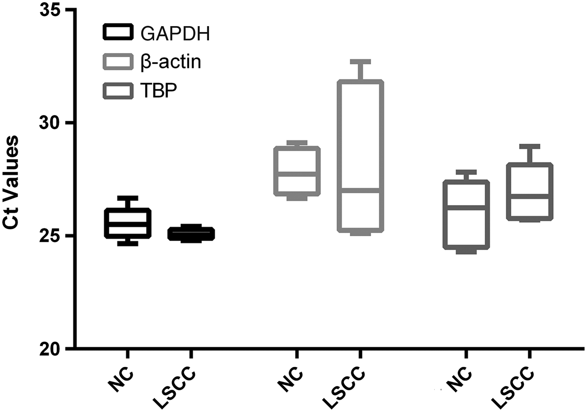
Ct values of reference genes. Raw Ct values of GAPDH, β-actin, and TBP (n = 10; 5 LSCC patients and 5 NC subjects). LSCC, lung squamous cell carcinoma; NC, negative control.
Selection and validation of candidate lncRNAs
A total of 20 lncRNAs, which are differentially expressed in LSCC, were selected for this study (Yang et al., 2014) (Supplementary Table S1). Differential lncRNAs were validated by real-time PCR in exosomes derived from 10 LSCC patients and 10 NC subjects. The detection rate of six lncRNAs was less than 75% in both LSCC exosome samples and NC exosome samples. Five lncRNAs (LOC152217, LOC730101, RP11-295G20.2, RNF157-AS1, and KDM5B-AS1) were significantly downregulated, and two lncRNAs (SOX2-OT and ENSG00000245648) were significantly upregulated (Fig. 3). SOX2-OT and ENSG00000245648 were validated by real-time PCR in plasma-derived exosomes of 75 LSCC patients and 79 NC subjects. Both of these lncRNAs were upregulated in LSCC samples (Fig. 4A, B). There were no significant differences between healthy controls and patients bearing cancer and lung tumors other than LSCC (Fig. 4C, D).
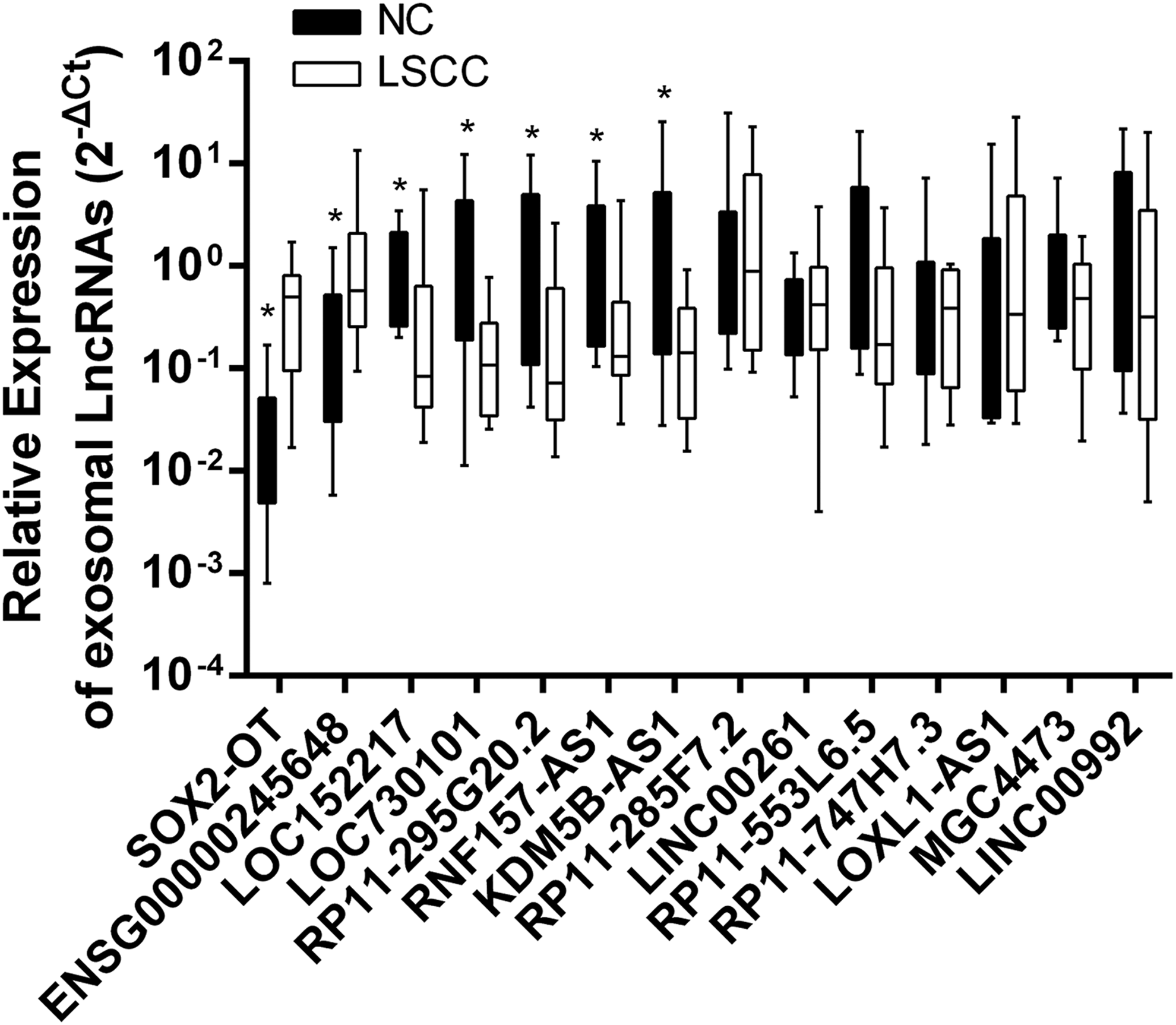
Expression of candidate exosomal lncRNAs. Expression levels of candidate exosomal lncRNAs in LSCC and NC samples (n = 20; n = 10 each). *p < 0.05. lncRNA, long noncoding RNA.
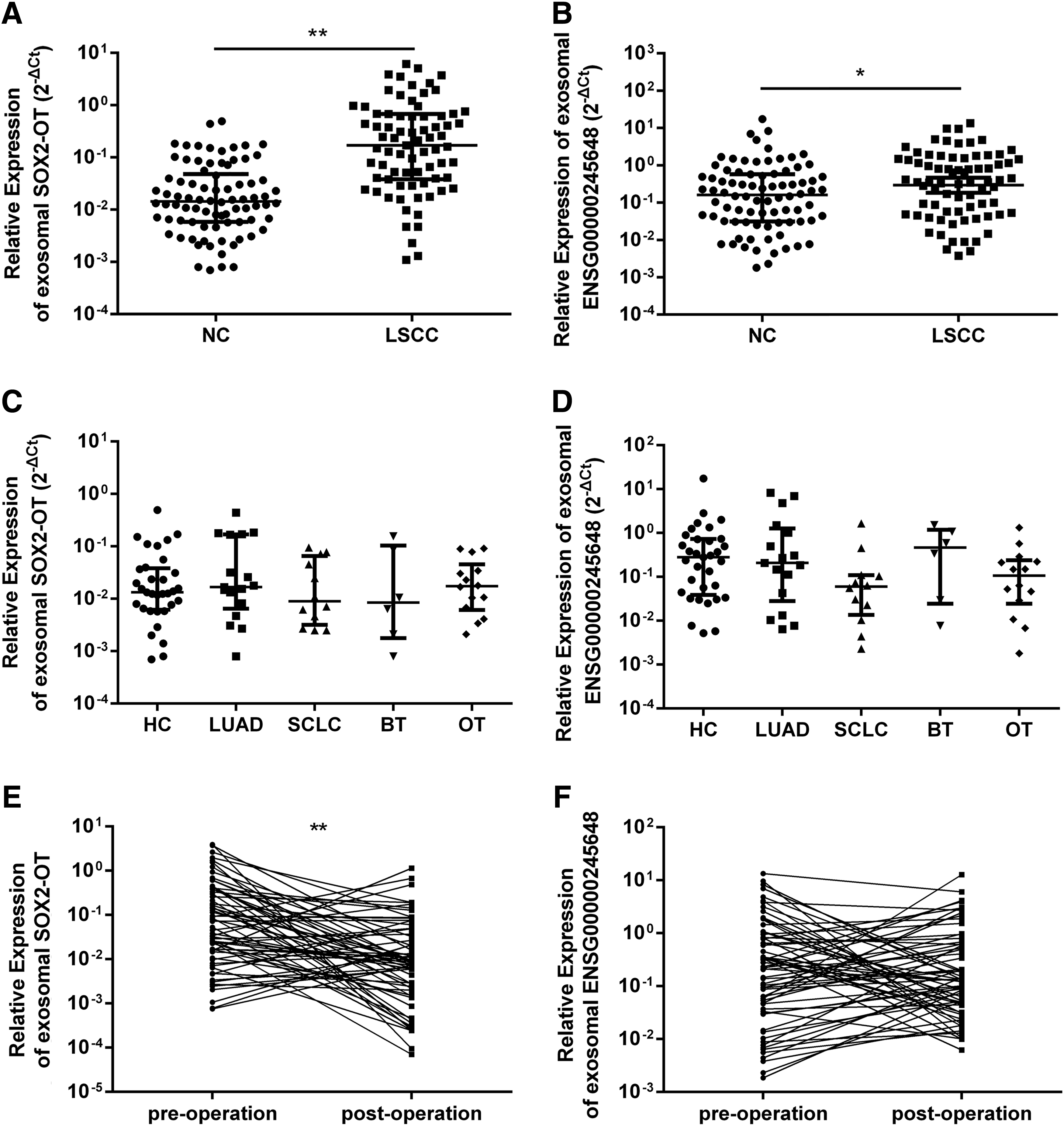
Expression levels of exosomal lncRNAs in validation set. Relative expression levels of SOX2-OT
We further measured the expression levels of exosomal SOX2-OT and ENSG00000245648 in 65 paired pre- and postoperative plasma samples from LSCC patients. Exosomal SOX2-OT expression levels were significantly reduced in postoperative plasma compared to preoperative plasma (Fig. 4E, F). These results indicate that SOX2-OT levels in plasma exosome might reflect expression in tumors. The PCR products were identified by the Sanger-based method.
Evaluation of SOX2-OT and ENSG00000245648 as exosomal biomarkers for diagnosing LSCC
ROC analysis was applied to evaluate the potential role of SOX2-OT and ENSG00000245648 as biomarkers for detecting LSCC. There were significant differences between the LSCC and NC groups, with an AUC of 0.815 (95% CI 0.748-0.882) for SOX2-OT and 0.611 (95% CI 0.522-0.699) for ENSG00000245648 (Fig. 5). The sensitivity and specificity of SOX2-OT were 76% and 73.17%, respectively.
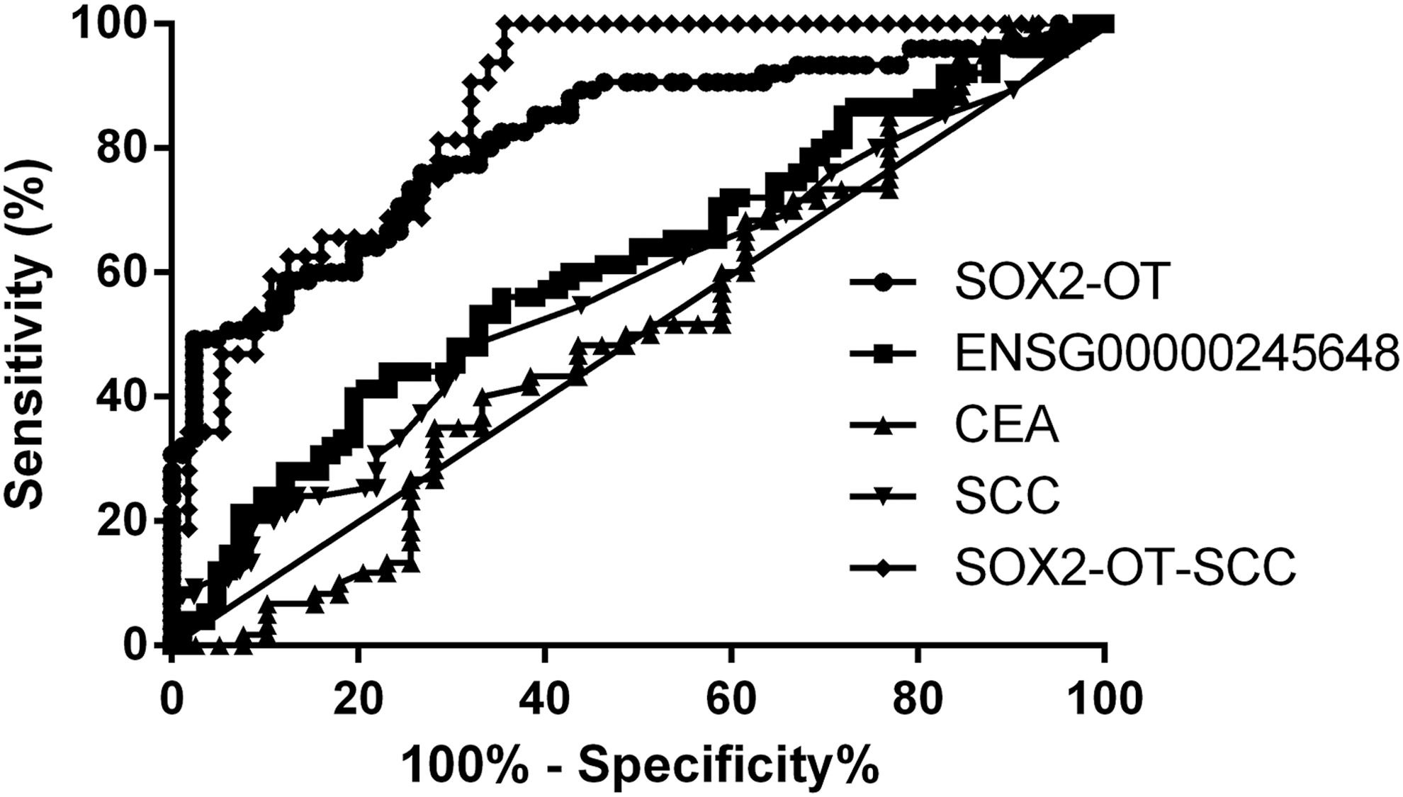
Diagnostic power of SOX2-OT, ENSG00000245648, and other tumor markers. To distinguish patients with LSCC from NC participants, diagnostic efficacy of SOX2-OT, ENSG00000245648, CEA, SCC, and SOX2-OT plus SCC was evaluated using ROC curve analysis. CEA, carcinoembryonic antigen; ROC, receiver operating characteristic; SCC, squamous carcinoma antigen.
The classic LSCC marker, SCC, had 49.09% sensitivity; however, the sensitivity of ENSG00000245648 was 44%. We combined SOX2-OT and ENSG00000245648 using binary logistic regression and found that the AUC was 0.816. Therefore, of the two lncRNAs in this analysis, SOX2-OT showed the best diagnostic power for discriminating LSCC from non-LSCC patients, suggesting that SOX2-OT could serve as a potential biomarker for detecting LSCC.
Correlations between exosomal SOX2-OT and clinical pathology characteristics
We examined the relationship between exosomal SOX2-OT levels with clinical pathology parameters, such as age, gender, smoking status, tumor size, TNM stage, lymph node metastasis, and distant metastases. Exosomal SOX2-OT expression correlated with tumor size, TNM stages, and lymph node metastasis; however, there were no differences between age and gender (Table 1).
Clinical Parameters of Plasma Exosomal SOX2-OT in Lung Squamous Cell Carcinoma Patients
p Values were estimated by Mann-Whitney U tests or Kruskal-Wallis tests.
LN, lymph node; LSCC, lung squamous cell carcinoma; NC, negative control.
Discussion
Real-time PCR is widely used for the quantitative detection of gene expression. However, with progression in exosomal lncRNA research, inclusion of a robust reference gene as an endogenous control is important for accurate results. As there was no established reference gene for exosomal lncRNAs, three housekeeping genes (GAPDH, TBP, and β-actin) were chosen as candidates for this study. BestKeeper, a common algorithm for reference gene evaluation, and GAPDH, endogenous control, were also selected; GAPDH has been selected in previous studies as an optimal reference gene to provide reliable results (Gezer et al., 2014; Zhang et al., 2016a; Xue et al., 2017).
Cargos in exosomes can be used as potential diagnostic and prognostic cancer biomarkers (Salehi and Sharifi, 2018). According to Tucci et al. (2018), patients with both PD-1 and CD28 upregulation in immune cell-derived exosomes have a better clinical response to ipilimumab. CoroMarker, which is stable in plasma and protected by exosomes, is a sensitive and specific biomarker for coronary artery disease (Yang et al., 2015). Therefore, exosomal RNAs could be potential biomarkers for cancer and other diseases.
In this study, we identified SOX2-OT and ENSG00000245648 as the only two lncRNAs stably detected and upregulated in plasma-derived exosomes. However, only SOX2-OT had the potential to discriminate LSCC from non-LSCC. The sensitivity and specificity of SOX2-OT were 76% and 73.17%, respectively, and it performed better than CEA and SCC, which are used currently in clinical practice; combining these two lncRNAs did not improve sensitivity and specificity. Furthermore, combining SOX2-OT and serum SCC could provide a more powerful differential diagnosis between LSCC patients and non-LSCC patients with an AUC of 0.864. These data suggest that plasma-derived exosomal SOX2-OT may serve as a valuable biomarker for detecting LSCC.
Additional analyses showed that exosomal SOX2-OT was higher in LSCC patients with bigger tumors or lymph node metastases. Exosomes are secreted from the cells of origin and fuse with membranes of receptor cells (Colombo et al., 2014; Kowal et al., 2014). In this way, exosomes can transfer RNAs and proteins; they therefore play important roles in cell-to-cell communication (Camussi et al., 2010). In addition, exosomes reach the target organ through the blood circulation (Zhang and Wang, 2015). As reported, integrins of tumor exosomes determine organotropic metastasis.
Tumor-derived exosomes taken up by organ-specific cells promote premetastatic niche formation. Exosomal integrins α6β4 and α6β1 have been associated with lung metastasis, while overexpression of αvβ5 in exosomes has been shown to promote liver metastasis (Hoshino et al., 2015). Moreover, serum exosome-derived long noncoding RNA MALAT-1 was shown to promote tumor growth and migration and to prevent apoptosis in lung cancer cell lines (Zhang et al., 2017b). As shown in this study, plasma exosomal SOX2-OT may play an important role in lung squamous carcinoma cell migration.
Recently, SOX2-OT expression levels were found to be significantly higher in hepatocellular carcinoma tissues compared with adjacent nontumor tissues (Sun et al., 2018). In addition, SOX2-OT was generally co-upregulated with SOX2 and OCT4 in esophageal squamous cell carcinoma (Shahryari et al., 2014; Wu et al., 2018). In this study, we found that SOX2-OT may serve as a biomarker for LSCC. We speculate that exosomal SOX2-OT may contribute to angiogenesis and metastasis of cancers, especially squamous cell carcinoma. To identify its tissue specificity, additional studies aimed at other patient populations are warranted.
In summary, our study showed that plasma-derived exosomal SOX2-OT was upregulated in LSCC samples and that SOX2-OT may serve as a novel biomarker for LSCC. In addition, exosomal SOX2-OT was shown to correlate with TNM stages and metastasis of LSCC patients. However, multicenter retrospective and prospective studies are warranted for further investigation.
Footnotes
References
Supplementary Material
Please find the following supplemental material available below.
For Open Access articles published under a Creative Commons License, all supplemental material carries the same license as the article it is associated with.
For non-Open Access articles published, all supplemental material carries a non-exclusive license, and permission requests for re-use of supplemental material or any part of supplemental material shall be sent directly to the copyright owner as specified in the copyright notice associated with the article.