




Abstract
Aims:
Metastasis is a significant obstacle to curing esophageal squamous cell carcinoma (ESCC). The CCAAT/enhancer binding protein β (C/EBPβ) and matrix metalloproteinase 3 (MMP3) are thought to play key roles in cancer invasion and metastasis. In this study, we aimed to detect whether C/EBPβ-mediated tumor invasion was dependent on MMP3. In addition, we determined whether C/EBPβ upregulation was associated with MMP3 levels and metastatic status in patients with ESCC.
Materials and Methods:
A total of 126 patients with ESCC were recruited for this study. The mRNA and protein levels of C/EBPβ and MMP3 in ESCC cell lines and specimens from ESCC patient were determined by reverse transcription-polymerase chain reaction and western blot, respectively. Tumor cell invasion was analyzed using an in vitro Matrigel Invasion Assay. The correlation between C/EBPβ and MMP3 expression was determined by Pearson's correlation analysis.
Results:
Both mRNA and protein levels of MMP3 were upregulated by C/EBPβ overexpression and downregulated by C/EBPβ siRNA in KYSE150 cell cultures. The promotion of ESCC cell invasion through C/EBPβ was inhibited by MMP3 siRNA. The level of C/EBPβ was correlated with MMP3 and metastatic status in patients with ESCC.
Conclusions:
C/EBPβ upregulation promoted tumor cell invasion in an MMP3-dependent manner in vitro and was associated with metastatic status in ESCC.
Introduction
Esophageal cancer is the eighth most prevalent cancer globally, which accounts for the sixth leading cause of cancer death worldwide (Torre et al., 2015). Esophageal cancer mainly comprises two different histopathological forms: esophageal squamous cell carcinoma (ESCC) and esophageal adenocarcinoma. The underlying molecular mechanism of these two forms is very distinct. In China, ESCC is the predominant form (∼90%) of tumor arising from esophageal epithelial cells (Torre et al., 2015; Siegel et al., 2016). Despite recent improvements, but as lack of specific symptoms and effective early diagnostic methods, esophageal cancer tends to be diagnosed at a late stage and its 5-year survival rate remains poor, which is <20% (Chen et al., 2016). Metastatic spread of cancer affords the greatest barriers to be overcome in curing ESCC. Therefore, it is critical to identify molecules associated with metastasis as biomarkers for early diagnosis, prognostic prediction, and therapeutic targets for developing effective therapy in ESCC.
CCAAT/enhancer binding protein β (C/EBPβ) belongs to a family of leucine-zipper transcription factors (Zahnow, 2002). Through heterodimerization or homodimerization with its relatives, C/EBPβ is recruited to target DNA to control gene transcription, which plays a key role in a range of physiological and pathophysiological processes, including cell proliferation/differentiation, survival and apoptosis, inflammation, metabolism, oncogene-induced senescence, and tumorigenesis (Zahnow, 2009). C/EBPβ is encoded by an intronless gene, named C/EBPβ, which can be expressed as three distinct protein isoforms (LAP1 and LAP2 as transcriptional activators mediating cell differentiation, and C/EBPβ LIP, which lacks the N-terminal trans-activating and central regulatory regions, as a dominant negative inhibitor of transcription with elevated expression in proliferative tissue) by using different in-frame translational start codons in its single exon (Rask et al., 2000; Zahnow, 2002). C/EBPβ was found to be aberrantly upregulated in multiple types of cancers, including colorectal cancer (Rask et al., 2000), ovary cancer (Sundfeldt et al., 1999), and breast cancer (Raught et al., 1996). In addition, C/EBPβ LIP isoform mediates epithelial growth factor-induced epithelial-mesenchymal transistion (EMT) and increases migration and invasion of esophageal cancer cells in a manner that is dependent on miR-203 inactivation (Li et al., 2014). It is believed that dysregulated expression of C/EBPβ is involved in regulation of tumor growth, angiogenesis, and tumor cell invasion and metastasis.
Tumor cell invasion and metastasis may involve controlled degradation of extracellular matrix (ECM), which is mediated by matrix metalloproteinases (MMPs), a large family of zinc-containing endopeptidases playing an important role in a number of pathological processes, including tumor metastasis (Shuman Moss et al., 2012; Brown and Murray, 2015). MMPs are usually expressed at low levels in tissues and upregulated when ECM degradation is required under both physiological and pathological conditions (Sternlicht et al., 2000). Similar to C/EBPβ, MMP upregulation is associated with tumor invasion and metastasis of different malignant tumors (Chambers and Matrisian, 1997). MMP3, also known as stromelysin-1, is an MMP family member involved in the breakdown of ECM during both normal tissue remodeling and pathological processes (Kessenbrock et al., 2010; Szarvas et al., 2011). Interestingly, it has been shown that the expression of MMP3 can be regulated by C/EBPβ in arthritis (Tsushima et al., 2012), breast cancer (Xia et al., 2015), and colorectal cancer (Ji et al., 2018). MMP3 has novel transcription-factor-like function, which can regulate the CTGF/CCN2 gene (Eguchi et al., 2008), and the intranuclear PEX domain of MMP involves proliferation, migration, and metastasis of aggressive adenocarcinoma cells (Okusha et al., 2018).
However, it is not clear whether C/EBPβ is also responsible for MMP3 dysregulation in ESCC and whether their upregulations regulate tumor cell migration and metastasis in ESCC. To address the questions, we investigated whether C/EBPβ induced MMP3 expression and mediated tumor cell migration in an MMP3-dependent manner in cultured ESCC cells. We further determined whether MMP3 upregulation is correlated with the level of C/EBPβ expression as well as with metastasis and survival in human patients with ESCC. Our study will facilitate the development of potential diagnostic markers and therapeutic targets against ESCC.
Materials and Methods
Patients
A total of 126 patients with primary sporadic ESCC who were admitted into Henan Province People's Hospital of Zhengzhou University (Zhengzhou, Henan, China) during January 2010 and June 2011 were enrolled in this study. All cancer samples were collected with patients' written content before any anticancer treatments and preserved in formalin-fixed paraffin-embedded (FFPE) blocks. This retrospective study was approved by our institutional ethics committee. All the specimens were obtained with full ethical approval from the institution committee.
Cell culture and DNA plasmid transfection
The human ESCC cell line KYSE150 and EC9706 were grown at 37°C under a humidified atmosphere of 95% of air and 5% CO2.The cells were maintained in Dulbecco's modified Eagle's medium (DMEM) supplemented with 10% fetal bovine serum (FBS), 50 mg/mL streptomycin, and 50 mg/mL penicillin.
For overexpression experiments, cells were transfected with pcDNA3.1 vector expressing C/EBPβ or MMP3 or empty control vector using Effectene reagent (Qiagen, Valencia, CA) according to the manufacturer's instruction. The stable cell lines were established after selection with 400 mg/mL of G418 (Invitrogen, Carlsbad, CA) and maintained with 200 mg/mL G418.
For knockdown experiments, cells were transfected with siRNA targeting C/EBPβ or MMP3 (SMARTpoolDharmacon, 10 nM; GE Dharmacon, Lafayette, CO), or negative control siRNA (siNTCDharmacon, 10 nM) mixed with DharmaFECT 1 transfection reagent (GE Dharmacon) per vendor's instruction.
RNA isolation and quantitative reverse transcription-polymerase chain reaction assay
We extracted total RNA from both cultured ESCC cells and FFPE tissue samples using RNeasy or RNeasy FFPE Kit (Qiagen) according to manufacturer's instruction. Before reverse transcription-polymerase chain reaction (RT-PCR), total RNAs were subjects for measuring RNA concentration and for quality check (a 260/A280 ratio of 1.8 or higher is required to pass the quality control). Quantitative real-time RT-PCR was performed on a 7500 RealTime PCR System (Applied Biosystems, Waltham, MA). Housekeeping gene GAPDH was used as an internal control. MMP3, forward 5′-TGTGGAGTTCCTGATGTTGGT-3′ and reverse 5′-CCTTCATACAGCCTGGAGAATG-3′; C/EBPβ, forward 5′-TTCTACTACGAGGCGGACTG-3′ and reverse 5′-GAGAAGAGGTCGGAGAGGAAG′; GAPDH, forward 5′-TTGGTATCGTGGAAGGACTCA-3′ and reverse 5′-CAGTAGAGGCAGGGATGATGT-3′. The RT-PCR was conducted under the following conditions: 42°C for 30 min, 94°C for 5 min, a 40 cycles of reaction (94°C for 20 s and 60°C for 1 min), and a final extension at 72°C for 10 min.
Western blotting
Cultured KYSE150 cells were collected with trypsin and subjected to protein lysis in prechilled lysis buffer (pH 7.5) containing Tris buffer (20 mM), NaCl (137 mM), EDTA (2 mM), Triton X (1% v/v), glycerol (10% v/v), NaF (50 mM), DTT (1 mM), and a protease inhibitor cocktail (Sigma, St. Louis, MO). Protein samples were then separated by electrophoresis (SDS-PAGE) and transferred onto PVDF membranes (Amersham, United Kingdom). After blocking in 5% nonfat milk in TBS with 0.1% Tween 20, the membrane was incubated with primary antibodies at room temperature for 1 h and then incubated with either HRP-tagged goat anti-mouse or rabbit IgG secondary antibodies. The following primary antibodies were used: MMP3, rabbit polyclonal (Abcam, United Kingdom); β-actin, mouse monoclonal (clone AC-74, A2228; Sigma-Aldrich). An Amersham ECL Western Blotting Detection Kit (Amersham) was used to visualize immunoreactive signals for MMP3 and the reference protein b-actin, which were quantitated using NIH ImageJ version 1.6.
Cell invasion assays
Cell invasion assays were conducted by using Matrigel-coated Transwell chambers (Corning, Inc., Corning, NY). In brief, 3 × 10⁴ transfected KYSE150 and EC9706 cells in 200 μL serum-free medium were plated into the upper chamber, and 500 μL of culture medium with 10% FBS was placed in the bottom chamber serving as a chemoattractant. After incubation at 37°C for 48 h, the cells that had invaded to the bottom chamber were fixed and stained with 0.1% crystal violet for 30 min. The invaded cells were photographed and counted in six random fields under microscope.
Statistical analysis
Statistical analyses were performed using Prism 6 analysis software
Results
To examine whether C/EBPβ regulates MMP3 expression in ESCC, we determined the MMP3 level in culture KYSE150 ESCC cells both upregulating and downregulating C/EBPβ. The mRNA and protein level of MMP3 are significantly higher in C/EBPβ-expressing cells than in control cells transfected with empty vectors (Fig. 1A). In contrast, MMP3 mRNA and protein are significantly lower in cells transfected with C/EBPβ siRNA than those with control siRNA (Fig. 1B).

MMP3 expression is upregulated by C/EBPβ overexpression and downregulated by C/EBPβ siRNA in ESCC KYSE150 cell line.
We next determined whether C/EBPβ-mediated ESCC tumor cell invasion was dependent on MMP3 expression. We transfected KYSE150 cells stably expressing C/EBPβ or control vectors with control or MMP3 siRNA for 24 h. Tumor cell invasion was then determined using Matrigel invasion assays. In vector-expressing cells, the number of invasive cells was significantly lower in MPP3 siRNA-transfected cells than in control siRNA-transfected cells (p < 0.043 by one-way ANOVA with Tukey's test, Fig. 2A, B). Meanwhile, in cells transfected with control siRNA, the number of invasive cells was significantly higher in C/EBPβ stably expressed cells than vector control cells (p < 0.029 by one-way ANOVA with Tukey's test, Fig. 2A, B). However, this upregulation was significantly reduced by MMP3 siRNA transfection (p < 0.037, column 3 vs. 4, by one-way ANOVA with Tukey's test, Fig. 2A, B). In contrast, MMP3 overexpression promoted KYSE150 cell invasion, which was demonstrated by the number of invasive cells in cells transfected with MMP3-encoding vectors were significantly higher (p < 0.05, Student's t-test) than that in cells transfected with control empty vectors (Fig. 2C). These results suggested a critical role of MMP3 in C/EBPβ-mediated tumor cell invasion.

C/EBPβ-mediated enhancement of ESCC invasion is dependent on MMP3.
To investigate whether MMP3 and C/EBPβ together have prognostic value in ESCC, we measured mRNA levels of both genes and determined their potential association with metastatic status and OS in 126 patients with ESCC. We found the expression of both C/EBPβ and MMP3 genes were significantly higher (p < 0.03 and p < 0.01, respectively, by Student's t-test) in metastatic patients than in patients without metastatic disease (Fig. 3A, B). We further found that there is a significant correlation (p < 0.01, Pearson's correlation) between the expression levels of C/EBPβ and MMP3 mRNA (Fig. 4A). Moreover, there was a significant correlation between the OS time and MMP3 mRNA levels (Fig. 4B). These results suggested that upregulation of both C/EBPβ and MMP3 was associated with metastatic status and an unfavorable prognosis in ESCC.

The expression of both MMP3 and C/EBPβ were upregulated in metastatic ESCC.
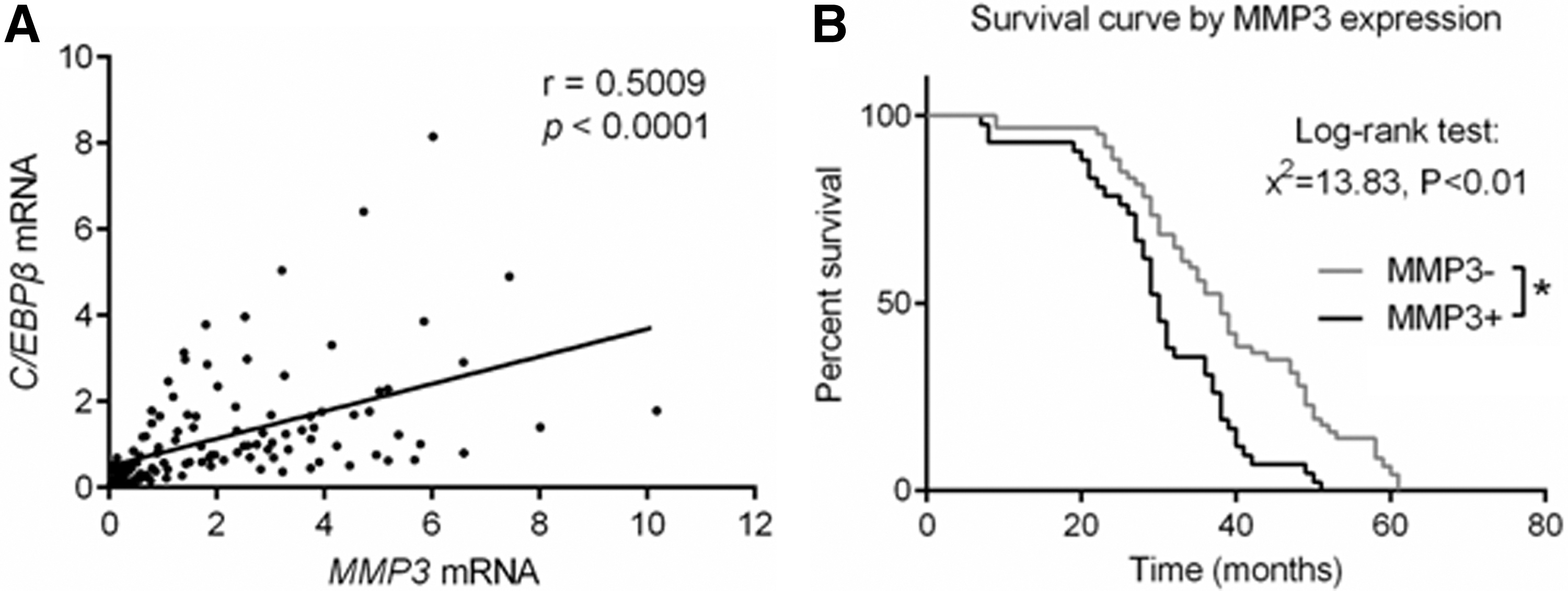
MMP3 expression level correlated C/EBPβ level and overall survival in patients with ESCC.
Discussion
Metastasis is one of the worst clinical manifestations and characteristics in malignant tumors. It is usually associated with unfavorable prognosis and poor survival in patients with colorectal cancer (Qin et al., 2016; Xu et al., 2018). In this study, we investigated the role of transcription factor C/EBPβ and MMP3 in the regulation of ESCC cell invasion in vitro and whether their expression levels are associated with tumor metastasis, unfavorable prognosis, and poor survival in human patients with ESCC. Our study provided strong evidence that both molecules are potential candidates as diagnostic markers and therapeutic targets in ESCC.
C/EBPβ is a transcription factor, which is important in a range of physiological and pathological processes, including cell differentiation, inflammation, metabolism, cell survival, and oncogene-induced senescence (Sebastian et al., 2005; Zahnow, 2009; Smink and Leutz, 2010; Huber et al., 2012). For example, it is well defined that C/EBPβ plays a key role in both mammary gland development and pathogenesis of breast cancer, especially during the regulation of angiogenesis associated with invasive phenotype (Grimm and Rosen, 2003; Kagan et al., 2003). It is verified that the decreased transcription activity mediated by C/EBPβ is closely associated with abnormal cell proliferation in cancer, whereas the increased transcription activity is usually associated with cellular differentiation (Grimm and Rosen, 2003; Zahnow, 2009). Previous study has indicated that LIP, the dominant-negative inhibitory isoform of C/EBPβ, is highly upregulated and associated with poorly differentiated tumor cells and poor prognosis in human breast cancer (Zahnow et al., 1997). Similarly, another publication has shown that LIP expression correlated with cell proliferation marker Ki67 and that high LIP/LAP ratio (transcriptional activity inhibition) was associated with tumor progression and poor prognosis in breast cancer (Milde-Langosch et al., 2003). In colorectal cancer, increased expression of C/EBPβ has been reported to be associated with tumor cell invasiveness (Rask et al., 2000). Recent studies have shown that C/EBPβ can maintain an open chromatin state by H3K79 methylation of multiple drug-resistance genes through recruiting the methyltransferase DOT1L, thereby augmenting the chemoresistance of ovarian cancer (Liu et al., 2018). Consistent with this report, we have demonstrated that C/EBPβ-expressing human ESCC cells exhibited enhanced migration. Moreover, we have shown that elevated level of C/EBPβ is associated with metastatic status and unfavorable prognosis in human patients with ESCC. Together, these results suggest that induction of C/EBPβ is likely a key step in initiating tumor cell invasion and metastasis in ESCC.
MMPs, including MMP3, are major proteolytic enzymes that mediate ECM breakdown (Johansson et al., 2000; John and Tuszynski, 2001; Sternlicht and Werb, 2001). The expression of MMPs was suppressed under normal conditions, but was upregulated when EMC remodeling or breakdown is required, which is involved in various physiological processes and numerous diseases, including colorectal cancer (Zucker and Vacirca, 2004). Previously published studies have shown that MMPs play a significant role in tumor growth, development, invasion, and metastasis (Szarvas et al., 2011; Shuman Moss et al., 2012). The MMP3-1612 5A/6A polymorphism was significantly associated with susceptibility to ESCC (Guan et al., 2014). Tumor suppressor gene leucine-rich glioma inactivated 1 (LGI1) acted through extracellular signal-regulated kinase (ERK1/2) signaling to downregulate MMP3 expression and subsequently suppressed metastasis of ESCC (Zhu et al., 2014). NRD1, which encodes nardilysin protein, promotes ESCC cell invasion through induction of MMP2 and MMP3 expression. Nardilysin-positive ESCC cases were more advanced in terms of T classification, N classification, and tumor stage than nardilysin-negative ESCC cases. Whereas the expression of MMP2 and MMP3 mRNA was significantly lower in NRD1-knockdown TE5 cells than in negative control siRNA-transfected cells (Uraoka et al., 2014). In line with these reports, we have shown that MMP3 is upregulated in cultured ESCC cells and MMP3 upregulation is associated with metastasis in patients with ESCC. Interestingly, it has be suggested that MMP3 expression can be regulated by C/EBPβ in arthritis (Tsushima et al., 2012) and in human breast cancer (Xia et al., 2015). Consistent with these findings, we have shown that MMP3 is upregulated in cultured ESCC cells overexpressing C/EBPβ, suggesting that MMP3 upregulation is mediated by C/EBPβ in ESCC. Moreover, the expression of MMP3 correlated with the expression of C/EBPβ and both the expression of MMP3 and C/EBPβ is independently associated with metastasis in human patients with ESCC, suggesting that C/EBPβ-induced MMP3 upregulation (likely through reduced transcriptional activity by increasing the inhibitory isoform LIP) is a key step to initiate ESCC migration and metastatic process.
In conclusion, we have demonstrated that MMP3 is upregulated in C/EBPβ-expressing ESCC cells and the upregulation is required in C/EBPβ-induced tumor cell migration in cultured ESCC cells. In a cohort of 126 patients with ESCC, the expressions of MMP3 and C/EBPβ correlate to each other and their upregulations are associated with metastasis and poor prognosis. These findings indicate that both MMP3 and C/EBPβ should be further investigated as potential biomarkers and therapeutic targets in ESCC.
Footnotes
Acknowledgment
This work was supported by a grant from the Major Project of Science and Technology in Henan Province (No. 161100311400).
Author Disclosure Statement
No competing financial interests exist.