




Abstract
Background:
HOX transcript antisense RNA (HOTAIR) and H19 are two long noncoding RNAs that play vital functions in the development of colorectal cancer (CRC).
Subjects and Methods:
The expression level of HOTAIR and H19 in the sera samples of Egyptian CRC patients along with normal controls was evaluated by quantitative real-time PCR. The possible correlations with the biochemical and clinicopathological characteristics were determined.
Results:
The expression of HOTAIR and H19 showed 7.55- and 11.38-fold increased levels, respectively, in CRC patients compared to the controls (p < 0.001). Furthermore, HOTAIR expression in CRC patients with regional lymph node metastasis was significantly higher when compared with CRC patients without regional lymph node metastasis (p = 0.034). HOTAIR and H19 expression showed no significant correlation with tumor site or carcinoembryonic antigen concentration. The sensitivity and specificity of HOTAIR and H19 in the detection of CRC cases were calculated as 92.9% and 100%, respectively.
Conclusion:
HOTAIR and H19 expression levels are upregulated in Egyptian CRC patients, and therefore can be considered noninvasive diagnostic biomarkers with high sensitivity and specificity.
Introduction
Colorectal cancer (CRC) is counted as the third prevalent cancer after breast and lung cancers with an incidence rate of 9.7% worldwide (Elshafei et al., 2017). More than 1 million cases develop CRC every year with a mortality rate reaching 33% in the developed countries (Shi et al., 2014). Cancer metastasis in the liver, which develops in 50% of CRC patients, represents the major cause of death in those patients (Hasselgren et al., 2015). Interestingly, some risk factors are known to increase the chances of CRC incidence such as alcohol, tobacco use, obesity, insufficient exercise, and chronic intestinal inflammation (Lizarbe et al., 2017). On the molecular level, CRC results from the accumulation of both genetic and epigenetic changes. The latter affects vital cellular processes such as proliferation, differentiation, migration, cell death, DNA repair, and stability (Bardhan and Liu, 2013).
Long noncoding RNAs (LncRNAs) are defined as LncRNAs with a length greater than 200 nucleotides and not translated into proteins (Shi et al., 2015). LncRNAs are expressed in different types of cancers and their functions are associated with many aspects of cancer biology such as differentiation, apoptosis, imprinting, and cell cycle (Chen et al., 2016). LncRNAs are classified into five major types: sense, antisense, bidirectional, intronic, and intergenic LncRNAs (Rejhova et al., 2017).
LncRNAs regulate the gene expression through several mechanisms, including scaffold, enhancer, decoy, and others (Melia et al., 2016). The binding of LncRNA with cellular proteins alters protein's activity and localization. Furthermore, LncRNA may be processed into smaller RNA, which can act as endo-small interfering RNA to target other RNAs, resulting in their degradation. LncRNA may also modulate the alternative splicing of pre-mRNAs at post-transcriptional processes (Gutschner and Diederichs, 2012). In particular, LncRNAs have vital functions in the regulation of CRC. They stimulate tumor growth, suppress apoptosis, enhance cell invasion, proliferation, and increase chromosomal instability (Han et al., 2014; Xu et al., 2014).
One of the LncRNAs is H19, which is defined as an oncogene involved in different cancer types. It is located on chromosome 11 in humans and it is a maternally expressed imprinted gene that has a biological function in the development of mammals (Wang et al., 2015; Han et al., 2016). Numerous studies indicated that H19 is upregulated in multiple cancer types, including CRC (Liang et al., 2015). H19 plays a critical role in cancer metastasis by promoting epithelial to mesenchymal transition. Moreover, H19 increases CRC cell proliferation through H19/miR-675/RB pathway (Tsang et al., 2009).
HOX transcript antisense RNA (HOTAIR) is an LncRNA located in the Homeobox C gene (HOXC) locus on chromosome 12q13.13. It has a length of 2158 nucleotides (Kogo et al., 2011). HOTAIR stimulates metastasis and proliferation in multiple cancers and it is considered a negative indicator for cancer prognosis (Zheng et al., 2015). Its expression level was significantly increased in hepatocellular carcinoma, breast cancer, and CRC (Ding et al., 2014).
Although LncRNAs, as tissue-based markers, show remarkable diagnostic and prognostic roles in different cancer types, they are viewed as invasive biomarkers characterized by a relative high cost, prone to sampling error, and may lead to possible complications. Several studies pointed to the advantageous role of the circulating (Plasma/serum) LncRNAs, which are derived from the tumor cells, as noninvasive cancer biomarkers to enhance screening, diagnosis, prognostic evaluation, following up of therapeutic efficiency, and monitoring of disease recurrence (Lindner et al., 2015; Zhou et al., 2015; Duan et al., 2016; Wang et al., 2016).
In particular, there is little information available on the gene expression of Egyptian patients due to the large population number and the high cost of genetic testing. Therefore, screening the expression level of LncRNAs in the Egyptian CRC patients as well as normal controls by using noninvasive and easily sampled biomarker will definitely have a positive impact on both the scientific and clinical settings.
This study aims to measure the expression level of HOTAIR and H19 LncRNAs in the serum of CRC patients from the Egyptian population and to investigate their correlations with the demographic, biochemical, and clinic investigatory findings of patients.
Subjects and Methods
Subjects
This study was performed on 56 CRC patients along with 60 normal controls. The patients attended the gastrointestinal colonoscopy at Al-kasr Al-Ainy Hospital, Cairo University, for CRC screening as they were complaining from different colonic symptoms, including CRC alarming symptoms.
Exclusion criteria
Patients who had the following criteria were excluded from the study:
Inflammatory bowel disease, patients with family history of CRC, CRC recurrence, and other cancer types, except metastases of CRC and CRC patients treated with radiotherapy or chemotherapy before the surgical interference.
Patients who were diagnosed with CRC based on pathological findings, colonoscopic and abdominal ultrasound findings as well as clinical decisions were recruited into the study. The study was carried out in agreement with the declaration of Helsinki principles as revised in 2000. The Ethics Review Committee of Faculty of Medicine at Cairo University approved this study. All subjects gave a written informed consent before participation in the study.
Serum total RNA extraction
Total RNA was extracted from sera samples by miRNeasy extraction kit (Qiagen). Briefly, 100 μL serum sample was added to 500 μL Qiazol lysis reagent and incubated at room temperature for 5 min. Addition of 100 μL chloroform, mixing by vortex for 15 s followed by incubation at room temperature for 3 min, and centrifugation again at 12,000 g for 15 min at 4°C were performed. The aqueous phase was moved to a new tube and 1.5 times its volume of absolute ethanol was added. From this mixture, 700 μL was added in RNeasy Mini spin column and centrifuged at 8000 g for 15 s at room temperature. When the mixture passed the column completely, 700 μL of buffer RWT was added to each column, and centrifuged at 8000 g for 15 s at room temperature. Five hundred microliters of buffer RPE was applied to the column and then centrifuged at 8000 g for 15 s at room temperature twice. Finally, the column was inserted into a 1.5 mL collection tube to elute RNA by applying 50 μL RNase-free water onto the column and then centrifuged at 8000 g for 1 min. Five microliters of the eluted RNA was utilized for the assessment of RNA purity and concentration by a nanodrop (Thermo scientific). When the ratio for 260/280 is ∼2.0, RNA was accepted as pure.
Reverse transcription into cDNA
In this step, reverse transcription (RT) was performed on 100 ng of total RNA in 20 μL final volume using the miRNeasy Reverse Transcription kit (Qiagen). RT master mix was prepared on ice by adding 4 μL miScript HiFlex buffer, 2 μL nucleotide mix, and 2 μL miScript reverse transcriptase mix, and then, 10 μL template RNA and 2 μL RNase-free water were added to produce a final volume of 20 μL. The reaction mixture was mixed, and incubated in thermal cycler (Biometra, Germany) for 60 min at 37°C, and 5 min at 95°C.
Quantitative RT-PCR analysis
Expression of HOTAIR and H19 LncRNAs, was evaluated by quantitative real-time PCR where RT2 SYBR green PCR kit and primer assays (Qiagen) were used. The housekeeping GAPDH was used as an endogenous reference RNA. For real-time PCR analysis of each LncRNA, 2.5 μL of diluted RT products (cDNA template) was added to 5 μL RNase free water, 12.5 μL RT2 SYBR Green PCR master mix, and 1 μL of the following assay primers:
HOTAIR: Forward primer: 5′-GGTAGAAAAAGCAACCACGAAGC-3′, reverse primer: 5′-ACATAAACCTCTGTCTGTGAGTGCC-3′ (Gupta et al., 2010).
H19: Forward primer: 5′-CCCACAACATGAAAGAAATGGTGC-3′, reverse primer: 5′-CACCTTCGAGAGCCGATTCC-3′ (Zhang et al., 2014).
GAPDH: Forward primer: 5′-CCGGGAAACTGTGGCGTGATGG-3′, reverse primer: 5′-AGGTGGAGGAGTGGGTGTCGCTGTT-3′ (Gupta et al., 2010).
Incubation was performed as follows: 15 min at 95°C, then 40 cycles, each cycle was carried out by three successive steps, including DNA denaturation for 15 s at 94°C, followed by annealing for 30 s at 55°C, and then extension for 30 s at 70°C. Quantitative Real-time PCR was carried out on a Rotor gene Q-Real Time PCR System (Qiagen).
Fold change of the circulating HOTAIR and H19 LncRNA
The expression levels of the two LncRNAs were evaluated using the Livak method (Livak and Schmittgen, 2001) as follows:
Where ΔCT is the difference in the CT for the target gene (X: HOTAIR/H19) and reference gene (R: GAPDH). ΔΔCT is obtained by subtracting the ΔCT value of the sample (q: CRC patient) from that of the calibrator (cb: Normal control).
Statistical analysis
The statistical SPSS version 25 (SPSS, Inc., Chicago, IL) was used for data analysis. Normality was checked by Shapiro Wilk test. Normally distributed data were expressed as mean ± standard deviation, whereas median and interquartile range were used for nonparametric data and relative frequencies were used for categorical variables. Comparisons between variables were performed using unpaired student t-test in normally distributed quantitative variables, while Mann-Whitney test was used for non-normally distributed quantitative variables such as HOTAIR and H19 (fold change). Chi-squared test was used for comparing categorical data. Fisher's exact test was performed instead when the expected frequency is less than 5. Spearman correlation coefficient was used to determine the correlations between different variables. Receiver operating characteristic (ROC) curve was constructed in which the sensitivity for the test of all possible cutoff points is plotted on the y-axis against 1-specificity on the x-axis, where the area under the curve (AUC) is a reflection of the diagnostic power of HOTAIR and H19 as CRC biomarkers. p-Values <0.05 were considered statistically significant.
Results
Demographic and clinical characteristics of the CRC patients
This study was performed on 56 CRC patients along with 60 normal controls. Male CRC patients represented 78.6%, while female CRC patients represented 21.4%. Obese CRC patients represented 28.5% of all cases with a body mass index (BMI) of 27.3 ± 3.4. On the other hand, male control subjects represented 53.3%, while female controls represented 46.7%, with 46.7% of controls being obese. According to the radiological and colonoscopic findings performed on the CRC patients, tumor site in cecum and ascending colon represented 25% of the cases and in transverse and flexures represented the same percentage (25%), whereas 57.1% of the cases presented with rectosigmoid CRC. Importantly, CRC patients with regional lymph node metastasis represented 34.6%, while CRC patients without regional lymph node metastasis represented 65.4%. Twelve (23.1%) of CRC patients showed liver metastasis. Other radiological, abdominal ultrasound, and colonoscopy findings are briefly represented in Table 1.
Demographic and Clinical Investigatory Findings of the Colorectal Cancer Patients
ABD US, abdominal ultrasound; CRC, colorectal cancer; CT, computed tomography; HFLs, hepatic focal lesions.
The biochemical characteristics of CRC patients with and without regional lymph node metastasis
To identify the effect of regional lymph node metastasis on our results, we divided the CRC patients into two categories: CRC patients with regional lymph node metastasis and CRC patients without regional lymph node metastasis. The mean age and BMI of CRC patients with lymph nodes were significantly higher compared with patients without lymph nodes. There were no significant differences in any of the other biochemical parameters between both categories (Table 2).
Biochemical Characteristics of Colorectal Cancer Patients, Colorectal Cancer Patients With Regional Lymph Node Metastasis, and Colorectal Cancer Patients Without Regional Lymph Node Metastasis (Parametric and Nonparametric Data)
Bold italic means significant value at p < 0.05.
ALB, albumin; ALK, alkaline phosphatase; ALT, alanine aminotransferase; AST, aspartate aminotransferase; BIL, bilirubin; BMI, body mass index; CA, cancer antigen; CEA, carcinoembryonic antigen; ESR, erythrocyte sedimentation rate; GGT, gamma-glutamyl transferase; HB, hemoglobin; INR, international normalized ratio of prothrombin; PLT, platelet; SD, standard deviation; TGs, triglycerides; TLC, total leukocyte count; WBC, white blood count.
It is worthy to mention that no significant differences were detected among CRC patients in any of the biochemical parameters based on the tumor site (Supplementary Table S1). Furthermore, comparison of CRC patients based on the regional lymph node metastasis or liver (distant) metastasis did not reveal any significant difference in the biochemical parameters (Supplementary Tables S2 and S3).
The expression level of HOTAIR and H19 in CRC patients
The gene expression analysis of HOTAIR and H19 levels in the sera samples of CRC patients and normal controls showed that, HOTAIR expression level was increased by 7.55-fold in CRC patients compared to the controls (p < 0.001). In addition, the expression level of H19 showed 11.38-fold increase in CRC patients compared to the controls (p < 0.001) (Fig. 1A). Interestingly, HOTAIR expression in the CRC patients with regional lymph node metastasis was significantly higher compared with the CRC patients without regional lymph node metastasis (27.10-fold vs. 5.14-fold, respectively, p = 0.034). However, H19 expression showed no significant difference between CRC patients with and without regional lymph node metastasis (p = 0.833) (Fig. 1B).
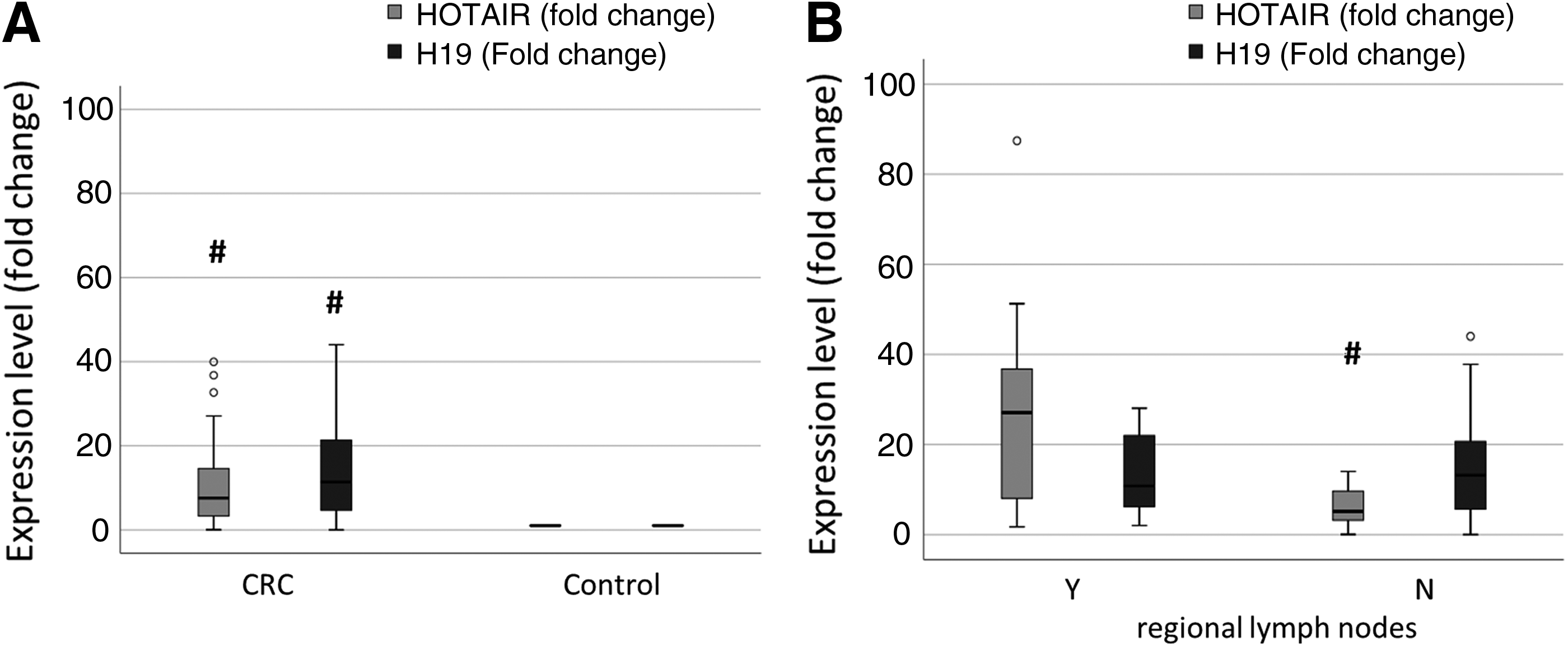
The expression level of HOTAIR and H19 among the subjects.
The sensitivity and specificity of HOTAIR and H19 as CRC diagnostic biomarkers
The ROC curve analysis was performed to compare the predictive power of HOTAIR and H19 as diagnostic biomarkers and to calculate the optimal cutoff values. The results showed that both HOTAIR and H19 have AUC = 0.929 with p-value <0.001. Both HOTAIR and H19 showed high specificity and sensitivity (100% and 92.9%, respectively) (Fig. 2).
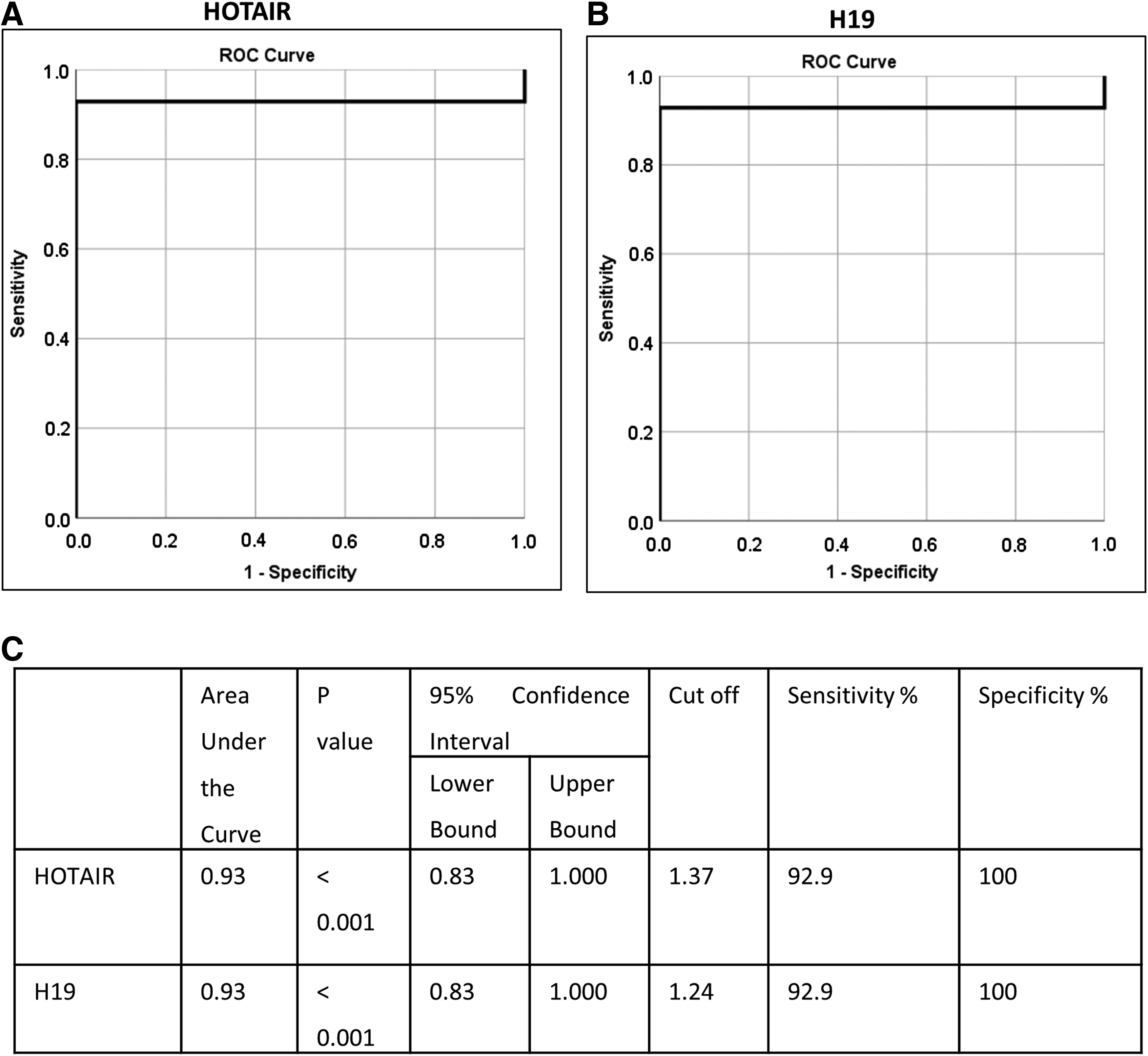
The sensitivity and specificity of HOTAIR and H19 for detection of CRC cases.
However, only HOTAIR showed a significant diagnostic power for the detection of CRC patients with regional lymph node metastasis with AUC = 0.76, sensitivity = 66.7%, and specificity = 88.2%, p = 0.03 (Fig. 3).
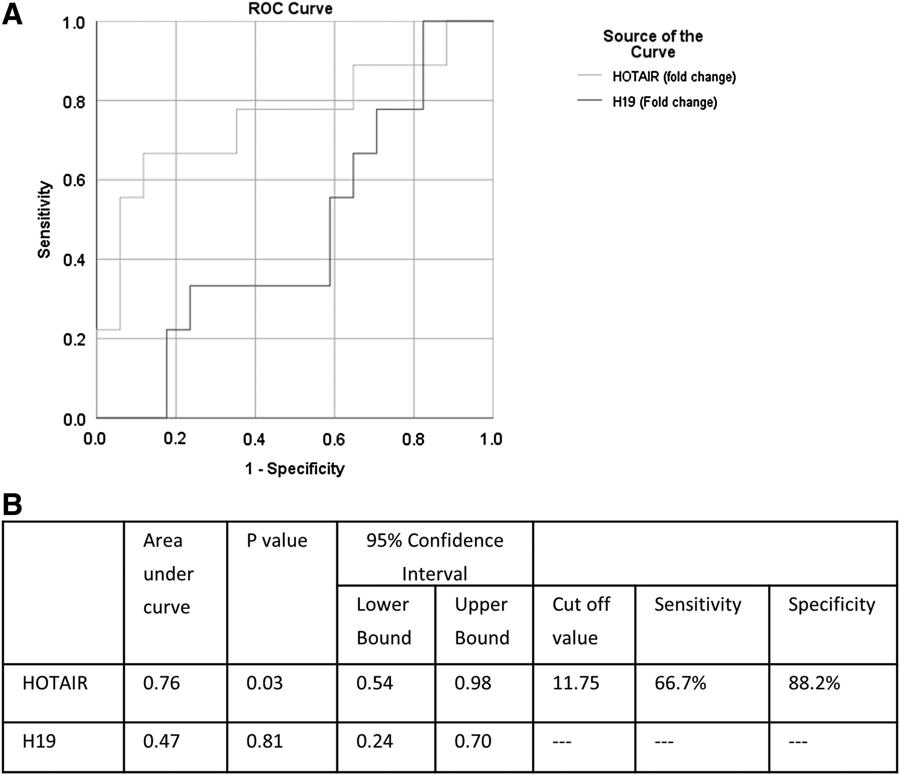
The sensitivity and specificity of HOTAIR and H19 for detection of CRC patients with lymph node metastasis.
The correlation between HOTAIR and H19 expression and the biochemical parameters
The correlation analysis between either HOTAIR or H19 expression levels and the biochemical parameters of CRC patients revealed that HOTAIR is inversely proportional with ESR (correlation coefficient = −0.69, p = 0.03) and alpha-fetoprotein (correlation coefficient = −0.90, p = 0.00), while directly proportional with ALT (correlation coefficient = 0.45, p = 0.02). Interestingly, H19 showed only a direct proportional correlation with GGT (correlation coefficient = 0.69, p = 0.01) (Table 3).
The Correlation Between HOTAIR and H19 Expression and the Biochemical Parameters of Colorectal Cancer Patients
Bold italic means significant value at p < 0.05.
HOTAIR, HOX transcript antisense RNA.
The correlation of HOTAIR and H19 expression with the clinic-investigatory findings
The correlations of the expression level of HOTAIR/H19 with the clinico-investigatory findings are summarized in Table 4. The results showed that HOTAIR expression was significantly higher in CRC patients with regional lymph node metastasis compared with CRC patients without regional lymph node metastasis (p = 0.03), as revealed by the computed tomography findings. Neither HOTAIR nor H19 expressions were significantly correlated with mass lesion or wall thickening. Interestingly, the expression level of H19 in CRC patients with fatty liver was significantly higher compared to CRC patients without fatty liver (p = 0.04), as revealed by abdominal ultrasound findings. No significant correlations were detected between HOTAIR/H19 expressions and patients with/without hepatic focal lesions or distension (Table 4).
The Correlations of HOTAIR and H19 Expression with the Clinico-Investigatory Findings
Bold italic means significant value at p < 0.05.
N, no; Y, yes.
Discussion
CRC is a human malignancy viewed as a life-threatening disease worldwide (Huang et al., 2019). It represents 8.5% of all cancer mortality in the world, which recalls for the development of early diagnostic biomarkers (Zhang et al., 2018a). LncRNAs are recognized as a class of noncoding RNA transcripts. They are novel research hotspots correlated to carcinogenesis and metastasis in various human cancers. In particular, LncRNAs play a dynamic function in the development and progression of CRC (Zheng et al., 2016). HOTAIR is one of these LncRNAs that are involved in multiple cancers such as hepatocellular, lung, breast, pancreatic, cervical, and esophageal, and CRCs (Geng et al., 2011; Ge et al., 2013; Kim et al., 2013; Sørensen et al., 2013; Xue et al., 2014; Lee et al., 2016). Our results demonstrated that HOTAIR expression was upregulated in CRC patients compared to the controls. These results are in agreement with the previous studies that demonstrated the upregulation of HOTAIR expression in CRC cells and pointed to the importance of measuring HOTAIR expression as a novel CRC prognostic biomarker (Xiao et al., 2018; Huang et al., 2019). Furthermore, Lin et al. (2018) suggested that downregulation of HOTAIR may inhibit the proliferation and invasion of CRC cells. Our study revealed that, HOTAIR expression was significantly higher in CRC patients with regional lymph node metastasis, which is compatible with the findings of Zhang et al. (2018b) who reported that elevated HOTAIR expression was associated and positively correlated with lymph node metastasis and other clinicopathological variables of CRC patients. Remarkably, our results showed an inverse relationship between AFP and HOTAIR expression, although Yang et al. (2011) found no significant correlation between AFP and HOTAIR expression in hepatocellular carcinoma.
LncRNA H19 exhibits multiple pathological functions in cancer development such as enhancing the tumor growth through recruiting and binding to the eukaryotic translation initiation factor 4A3 (eIF4A3) (Wang et al., 2018). Furthermore, it was previously found that H19 exhibited a role in CRC metastasis by enhancing RAS-MAPK signaling pathway (Yang et al., 2018). Wang et al. (2018) demonstrated that overexpression of H19 had a significant correlation with CRC differentiation and depth as well as recurrence. H19 knockdown blocked G1-S transition, inhibited cell proliferation, and reduced cell migration (Ohtsuka et al., 2016). Hence, H19 expression was found to be upregulated in bladder, breast, hepatocellular, esophageal, and CRCs (Hibi et al., 1996; Ariel et al., 1998; Berteaux et al., 2005; Matouk et al., 2007; Luo et al., 2013).
Multiple studies demonstrated the relevance between obesity and increased risk of CRC incidence; however, the mechanism of action is not fully illustrated. Increased BMI is correlated with increased insulin resistance, which in turn enhances the activity of insulin-like growth factor I (IGF-I). The increased circulating levels of IGF-I was found to promote cell proliferation and suppress apoptosis (Meyerhardt et al., 2003). This may explain the observed significant correlation between H19 expression and fatty liver in our study. However, others found that increased BMI in CRC patients was not significantly associated with increased risk of CRC recurrence or death (Meyerhardt et al., 2008). Interestingly, our study showed no significant correlation between the expression of HOTAIR or H19 and the tumor site. This is in agreement with Lin et al. (2018), who demonstrated that there was no significant correlation between HOTAIR expression and tumor location. In addition, our results showed no correlation between CEA and the expression level of the studied LncRNAs. CEA is a glycoprotein produced by the intestinal mucosal tissue and its blood level is mostly elevated in CRC patients (El-taher et al., 2016). The current results are compatible with Tatangelo et al. (2018), who found that the elevated CEA levels in CRC patients were associated with lymph node metastasis, while no significant correlation with HOTAIR expression was detected (Tatangelo et al., 2018).
The most remarkable limitation of this study is the relatively small number of subjects. Therefore, large-scale samples from the Egyptian population are recommended for verification of these findings. In addition, future studies are required to ensure that circulating HOTAIR and H19 expression levels are matched with that of CRC tissues.
Conclusions
This study demonstrated the upregulation of HOTAIR and H19 expression in the Egyptian CRC patients, and therefore their relevance as noninvasive diagnostic biomarkers with high sensitivity and specificity. Furthermore, there was no correlation between HOTAIR or H19 expression with the tumor site or CEA concentration.
Footnotes

References
Supplementary Material
Please find the following supplemental material available below.
For Open Access articles published under a Creative Commons License, all supplemental material carries the same license as the article it is associated with.
For non-Open Access articles published, all supplemental material carries a non-exclusive license, and permission requests for re-use of supplemental material or any part of supplemental material shall be sent directly to the copyright owner as specified in the copyright notice associated with the article.
