




Abstract
Objective:
Carotid atherosclerosis is one of the major risk factors for ischemic stroke. The presence of carotid plaque has been widely used to assess the risk of clinical atherosclerotic disease. Lectin-type oxidized LDL (low-density lipoprotein) receptor 1 (LOX-1), lysosomal acid lipase (LAL), and acyl-CoA:cholesterol acyltransferase 1 (ACAT1) are important for lipid accumulation in atherosclerosis. The objective of this study was to investigate the relationship between single nucleotide polymorphisms (SNPs) in the LOX-1, LAL, and ACAT1 genes and the presence of carotid plaque in a Northern Chinese population.
Methods:
Three polymorphisms in LOX-1 (rs1050286), LAL (rs11203042), and ACAT1 (rs11576517) were identified and genotyped in 215 patients with carotid plaque and 252 controls using the polymerase chain reaction with high-resolution melting analysis.
Results:
The LOX-1 (rs1050286) AA and LAL (rs11203042) TT genotypes were significantly associated with increased risk of carotid plaque, whereas a ACAT1 (rs11576517) TT genotype was shown to be protective against carotid plaque in a Northern Chinese population (p < 0.05). Even after the Bonferroni correction, the LAL (rs11203042) TT genotype (odds ratio = 3.838, 95% confidence interval = 1.748-8.426, p < 0.001) was still associated with an increased risk for carotid plaque.
Conclusions:
These results suggest that the LAL (rs11203042) TT genotype is associated with increased risk for carotid plaque in a Northern Chinese population, and that the LOX-1 (rs1050286) AA genotype shows a nonstatistically significant trend towards association. However, no association was found between the ACAT1 (rs11576517) polymorphisms and carotid plaque presence.
Introduction
Atherosclerosis is a chronic vascular inflammatory disease characterized by excessive accumulation of foam cells in the arterial wall, plaque formation, and ultimate stenosis of blood vessels (Sini et al., 2018). Carotid atherosclerosis, which is a manifestation of atherosclerotic disease in the cervical arteries, leads to obstruction of blood supply to the brain and has been recognized as a risk factor for ischemic stroke (Forgo et al., 2018). Presence of carotid plaque has been widely used to assess the risk of clinical atherosclerotic disease (Bonanno et al., 2017). Patients with bilateral carotid plaques are more likely to develop coronary artery disease or other fatal diseases (Jashari et al., 2013). Several studies have shown that heritability of carotid plaque is more than 20%, indicating that genetic factors play an important role in the pathogenesis of carotid plaque (Zivotic et al., 2016).
The foam cell is a hallmark of atherosclerotic disease. Macrophages serve as the main source of foam cells (Chistiakov et al., 2015), and cholesterol ester levels in macrophages may influence the development of foam cells (Hutchins and Heinecke, 2015). Cholesterol ester levels are regulated by many factors. Macrophages and endothelial cells express lectin-type oxidized LDL (low-density lipoprotein) receptor 1 (LOX-1) receptors that sense, transport, or take up LDL, especially ox-LDL (Pothineni et al., 2017). Cellular concentration of cholesterol/cholesterol ester is determined by enzymes, such as lysosomal acid lipase (LAL) and acyl-CoA:cholesterol acyltransferase 1 (ACAT1). LAL digests cholesteryl esters, thereby releasing free cholesterol in lysosome where ACAT1 enzymes transform cholesterol to cholesteryl esters in the endoplasmic reticulum. Changes in enzyme activity and content can alter the process, break the dynamic balance of lipids, and drive the formation of foam cells (Chistiakov et al., 2016) (Fig. 1).
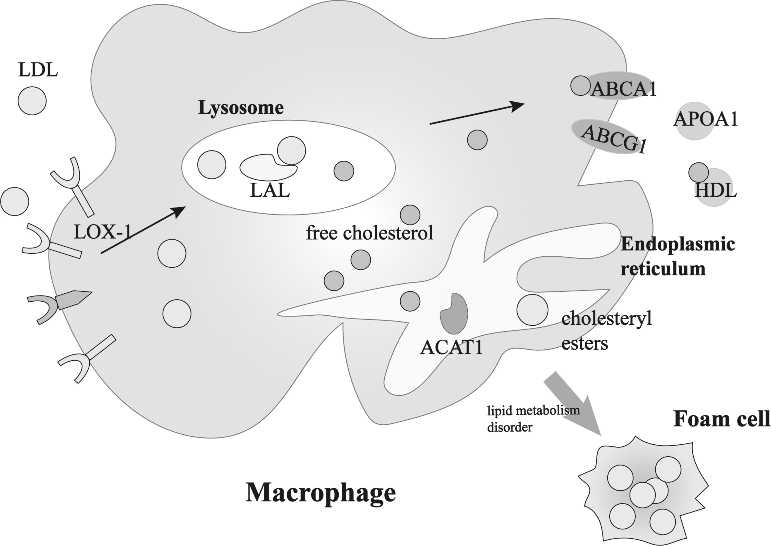
Mechanisms of LOX-1, LAL, and ACAT1 in macrophages. ABCA1, ATP binding cassette subfamily A member 1; ABCG1, ATP binding cassette subfamily G member 1; ACAT1, acyl-CoA:cholesterol acyltransferase 1; APOA1, apolipoprotein A1; HDL, high-density lipoprotein; LAL, lysosomal acid lipase; LDL, low-density lipoprotein; LOX-1, lectin-type oxidized LDL receptor 1.
Single nucleotide polymorphisms (SNPs) have been found to influence the expression of receptors or enzymes at the gene level, thereby affecting the disease state. Morini et al. (2016) found that the LOX-1 (rs1050286) variant AA could upregulate LOX-1 expression in human HeLa and HepG2 cell lines. Dehghan et al. (2016) found that LAL (rs11203042) may increase the risk of coronary atherosclerotic heart disease through the Genome Wide Association Study. ACAT1 (rs11576517) is located in the exon. Exon site mutations may have more important effects on ACAT1 expression, but no relevant studies on ACAT1 (rs11576517) have been reported. These genes are involved in lipid metabolism (Liang et al., 2013), atherosclerosis (Li et al., 2006), and Alzheimer's disease (Shibuya et al., 2015). However, little is known about the relationship between SNPs in these genes and carotid plaque in a Northern Chinese population.
In this study, we used polymerase chain reaction with high-resolution melting analysis (HRM-PCR) to investigate the association of LOX-1 (rs1050286), LAL (rs11203042), and ACAT1 (rs11576517) with carotid plaque presence in a Northern Chinese population.
Materials and Methods
Subjects
We designed a case-control study involving 215 patients with carotid plaque (129 males and 86 females) from The First Affiliated Hospital of China Medical University. All patients presented with carotid plaque when examined by carotid color Doppler ultrasonography. Carotid plaques were defined as an intima-media thickness ≥1.5 mm, and the properties of carotid plaque, such as size, morphology, and echogenic features, were evaluated by Doppler ultrasonography (Saba et al., 2010). Patients with cancer, HIV, or any type of heart, liver, and kidney diseases were excluded from enrollment. We recruited 252 healthy individuals (134 males and 118 females) from the Medical Examination Center to serve as controls. Subjects in the control group showed no carotid atherosclerotic changes. Both cases and controls were of Chinese descent and were younger than 65 years. This study was approved by the Ethics Committee of The First Affiliated Hospital of China Medical University ([2015]23). The consent was obtained from each participant. Study population characteristics are summarized in Table 1.
Demographic Characteristics of the Study Population
p < 0.05, statistical significance.
Alb, albumin; Cys-C, cystatin C; Glu, glucose; Hcy, homocysteine; HDL, high-density lipoprotein; LDL, low-density lipoprotein; TC, total cholesterol; TG, triglycerides.
DNA extraction
DNA was extracted from peripheral blood using the QIAamp DNA Micro Kit (QIAGEN, Hilden, Germany) following the manufacturer's instructions. Extracted DNA was stored in a −80°C freezer (Thermo Fisher Scientific, Bartlesville, OK).
SNPs selection and genotyping
SNP sites were selected based on the following criteria: (1) targeted SNPs LOX-1 (rs1050286) and LAL (rs11203042) were selected based on previous studies; SNP site rs11576517 in the exon region of ACAT1 was selected from NCBI dbSNP database, as there has been no research reported on rs11576517; (2) all SNPs had a minor allele frequency >0.05; (3) the two ends of SNP sites can be used to design primers for HRM-PCR.
We designed primers with annealing temperatures ranging from 55°C to 65°C and produced amplicons from 100 to 200 bp in length using Primer Premier 6 software (Table 2). The Light Cycler® 480 System (Roche Applied Sciences, Penzberg, Germany) was used to perform HRM analyses. PCR systems were as follows: Master Mix (5 μL; Roche Applied Sciences) containing FastStart Taq DNA polymerase, reaction buffer, dNTPs and HRM dye, forward and reverse primers (0.5 μL, 4 μM; Sangon Biotech), magnesium chloride (1.0 μL, 25 mM; Roche Applied Sciences), genomic DNA (0.5 μL, 30 ng/μL), and double-distilled water (2.5 μL). PCR conditions were as follows: initial denaturation for 4 min at 95°C, then 45 cycles of 95°C for 10 s, annealing in the range of 53-65°C for 30 s, and 72°C for 10 s. HRM-PCR (Roche Applied Science, Mannheim, Germany) was used for genotyping. We used Gene Scanning Module to analyze the data and obtained three sets of curves: wild homozygous type, mutant homozygous type, and heterozygous type. Twenty PCR products were randomly selected from the three genotypes to verify sequencing using an ABI7000 sequence detection system (Applied Biosystems, Foster City, CA) (Fig. 2). If the sequencing results were consistent with the genotype obtained by the HRM-PCR method, three groups of standards were obtained: wild homozygous, mutant homozygous, and heterozygous, and the remaining samples were tested by HRM-PCR to obtain the genotype results.
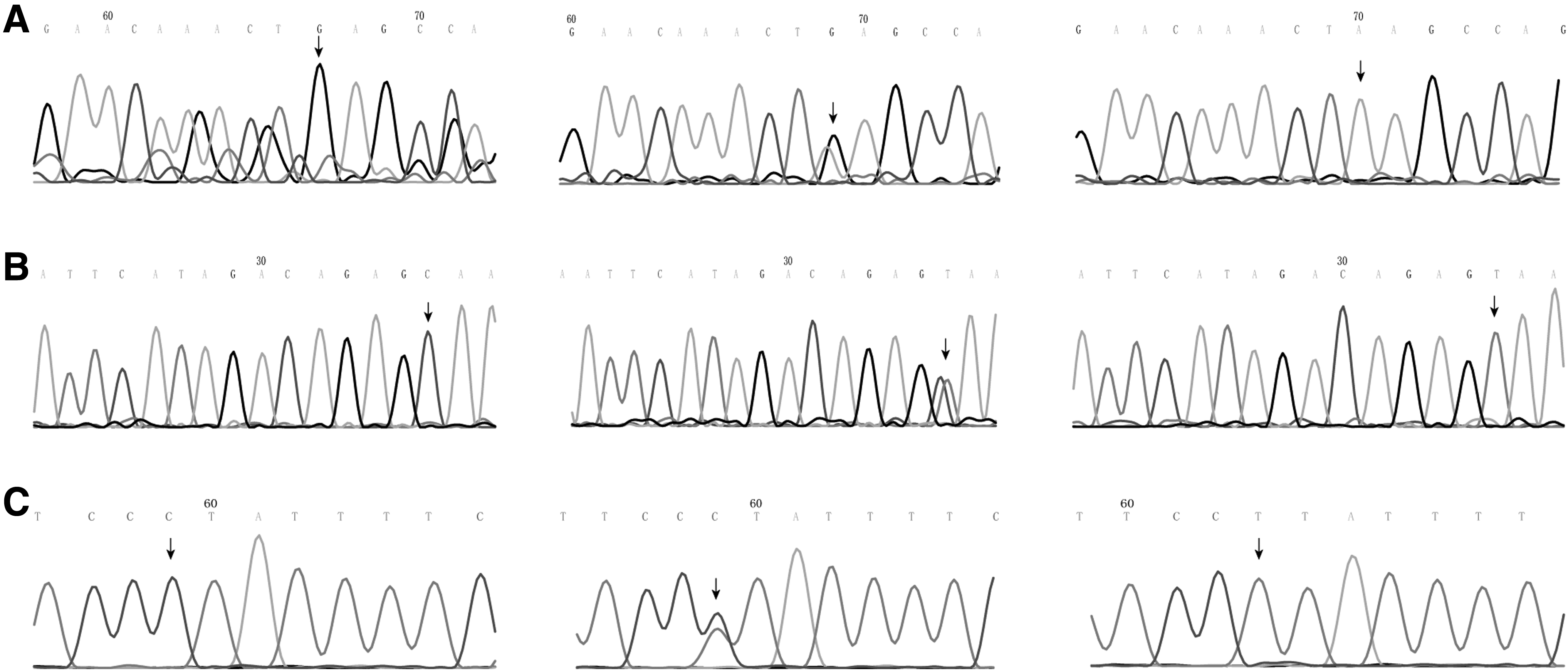
The arrows are pointing to
Polymerase Chain Reaction Primers and Amplicons
SNP, single nucleotide polymorphism.
Statistics
Statistical analysis was performed using SPSS 24.0 statistical software (SPSS, Inc., Chicago, IL). Student's t-test was used to compare normally distributed data. The Mann-Whitney U test was used to analyze data with a nonnormal distribution. Chi-square test was used to assess the Hardy-Weinberg equilibrium (HWE) and compare genotypic distributions of LOX-1 (rs1050286), LAL (rs11203042), and ACAT1 (rs11576517) between cases and controls. All tests were two-tailed, and a p-value of <0.05 was considered statistically significant. The Bonferroni correction was used for multiple comparisons. It adjusts the critical level of significance for each test by dividing an alpha of 0.05 by the number of significance tests performed (Sedgwick, 2014).
Results
Correlation between SNPs and carotid plaque
To determine the associations between SNPs in LOX-1 (rs1050286), LAL (rs11203042), and ACAT1 (rs11576517) and carotid plaque presence, we successfully genotyped case and control subjects using HRM-PCR. In the control group, none of the SNPs deviated from the HWE: LOX-1 (rs1050286; p = 0.240), LAL (rs11203042; p = 0.062), and ACAT1 (rs11576517; p = 0.155). The results of LOX-1 (rs1050286), LAL (rs11203042), and ACAT1 (rs11576517) polymorphism analysis for both case and control groups are shown in Table 3.
Genotype and Allele Frequencies of LOX-1 (rs1050286), LAL (rs11203042), and ACAT1 (rs11576517) Single Nucleotide Polymorphisms in Case and Control Groups
p < 0.05/4 = 0.013 was considered to indicate a statistical significance after the Bonferroni multiple testing.
95% CI, 95% confidence interval; OR, odds ratio.
Genotype distributions of LOX-1 (rs1050286), LAL (rs11203042), and ACAT1 (rs11576517) in the case and control groups were significantly different. The AA genotype of LOX-1 (rs1050286) and TT genotype of LAL (rs11203042) were found to be associated with increased risk of carotid plaque (odds ratio [OR] = 2.380, 95% confidence interval [CI] = 1.138-4.979, p = 0.019; OR = 3.838, 95% CI = 1.748-8.426, p < 0.001, respectively), whereas the ACAT1 (rs11576517) TT genotype was shown to be protective against carotid plaque (OR = 0.514, 95% CI = 0.268-0.984, p = 0.043). Since we performed four significance tests for each SNP, the Bonferroni correction should be 0.05/4 = 0.013. Therefore, a p-value of <0.013 was considered to indicate a statistical significance; the LAL (rs11203042) TT genotype (p < 0.001) was still associated with carotid plaque.
Combined effect of LOX-1 (rs1050286) and LAL (rs11203042) gene polymorphisms on carotid plaque
To determine the combination effect of the two SNPs LOX-1 (rs1050286) and LAL (rs11203042) on carotid plaque presence, nine genotypes were analyzed (Table 4). Compared with rs1050286 GG and rs11203042 CC, rs1050286 GG and rs11203042 TT, rs1050286 AG and rs11203042 TT, and rs1050286 AA and rs11203042 CT were higher in the case group than in the control group (p = 0.038, p = 0.038, and p = 0.021, respectively). Compared with rs1050286 GG and rs11203042 CC, patients who carried these three combinations were 6.400-fold, 6.400-fold, and 3.840-fold more likely to have carotid plaque, respectively. However, none of the combinations showed statistical significance after the Bonferroni correction (corrected p < 0.05/8 = 0.006).
Distribution of Combinations of LOX-1 (rs1050286) and LAL (rs11203042) Genotype Frequencies in Case and Control Groups
Continuity correction was performed in groups with fewer than five subjects.
p < 0.05/8 = 0.006 was considered to indicate a statistical significance after the Bonferroni multiple testing.
Influence of LOX-1 (rs1050286), LAL (rs11203042), and ACAT1 (rs11576517) polymorphisms on serum lipid levels
To explore whether LOX-1 (rs1050286), LAL (rs11203042), and ACAT1 (rs11576517) are related to carotid plaque by affecting serum lipid levels, we analyzed the relationship between SNPs and lipids. We found that patients with the LAL (rs11203042) TT variant had lower triglyceride levels and higher high-density lipoprotein (HDL) levels after the Bonferroni correction (corrected p < 0.05/2 = 0.025) (Table 5).
Intergenotypic Comparison Between Triglycerides, Total Cholesterol, Low-Density Lipoprotein, and High-Density Lipoprotein in LOX-1 (rs1050286), LAL (rs11203042), and ACAT1 (rs11576517) in Cases and Controls
Compared with the CC genotype in the case group, CT genotype p < 0.01.
Compared with the CC genotype in the case group, TT genotype p < 0.001.
Compared with the CC genotype in the case group, TT genotype p < 0.01.
Discussion
This study represents the first analysis describing the association between LOX-1 (rs1050286), LAL (rs11203042), and ACAT1 (rs11576517) and the presence of carotid plaque in a Northern Chinese population.
LOX-1 is widely expressed in endothelial cells, macrophages, and smooth muscle cells. Low expression under normal circumstances is typical, but levels can increase rapidly in case of oxidative stress, inflammation, and atherosclerosis (Pothineni et al., 2017). Genetic variations, such as SNPs, can influence the expression of LOX-1 and its role in diseases. LOX-1 (rs1050286) is located in the 3′-untranslated region, and although it does not encode proteins, the SNP can extensively bind microRNA and other regulatory molecules and play a regulatory role in gene expression (Cipollini et al., 2014). Morini et al. (2016) reported that LOX-1 (rs1050286) could change LOX-1 expression by modifying the miR-24 binding site. Because miR-24 targets the G, when a single nucleotide changes from G to A, miR-24 cannot inhibit LOX-1 transcription because it cannot bind to LOX-1, leading to the upregulation of LOX-1 expression. The overexpression of LOX-1 enhances lipid deposition and inflammation and leads to atherosclerotic plaque formation (Akhmedov et al., 2014). However, in the present study, patients with allele A were not found to have higher lipid levels, and no correlation between LOX-1 (rs1050286) polymorphism and carotid plaque was found after the Bonferroni correction.
LAL is located within lysosomal compartments where it digests cholesterol esters and triglycerides release free cholesterol, thereby reducing the concentration of cholesterol esters and triglycerides in the cell (Maciejko, 2017). Zschenker et al. (2006) showed that LAL deficiency was the cause of lipid accumulation in cells in atherosclerosis. When LAL activity is reduced, triglycerides and cholesterol esters cannot be fully hydrolyzed. Excessive accumulation of triglycerides and cholesterol esters in macrophages can drive the formation of foam cells (Yuan et al., 2000). An animal model indicated that LAL has anti-atherosclerotic effects. Injecting LAL into mice with atherosclerosis eliminated early aortic and coronary artery lesions as well as reducing the lesional size in advanced disease (Du and Grabowski, 2004). LAL plays an important role in lipid metabolism of atherosclerosis and could be a potential target for anti-atherosclerosis therapy. Many factors can affect the content and activity of LAL, such as component changes in the cells, SNPs, and other genetic changes (Dubland and Francis, 2015). Dehghan et al. (2016) confirmed that the rs11203042 TT variant was associated with a higher risk of coronary heart disease. In the present study, we observed significant association between LAL (rs11203042) and carotid plaque presence. The rs11203042 TT genotype may reduce the activity of LAL enzyme, thereby increasing the risk for carotid plaque. Although rs11203042 cannot alter protein expression by direct alteration of the gene sequence due to its location in the intron region, it can be used as a binding site for other molecules that regulate gene expression (Jacob and Smith, 2017). In addition, we found that the rs11203042 TT variant group had lower triglyceride and higher HDL levels. This phenomenon could most probably be caused by the relatively small sample size of each group after grouping by genotypes. Therefore, further studies with larger sample size are required to confirm the potential association.
ACAT1 is a key factor in cellular cholesterol homeostasis signaling pathways (Chen et al., 2018). It is localized mainly in the endoplasmic reticulum and is expressed in nearly all human tissues. ACAT1 overproduction results in enhanced accumulation of cholesterol esters, followed by foam cell formation (Lei et al., 2009). Studies have shown that other ACAT1 SNPs, such as rs1044925, are not only associated with reduced risk of coronary heart disease and ischemic stroke but are also linked with increased HDL levels (Wu et al., 2014). In this study, we did not find a correlation between ACAT1 (rs11576517) and serum lipids. We only found that the ACAT1 (rs11576517) TT variant could be a protective factor for carotid plaque. However, there was no statistically significant difference after Bonferroni corrections. ACAT1 (rs11576517) is a synonymous mutation in the exon, which does not affect protein function directly by altering the ACAT1 protein sequence. However, the results of the present study imply that synonymous mutations can affect the formation and function of proteins by influencing the rate of translation (Siller et al., 2010), protein levels (Lu et al., 2007), protein stability (Turgeon et al., 2001), and protein conformation (Sauna and Kimchi-Sarfaty, 2011).
The present study provides a theoretical basis for future studies to better explain the influence of genetic changes in LOX-1, LAL, and ACAT1 on carotid plaque. In the future, we can test the activity or content of LOX-1, LAL, and ACAT1 and using a larger sample size to provide a more scientific basis for risk assessment of carotid plaque.
Conclusions
We report that LAL (rs11203042) is associated with carotid plaque in a Northern Chinese population with the TT genotype of LAL (rs11203042) increases the risk for carotid plaque. However, no association between LOX-1 (rs1050286) and ACAT1 (rs11576517) polymorphisms and carotid plaque was found in a Northern Chinese population.
Footnotes
Acknowledgments
We thank all patients and researchers involved in this study. We also appreciate valuable advice from other individuals in our laboratories.
Author Disclosure Statement
No competing financial interests exist.
Funding Information
This work was supported by the National Natural Science Foundation of China (81501801). The funder had no role in study design, data collection and analysis, decision to publish, or preparation of the article.