




Abstract
Background:
The metastasis-associated lung adenocarcinoma transcript 1 (MALAT1) gene's rs664589 locus is located within the binding site of the miRNA hsa-miR-194-5p for its cognate long-chain noncoding RNA (lncRNA) MALAT1. The aim of this study was to investigate the association between the rs664589 single nucleotide polymorphism (SNP) and the risk and prognosis of colorectal cancer (CRC) in the Chinese Han population.
Methods:
A cohort of 340 patients with CRC who underwent surgical resection and another group of 340 healthy subjects were enrolled in this study and analyzed for their rs664589 genotypes. In addition, quantitative real-time-polymerase chain reaction was used to detect the expression levels of the lncRNA MALAT1 and the hsa-miR-194-5p in cancer tissues and paracancerous normal tissues of CRC patients.
Results:
The risk of CRC in subjects carrying the G allele at the rs664589 locus in the 3′ untranslated region of the MALAT1 gene was 1.81 times higher than for C allele carriers. The expression levels of the lncRNA MALAT1 in cancer tissues was significantly higher than that in paracancerous tissues, while the hsa-miR-194-5p expression level was significantly lower in cancerous tissues compared to cognate paracancerous tissues. The progression-free survival (PFS) rate for patients with the MALAT1 gene rs664589 locus GG genotype was significantly lower than that of CG genotype patients. Moreover, lncRNA MALAT1 inhibited the expression of hsa-miR-194-5p.
Conclusion:
The risk of CRC was relatively higher among MALAT1 rs664589 G allele carriers, and the CRC patients with a G allele had a lower PFS. The likely mechanism underlying these observations is that the rs664589 SNP affects the binding efficiency between the lncRNA MALAT1 and the miRNA has-miR-194-5p, although this awaits laboratory confirmation.
Introduction
Colorectal cancer (CRC) manifests as a common tumor in the digestive system. It ranks as the second- most common malignant tumor in women, and the third in men (Torre et al., 2015, 2017). In China, CRC has become the most common cause of malignant tumors of the digestive tract due to changes in people's lifestyle and diet structure, which seriously threatens people's health and quality of life (Li et al., 2013).
Radiotherapy is an important method for comprehensive CRC treatment. The application of preoperative radiotherapy significantly improves the quality of life of patients with early-stage rectal cancer and some patients with advanced stage of rectal cancer (Ludmir et al., 2017). Due to the presence of radiotherapy resistance, failure of preoperative radiotherapy might delay the timing of surgery and treatment (Liu and Cai, 2019). Hence, to improve the effect of preoperative radiotherapy, expand the indications for surgery, and improve the quality of life of patients, it is exceedingly important to search new tumor molecular markers and therapeutic targets for CRC, and to confirm the association of the sensitivity of radiotherapy to CRC.
Long-chain noncoding RNA (lncRNA) is a family of RNA with more than 200 nucleotides in length and does not code to a protein. It was found that the expression of metastasis associated lung adenocarcinoma transcript 1 (MALAT1) in diverse types of tumors was dysregulated (Feng et al., 2019; Stone et al., 2019). For instance, Schmidt et al. (2011) found that high MALAT1 RNA expression in lung squamous cell carcinoma was associated with poor prognosis. Lai et al. (2012) found that the expression of MALAT1 was associated with tumor recurrence and lymph node metastasis in patients with liver cancer, suggesting that MALAT1 might have a predictive meaning in tumor metastasis and recurrence. Although previous studies have shown that MALAT1 was closely related to the occurrence and invasion of CRC, the underlying mechanisms of MALAT1 regulating the occurrence and metastasis of CRC remain to be clearly determined.
The MALAT1 gene is located on the 11q13.1 chromosome. Multiple variant sites of this gene have been found to be involved in the development of diseases. For example, lncRNA MALAT1 rs664589 locus mutation was associated with the occurrence of recurrent abortion (Che et al., 2019), and rs3200401 single nucleotide polymorphism (SNP) of lncRNA MALAT1 was associated with prognosis in patients with advanced lung adenocarcinoma (Wang et al., 2017). Among these SNPs, rs664589 is located in the 3′ noncoding region (untranslated region [UTR]) of the MALAT1 gene. Analysis conducted using the lncRNASNP2 database indicates that rs664589 is located in the binding site of lncRNA MALAT1 and hsa-miR-194-5p. Furthermore, we found that hsa-miR-194-5p is highly expressed in CRC through searching in the human microRNA (miRNA) expression database (Experiment ID: EXP00387 and EXP00391). The hsa-miR-194-5p is a regulator of various cancers. Previous studies showed that hsa-miR-194-5p is upregulated in tumor tissues of patients with CRC, while hsa-miR-194-5p overexpression promotes cell migration and invasion in CRC cell lines, acting as a tumor promoter in CRC (Cai et al., 2017). Therefore, we hypothesized that the rs664589 locus gene polymorphism might affect the targeted binding of lncRNA MALAT1 to hsa-miR-194-5p, which in turn affects the expression of hsa-miR-194-5p, before finally participating in the development of CRC.
Here, we adopted a case-control study to analyze the association between rs664589 locus polymorphism and the risk of CRC, and to study its mechanism by in vitro experiments.
Materials and Methods
Subjects
A cohort of 340 patients with CRC who underwent surgical resection of tumor tissue were recruited from the Third People's Hospital of Hangzhou. All enrolled CRC patients were diagnosed by two experienced pathologists from our hospital, aged between 34 and 88 years. The inclusion criterion was as follows: all patients were diagnosed with CRC through case or CT diagnosis. And the exclusion criterion was as follows: patients who received chemotherapy and radiation before surgery. Another cohort of 340 healthy subjects who had no history of cancer were recruited as health control, aged between 35 and 87 years. Demographic data of all subjects were collected using our self-made questionnaire. The CRC group was genetically unrelated to the control group, which was confirmed by a demographic survey. The study was approved by the Medical Ethics Committee of the Third People's Hospital of Hangzhou and all subjects signed their informed consent. The recruitment procedure was in accordance with the principles of the World Medical Association Declaration of Helsinki.
Genotyping analysis
Genomic DNA was extracted from 2 mL peripheral blood samples using the QIAamp DNA Mini Kit (Qiagen). The DNA fragment with the SNP sites was amplified by polymerase chain reaction (PCR) using the extracted genomic DNA as a template. The primers were 5′-TTT ATT AAA GGG GAG GGG CAA A-3′ (forward); 5′-ATT ACC TAA ACC CAC CCC ACC-3′ (reverse). The 20 μL PCR mixture contained 1.5 U Taq DNA polymerase (Takara), 0.5 μL of each primer, 2 μL of 2.5 mM dNTPs, and 100 ng of genomic DNA, and the addition of sterile water to a final volume of 20 μL. The PCR conditions were 95°C, 3 min; 30 cycles of 95°C, 30 s; 60°C, 30 s, 72°C, 30 s, and then 72°C extension for 10 min. After the completion of PCR, Sanger sequencing was performed, and the results were compared with the sequences in the NCBI database.
Real-time quantitative PCR
Total RNA was extracted from surgically resected tumor tissues and paracancerous normal tissues of 340 CRC patients using the TRIzol reagent (Invitrogen), according to the manufacturer's instructions. Real-time quantitative PCR (qRT-PCR) of lncRNA MALAT1 and hsa-miR-194-5p was conducted on a Bio-Rad CFX96 thermocycler using SYBR Green Q-PCR Master Mix (Takara, Dalian, China). The primers for lncRNA MALAT1 were 5′-GGC GGA ATT GCT GGT AGT TT-3′ (forward) and 5′-AGC ATA GCA GTA CAC GCC TT-3′ (reverse). The primers for hsa-miR-194-5p were 5′-GCG GCG GTG TAA CAG CAA CTC C-3′ (forward) and 5′-ATC CAG TGC AGG GTC CGA GG-3′ (reverse). And the primers for U6 were 5′-GCT TCG GCA CAT ATA CTA AAA T-3′ (forward) and 5′-CGC TTC ACG AAT TTG CGT GTC AT-3′ (reverse). lncRNA MALAT1 and hsa-miR-194-5p were normalized to the expression level of U6, and each sample was tested three times.
Follow-up
All patients with CRC were followed up for 3 years, from May 2014 to January 2019, and the progression-free survival (PFS) was recorded.
Transfection
lncRNA MALAT1, si-MALAT1 (5′-CUU AUC AAU UCA CCA AGG ATT dTd T-3′ and 3′-dTd TUC CUU GGU GAA UUG AUA AGT A-5′), si-MALAT1 negative control (NC) (5′-CTC TGC TCT TAA AGA TAA TTT-3′), and NC (5′-UGU CUG CCC GCA UGC CUG CCU CU-3′) were synthesized by Shanghai GenePharma Co. Ltd. (Shanghai, China). The CRC cell line HT-29 was purchased from the Shanghai Cell Bank of the Chinese Academy of Sciences (Shanghai). HT-29 cells were seeded into a six-well plate at a density of 1 × 10⁵/well and cultured overnight before transfection. Then, lncRNA MALAT1, si-MALAT1 NC, si-MALAT1, and NC were transfected into HT-29 cells using Lipofectamine 2000 (Invitrogen, CA) according to the manufacturer's instructions, and cultured for 48 h in a 37°C, 5% CO2 incubator.
Cell proliferation
The HT-29 cells transfected with lncRNA MALAT1, si-MALAT1 NC, si-MALAT1, and NC were cultured to the logarithmic phase, after which 0.25% trypsin was used to prepare a single-cell suspension. A 100 μL solution with a density of 1 × 10⁴ cells/mL was seeded into each well of a 96-well plate, and three replicate wells were set in each group, which was then cultured for 24 h at 37°C in a 5% CO2 incubator. A 100 μL medium containing 10% CCK-8 was added to each well on the first, second, third, fourth, and fifth day after the cells were seeded into 96-well plates and continued being cultured for 1-4 h. Finally, the absorbance (optical density) of each well at a wavelength of 450 nm was measured by a microplate reader with 3 wells/group.
Luciferase reporter gene assay
A complementary DNA fragment of lncRNA MALAT1 wild type (WT) and mutant type (MT) containing the hsa-miR-194-5p binding site was amplified and then subcloned into the pmirGLO vector (Promega, Madison, WI). To detect luciferase activity, human embryonic kidney (HEK) 293 cells were seeded into a 48-well plate 24 h before the transfection. Ten nanograms of pmirGLO-KLF4 recombinant vector and 50 nM hsa-miR-194-5p mimics, hsa-miR-194-5p inhibitor, or hsa-miR-194-5p NC were cotransfected into cells using Lipofectamine 2000, and cultured for 24 h. The luciferase intensity was measured using a dual luciferase assay system (Promega) according to the manufacturer's instructions.
Statistical analyses
First, statistical analyses of the data in this study were conducted using SPSS 22.0. Continuous variables were expressed as mean ± standard deviation, followed by further statistical analyses using one-way ANOVA or t-test. Categorical variables were expressed in n (%), which was followed by a chi-square test to detect the Hardy-Weinberg equilibrium (HWE) of the rs664589 locus genotype in the 3′ UTR of the MALAT1 gene. Binary logistic regression analysis was conducted to detect the association between rs664589 locus genotype and the risk of CRC. The PFS time was estimated using the Kaplan-Meier method. The correlation between rs664589 locus genotype and PFS was analyzed by Cox proportional hazard regression model. All tests were two sided, with p < 0.05 considered statistically significant.
Results
Clinical characteristics of subjects
The general characteristics of the 340 CRC patients and 340 control subjects participating in this study are presented in Table 1. The age of CRC patients was 34-88 years, with an average of 61.31 ± 10.79 years, and the control group was 35-87 years, with an average of 62.09 ± 11.66 years. There was no significant difference in the general characteristics of age, gender, body mass index (BMI), smoking status, and drinking status between CRC patients and controls (p > 0.05). The proportion of CRC patients with CRC family history was significantly higher than that in the control group (p < 0.05). Among the recruited CRC patients, there were 33 cases of TNM stage I, 153 cases of stage II, 119 cases of stage III, and 35 cases of stage IV. There were 187 patients with colon cancer and 153 patients with rectal cancer.
Comparison of General Characteristics Between Colorectal Cancer Patients and Controls
p < 0.05.
BMI, body mass index; CRC, colorectal cancer; PFS, progression-free survival; SD, standard deviation.
Association of MALAT1 gene 3′ UTR rs664589 locus polymorphism with the risk of CRC
The correlation between genotype and allele frequency of rs664589 locus in the 3′ UTR of MALAT1 gene and the risk of CRC is shown in Table 2. The genotype frequency distribution of rs664589 site was consistent with the HWE (p = 0.08). With the CC genotype as a reference, the risk of CRC in subjects with CG genotype was not significantly altered (odds ratio [OR] = 1.29, 95% confidence interval [CI]: 0.89-1.87, p = 0.18); however, the risk of CRC in subjects with GG genotype was significantly increased (OR = 4.05, 95% CI: 1.89-8.66, p < 0.001). The risk of CRC was slightly increased in the additive model, but it was not significant (OR = 1.14, 95% CI: 0.90-1.44, p = 0.27), whereas the risk of CRC was significantly increased in both the dominant and recessive models (OR = 1.61, 95% CI: 1.15-2.27, p = 0.01; OR = 3.82, 95% CI: 1.80-8.14, p < 0.001, respectively). The risk of CRC in G allele carriers of the rs664589 locus in the 3′ UTR of the MALAT1 gene was 1.81 times higher than the C allele carriers (95% CI: 1.35-2.42, p < 0.001).
The Correlation Between Genotype and Allele Frequency of MALAT1 Gene 3′ Untranslated Region rs664589 Locus and the Risk of Colorectal Cancer
Adjusted by age, sex, BMI, smoking status, alcohol status, and family history in logistic regression analysis.
p < 0.05, ***p < 0.001.
CI, confidence interval; HWE, Hardy-Weinberg equilibrium; OR, odds ratio.
Stratified analysis of rs664589 locus in the 3′ UTR of MALAT1 gene
We further demonstrated the effects of the rs664589 genotype, stratified according to the demographic characteristics, as shown in Table 3, including age, gender, BMI, smoking status, drinking status, and family history of CRC. In this analysis, we defined individuals <60 years as young subjects and individuals ≥60 years as elderly subjects. A BMI <24.5 kg/m2 (average BMI was 24.5 kg/m2 for all subjects in this study) could be defined as a nonobese subject, and a BMI ≥24.5 kg/m2 was defined as an obese subject. We found that the risk of CRC was significantly elevated in elderly subjects (adjusted OR = 2.04, 95% CI = 1.28-3.24, p = 0.002), female (adjusted OR = 1.67, 95% CI = 1.01-2.78, p = 0.047), obese (adjusted OR = 1.74, 95% CI = 1.03-2.95, p = 0.038), nonsmoking (adjusted OR = 1.59, 95% CI = 1.06-2.39, p = 0.02), drinking (adjusted OR = 1.88, 95% CI = 1.08-3.27, p = 0.02), or no family history of CRC (adjusted OR = 1.70, 95% CI = 1.18-2.44, p = 0.004) subjects carrying the CG ± GA genotype.
Stratified Analysis of MALAT1 Gene rs664589 Locus Polymorphism and the Risk of Colorectal Cancer
Adjusted by age, sex, BMI, smoking status, alcohol status, and family history in logistic regression analysis.
p < 0.05, **p < 0.01.
Bold indicates significant difference.
lncRNA MALAT1 was upregulated, while hsa-miR-194-5p was downregulated, in cancer tissues of CRC patients
We performed RT-PCR to detect the expression levels of lncRNA MALAT1 and hsa-miR-194-5p in cancer tissues and paracancerous tissues of 340 patients with CRC. The results showed that the expression level of lncRNA MALAT1 in cancer tissues was significantly higher than that in paracancerous tissues (Fig. 1A), while hsa-miR-194-5p was significantly lower than that in paracancerous tissues (Fig. 1B; p < 0.05).
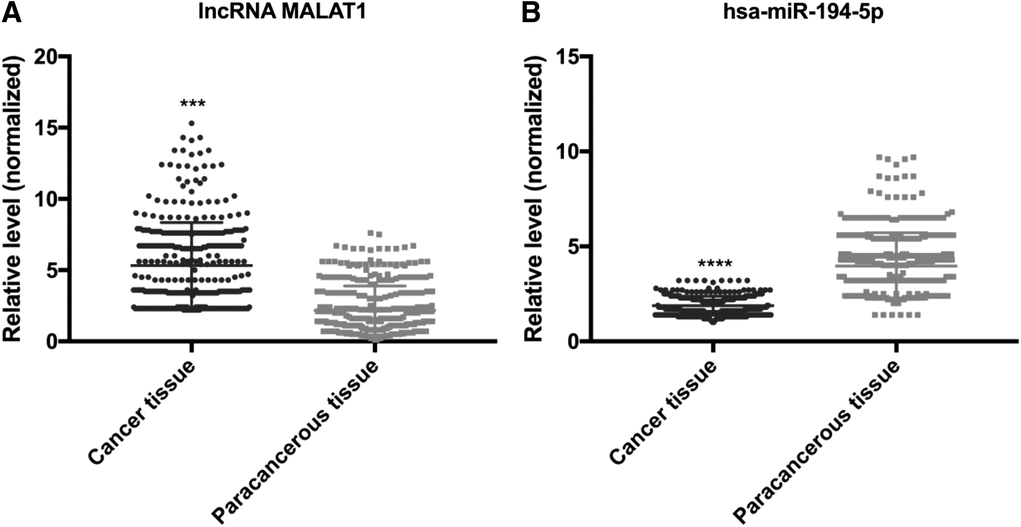
Expression levels of lncRNA MALAT1 and hsa-miR-194-5p in cancer tissues and paracancerous tissues.
Association of MALAT1 gene rs664589 locus SNP with levels of lncRNA MALAT1 and hsa-miR-194-5p
We detected the levels of lncRNA MALAT1 and hsa-miR-194-5p in cancer tissues and paracancerous tissues of 340 patients with CRC. The results showed that the level of lncRNA MALAT1 in cancer tissues was significantly higher than that in paracancerous tissues of CRC patients with the identical genotype, and the level of hsa-miR-194-5p in the cancer tissues was significantly lower than that in the paracancerous tissues (p < 0.05). In both cancer and paracancerous tissues, the level of lncRNA MALAT1 in cancer tissues and paracancerous tissues of patients with C genotype was significantly lower than that of CG genotype, and the GG genotype was the highest (p ≤ 0.05; Fig. 2A). However, the hsa-miR-194-5p level in cancer tissues was the highest in CC tissues, followed by CG genotypes, and lowest in GG genotypes (p ≤ 0.05; Fig. 2B).
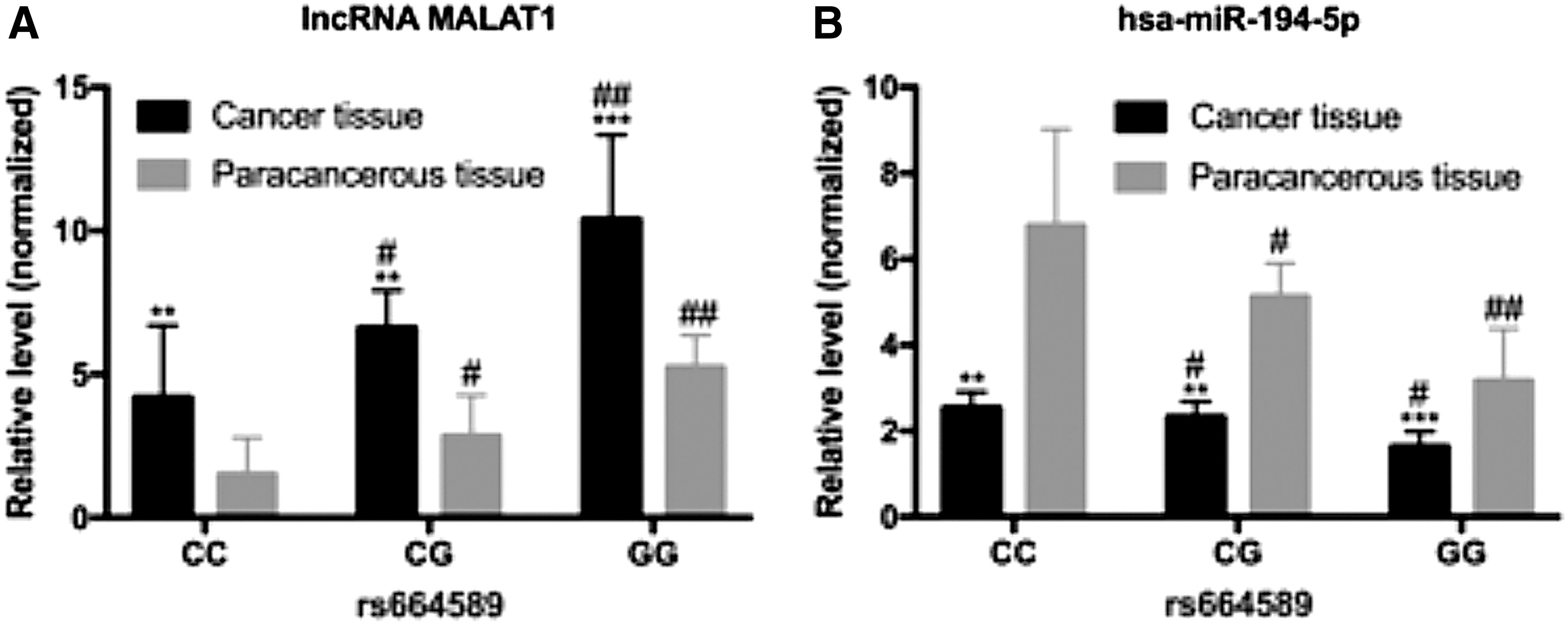
Expression levels of lncRNA MALAT1 and hsa-miR-194-5p in cancer tissues and paracancerous tissues of different genotypes of rs664589 locus in CRC patients.
MALAT1 gene rs664589 locus C > G mutation reduced PFS in CRC patients
We investigated the 3-year PFS outlook of CRC patients; all 340 patients with CRC completed the follow-up studies. The results revealed that patients with MALAT1 gene rs664589 GG genotype had significantly lower PFS (11.5 months) than CG genotype patients (13.4 months). The PFS of patients with CC genotype was the highest (15.5 months) (p < 0.05; Fig. 3).

Comparison of 3-year PFS of CRC patients with different rs664589 genotypes. The dark gray line represents the 3-year PFS outlook of CC genotype CRC patients; the black line represents the 3-year PFS outlook of CG genotype CRC patients; the light gray line represents the 3-year PFS outlook of GG genotype CRC patients. PFS, progression-free survival.
Relationship between expression levels of lncRNA MALAT1 and hsa-miR-194-5p and PFS
We analyzed the expression levels of lncRNA MALAT1 and hsa-miR-194-5p in 340 CRC patients with cancer tissues, where we defined a high expression level above the mean and a low expression level below the mean. In the present study, lncRNA MALAT1/U6 ≥ 5.32 was defined as a high expression level, lncRNA MALAT1/U6 < 5.32 was defined as a low expression level; hsa-miR-194-5p/U6 ≥ 1.88 was defined as a high expression level, hsa-miR-194-5p/U6 < 1.88 was defined as a low expression level. The results showed that PFS was shorter in CRC patients with a high level of lncRNA MALAT1 (Fig. 4A), whereas PFS was relatively long in CRC patients with a high level of hsa-miR-194-5p (Fig. 4B).
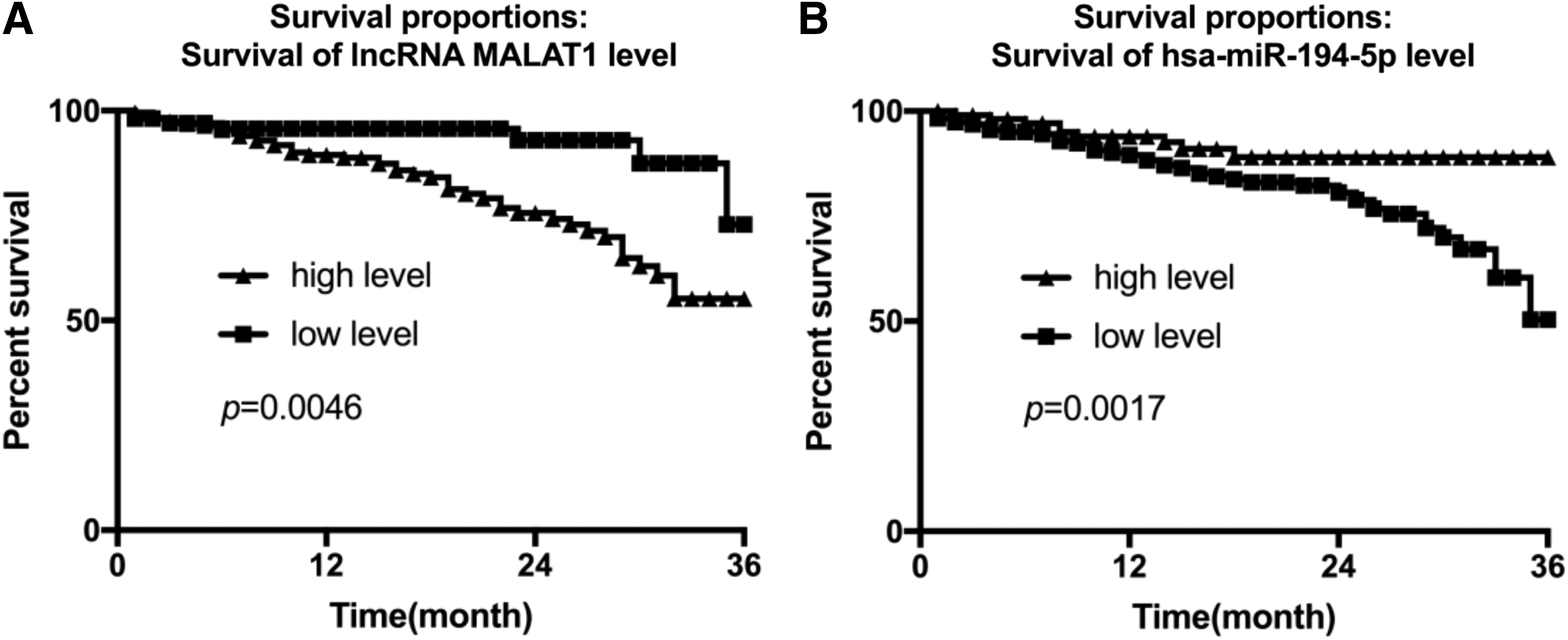
Kaplan-Meier plot for CRC patients (n = 340) stratified by lncRNA MALAT1 and hsa-miR-194-5p expression levels in cancer tissues.
lncRNA MALAT1 promoted the proliferation of CRC cell line HT-29 and inhibited the expression of hsa-miR-194-5p
The results of cell transfection experiments showed that the level of lncRNA MALAT1 was significantly higher in cells transfected with si-MALAT1 NC and untransfected group (NC) than in cells transfected with si-MALAT1, while lower than cells transfected with lncRNA MALAT1 (Fig. 5A). The level of hsa-miR-194-5p was significantly lower than that of cells transfected with si-MALAT1, while higher than that of cells transfected with the lncRNA MALAT1 group (Fig. 5C). Transfection of lncRNA MALAT1 promoted proliferation of CRC cell line HT-29, whereas transfection of si-MALAT1 inhibited proliferation of CRC cell line HT-29 (Fig. 5B).
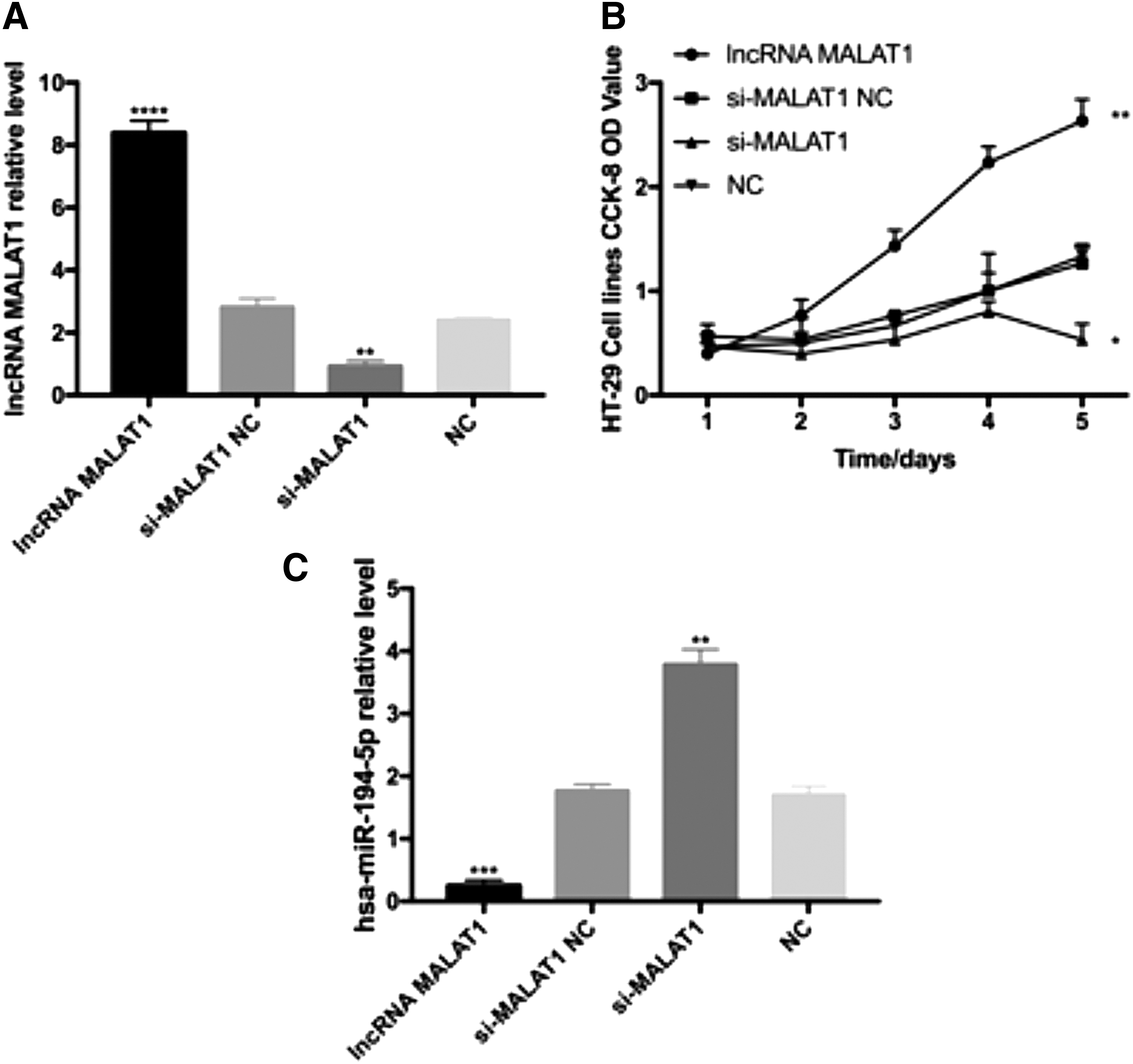
CRC cell HT-29 transfection.
hsa-miR-194-5p targeted binding to the rs664589 locus of the 3′ UTR of the MALAT1 gene
To further demonstrate whether hsa-miR-194-5p could bind to the rs664589 locus of the 3′ UTR of the MALAT1 gene, we conducted a dual luciferase reporter gene system. The 3′ UTR of the MALAT1 gene containing a 50 bp fragment upstream and downstream of the rs664589 locus was cloned to the downstream of the luciferase reporter gene in the reporter vector PmirGLO, resulting in a WT reporter vector (Fig. 6A). In addition, an MT reporter vector was also constructed, and the sequence of the reporter vector was confirmed by sequencing. We cotransfected hsa-miR-194-5p mimics and a reporter vector containing MALAT1 3′ UTR in HEK 293T cells, then detected the influence of hsa-miR-194-5p on the activity of luciferase of the MALAT1 3′ UTR reporter vector using a dual luciferase reporter system. The results showed that after overexpression of hsa-miR-194-5p, the luciferase activity of the WT reporter vector was significantly inhibited, while the MT reporter vector showed no significant change in luciferase activity (Fig. 6B).
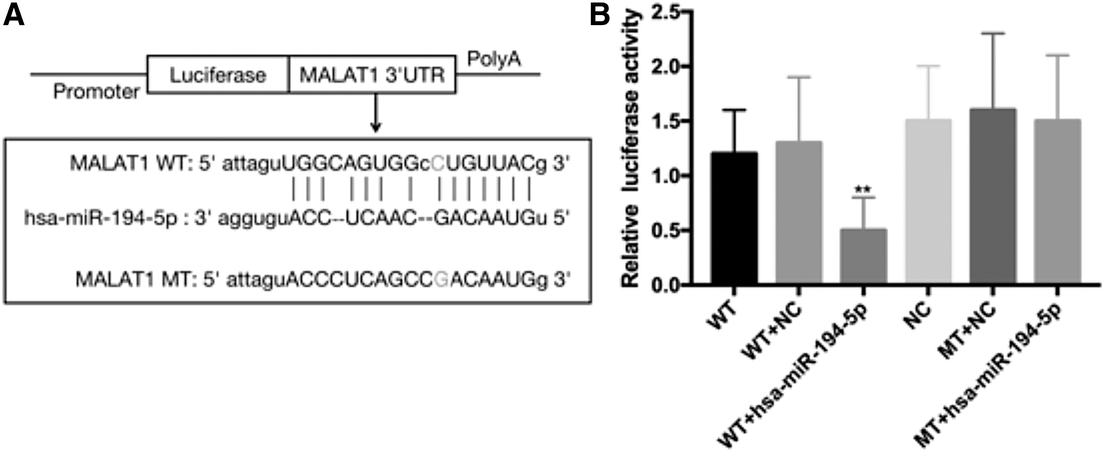
Dual luciferase reporter gene assay.
Discussion
In the present study, we found that the rs664589 locus SNP in the 3′ UTR of MALAT1 gene was associated with the risk of CRC in the Chinese Han population by a case-control study. The risk of CRC in MALAT1 rs664589 G allele carriers was increased, and the PFS in CRC patients with the G allele was reduced. Furthermore, in vitro studies revealed that the lncRNA MALAT1 expressed by the C allele of the rs664589 locus in the 3′ UTR of the MALAT1 gene was a target of hsa-miR-194-5p, whereas the product of the G allele was not a target of hsa-miR-194-5p, suggesting that the SNP of rs664589 locus in the 3′ UTR of MALAT1 gene might affect the binding efficiency of lncRNA MALAT1 and hsa-miR-194-5p.
The MALAT1 gene is located on the 11q13.1 chromosome, which produces a precursor transcript from which a small noncoding RNA is derived by cleavage of a tRNA-like small ncRNA from its 3′ end by RNase P (Li et al., 2019). It was reported that high expression of MALAT1 in patients with stage I nonsmall-cell lung cancer (NSCLC) was associated with worsening survival, with 9% of patients with low expression dying at 5 years of follow-up, while patients with a high expression of MALAT1 had a mortality rate of 40% (Ji et al., 2003). Hence, it is possible to predict disease progression early and predict metastasis of NSCLC by the expression level of MALAT1. A study conducted by Schmidt et al. (2011) revealed that high expression of MALAT1 in squamous cell lung cancer was associated with poor prognosis in patients by NSCLC tissue in situ hybridization. In addition, the expression of MALAT1 in NSCLC was upregulated compared with adjacent tissues. Moreover, the migration of tumor cells and the tumorigenic ability in nude mice were decreased significantly after siRNA knockdown, suggesting that MALAT1 could promote the formation of tumor cells. Tano et al. (2017) found that MALAT1 promotes the migration of lung cancer cells. In CRC, MALAT1 also plays an important role in promoting cell proliferation and enhancing tumor invasion and migration ability (Cho et al., 2014; Li et al., 2014a; Okugawa et al., 2014; Zheng et al., 2014; Yang et al., 2015). For example, a study by Wu et al. [PMID: 29226325] and other studies have shown that the lncRNA MALAT1/has-miR-129-5p/HMGB1 axis plays an important role in the occurrence and development of CRC. For example, Wu et al. (2018) showed that the lncRNA MALAT1/has-miR-129-5p/HMGB1 axis plays an important role in the occurrence and development of CRC. Another study by Wu et al. (2019) using the multivariate Cox regression analysis showed that lncRNA MALAT1 is a poor prognostic factor for CRC. The rs664589 G allele altered the binding of MALAT1 to has-miR-129-5p, resulting in increased expression of lncRNA MALAT1. With the rs664589 G allele, the binding of lncRNA MALAT1 to has-miR-129-5p in the nucleus was changed, resulting in increased expression of lncRNA MALAT1 and an increased risk of CRC, which is consistent with the results of the present study. However, the role of the regulatory axis formed by lncRNA MALAT1 and hsa-miR-194-5p and their target genes in CRC needs to be further confirmed.
In the present study, we found that the expression level of lncRNA MALAT1 in tumor tissues of CRC patients was significantly higher than that in paracancerous tissues, confirming that lncRNA MALAT1 plays a cancer-promoting role in CRC. In addition, patients with higher expression levels of lncRNA MALAT1 in cancer tissues also had longer PFS, suggesting that lncRNA MALAT1 also has a predictive effect on the prognosis of patients with CRC. Zheng et al. (2014) also showed that the expression of lncRNA MALAT1 was upregulated in CRC tissues, and CRC patients with higher expression of lncRNA MALAT1 had a worse prognosis.
miRNA is a small noncoding RNA that binds to complementary sequences in the 3′ UTR of mRNA, causing degradation or inhibition of translation, and ultimately leading to gene silencing (Bartel, 2004). It has been reported that hsa-miR-194-5p plays an important tumor suppression role in tumorigenesis, such as in gastric cancer (Li et al., 2014b), renal cell carcinoma (Khella et al., 2013), lung cancer (Wu et al., 2014), and CRC (Wang et al., 2015). In the present study, we found that the level of hsa-miR-194-5p in CRC tumor tissues was significantly lower than that in paracancerous normal tissues, suggesting that hsa-miR-194-5p plays a tumor suppressor role in CRC, which was consistent with a previous study (Wang et al., 2015). Interestingly, previous studies have shown that hsa-miR-194-5p is upregulated in CRC tumor samples, while hsa-miR-194-5p overexpression promotes cell migration and invasion in CRC cell lines and plays as a tumor promoter in CRC (Cai et al., 2017). We consider that in the occurrence and development of CRC, hsa-miR-194-5p alone has a limited role, and may play divert roles in different regulatory pathways. This also reflects the complexity of tumorigenesis and development. To support this point, we can find in the dbDEMC2.0 database that the same miRNA hsa-miR-194-5p may be upregulated or downregulated in the same tumor. In addition, we found that CRC patients with high hsa-miR-194-5p expression levels had a relatively high PFS, indicating that hsa-miR-194-5p was associated with prognosis in patients with CRC, and patients with high hsa-miR-194-5p expression levels had a better prognosis.
By bioinformatic prediction, we found that lncRNA MALAT1 and hsa-miR-194-5p have a binding site, and that there is an SNP locus in the binding site—rs664589. In this case-control study, we found that the risk of CRC in G allele carriers of the rs664589 locus in the 3′ UTR of the MALAT1 gene was 1.81 times higher than the C allele carriers (95% CI: 1.35-2.42, p < 0.001). Until now, no study had confirmed the correlation between rs664589 SNP and the risk of CRC in the Chinese Han population. In the present study, we found that the minor allele frequency of the control group was 0.1265, indicating that there are a considerable number of people in this group, thus underscoring the urgency for more serious clinical research to be conducted on the matter. Furthermore, we found that patients with rs664589 locus GG genotype had a significantly lower PFS than patients with CG genotype, and CC genotype had the highest PFS, suggesting that rs664589 SNP is associated with prognosis in patients with CRC.
Interestingly, the risk of CRC occurrence was significantly increased in nonsmokers and those without a family history of CRC. We know that smoking and family history of CRC are risk factors for CRC (Yahagi et al., 2017; Chen et al., 2018), but it seems that there is no correlation between rs664589 locus SNP and the risk of CRC in this subpopulation. We consider the reason for this is that the number of included smokers and those with a family history of CRC was small, and further validation was needed in a larger number of smokers and those with a family history of CRC.
In addition, by cell transfection experiments, we confirmed that lncRNA MALAT1 has a negative regulatory effect on hsa-miR-194-5p expression. Furthermore, we revealed that lncRNA MALAT1 expressed by the C allele of the rs664589 locus of the MALAT1 gene is a target of hsa-miR-194-5p, whereas the G allele is not, indicating that the rs664589 locus SNP affects the binding efficiency of lncRNA MALAT1 and hsa-miR-194-5p.
From the results of this study, it was shown that that there are significant differences in the risk of CRC among populations with different genetic backgrounds, suggesting that we should further explore its underlying mechanisms in clinical researches, investigate the nature of gene polymorphisms and clinical phenotype diversity, and develop individualized clinical treatments.
However, we did not study the mechanism of hsa-miR-194-5p in the development and progression of CRC, leaving the question to be resolved in future studies.
Conclusion
In the present study, we reported that the risk of CRC in MALAT1 gene rs664589 locus G allele carriers was relatively high. Patients with G allele had a lower PFS and poor prognosis. One of the possible underlying mechanisms might be that rs664589 SNP affected the binding efficiency of lncRNA MALAT1 and hsa-miR-194-5p. However, further studies are needed to pinpoint the specific mechanism responsible.
Footnotes
Author Disclosure Statement
No competing financial interests exist.
Funding Information
This work was supported by the Zhejiang Provincial Medicine Health Science and Technology Program (2020KY721).