




Abstract
Aims:
The aim of this study was to examine the RNA and protein expression levels and clinical significance of the pore membrane protein 121 kDa (POM121) in lung cancer.
Materials and Methods:
Paired lung cancer and adjacent nontumor tissues were obtained from lung cancer patients to measure the expression of POM121 by quantitative reverse transcription-polymerase chain reaction and immunohistochemistry. Patient clinical and pathological data were collected to analyze their relationships with POM121 protein expression levels by chi-square test and log-rank test, respectively.
Results:
POM121 mRNA and protein expression were both upregulated in lung cancer tissues. POM121 protein expression was observed in 48.00% (36/75) of lung cancer tissues and 25.33% (19/75) of adjacent nontumor tissues. A chi-square analysis indicated that this difference was statistically significant (p < 0.05). Furthermore, we found that POM121 protein expression was correlated with gender, tumor node metastasis stage, and lymphatic metastasis (p < 0.05). In addition, we found a significant relationship among POM121 expression, gender, and metastasis based on a multivariate logistic regression analysis. A Kaplan-Meier survival analysis indicated that lung cancer patients with POM121 expression had a poorer prognosis than those without POM121 expression (p < 0.05).
Conclusion:
POM121 protein expression is associated with lung cancer metastasis and is a potential prognostic biomarker for lung cancer patients.
Introduction
As the most common cancer, lung cancer had 2.09 million new cases and 1.76 million deaths in 2018, resulting in lung cancer to be the leading cause of cancer-related deaths worldwide. In Asia, lung cancer is the most common cause of death in men more than women (Pakzad et al., 2015). And in China, the incidence of lung cancer is increasing year by year, with an annual growth rate of 1.63% (Cao and Chen, 2019). Although great progress has been made in the diagnosis and treatment of malignant tumors, 90% of deaths in patients with advanced cancer can be attributed to metastasis (Lambert et al., 2017). Therefore, it is of great significance to identify effective biomarkers for improving the prognosis of lung cancer.
Nuclear pore complexes (NPCs) are the largest and most complicated protein complexes in human cells with an estimated mass of 60-125 MDa (Mega dalton) in vertebrates, constructed from multiple copies of about 30 different nucleoporins. NPCs are the only channels that regulate the transport of macromolecules through the nuclear membrane (NE). In addition, studies have shown that NPCs can regulate mitosis, transcription activation, RNA processing, gene silencing, and heterochromatin modulation (Beck and Hurt, 2017).
Pore membrane protein 121 kDa (POM121) is a critical component of NPCs, and its coding gene is located on human chromosome 7q11.23. It has been reported in the literatures that POM121 participates in NE assembly and nuclear transport and anchors NPCs to the NE by interacting with other NPC subunits (Antonin et al., 2005). Most previous studies have focused on the cell biological functions of POM121, with few research on its relationship with tumors. A study showed that the increased expression of POM121 can promote the nuclear import of MYC (MYC proto-oncogene protein), E2F1 (E2F transcription factor 1), AR (androgen receptor), and GATA2 (GATA binding protein 2), thus promoting the tumor initiating, proliferation, and survival properties of prostate cancer cells (Rodriguez-Bravo et al., 2018). Recent studies have shown that POM121 is overexpressed in colorectal cancer and oral squamous cell cancer tissues and is associated with poor prognosis of patients (Ma et al., 2019; Wang et al., 2019), which attracted our attention to explore the role of POM121 in lung cancer.
The aim of this study was to determine the expression of POM121 and its clinical significance in lung cancer. We detected the protein expression of POM121 in 75 paired lung cancer and adjacent nontumor tissues. We found that POM121 protein was overexpressed in lung cancer tissues and correlated with the metastasis and survival of lung cancer patients, suggesting a potential role for POM121 in lung cancer progression.
Materials and Methods
Tissue samples
Paired lung cancer and adjacent nontumor tissues were obtained for immunohistochemistry (IHC) from 75 lung cancer cases that were diagnosed by both pathological and clinical methods in the Affiliated Hospital of Guilin Medical University between July 2005 and December 2011. Inclusion criteria: were Chinese Han nationality; obtained complete clinicopathological data before operation or during follow-up; and received surgery without radiotherapy or chemotherapy before. Exclusion criteria: had severe organ dysfunction or mental disorders; lung cancer with other malignancies; hospitalized with recurrence, metastasis, or other complications; were difficult to follow-up, unable to cooperate, refused to participate; and vulnerable groups, such as pregnant women, elderly people, and so on.
Another 40 pairs of fresh lung cancer and adjacent nontumor tissues were collected for quantitative reverse transcription-polymerase chain reaction (qRT-PCR) detection. All specimens and clinicopathological data were obtained with informed consent and approved by the Ethics Committee of Guilin Medical University.
Immunohistochemistry
The IHC assay was performed to detect the expression of POM121. A tissue microarray that contains 75 paired paraffin-embedded lung cancer tissues was made by Fanpu Company (Guilin, China) and treated as described previously (Luo et al., 2016), including baking, dewaxing, antigen repair, peroxidase blocking, and goat serum incubation. And then the tissue microarray was incubated with rabbit polyclonal antibody against POM121 (GTX102128; GeneTex) at a dilution for 1:500 overnight at 4°C and secondary anti-rabbit antibody for 30 min at 37°C. After that, 3,3′-aminobenzidine tetrahydrochloride was given to treat the tissues for 5 min to appear an appropriate color. Finally, tissues were counterstained with hematoxylin, dehydrated, transparentized, and fixed.
All sections were evaluated by three professional pathologists. Each specimen had five random fields selected for analysis. The percentage of positively stained cells and average staining intensity were counted for evaluation. The scores for the percentage of positively stained cells were 0 for ≤5%, 1 for 6-25%, 2 for 26-50%, 3 for 51-75%, and 4 for >75%, respectively. And the scores for staining intensity were 0 for no staining, 1 for light brown, 2 for medium brown, and 3 for dark brown. The product of the percentage of positive stained cells and staining intensity scores was used as the final POM121 staining score. Positive and negative expression of POM121 were defined as scores >4 and ≤4, respectively.
Quantitative reverse transcription-polymerase chain reaction
After the fresh tissues were powdered, total RNA was isolated using TRIzol RNA isolation reagent (Life Technologies, Carlsbad, CA). Then the RNA integrity and concentration were determined on Agilent RNA 6000 Nano Kit and Agilent 2100 Bioanalyzer (Agilent, Santa Clara, CA). TaqMan™ Reverse Transcription Reagents (Thermo Fisher Scientific, Waltham, MA) were used to reverse RNA to cDNA. Then, PCR was started after SYBR Green Mixture (Tiangen, Beijing, China) was added according to the manufacturer's procedure. qRT-PCR primers were as follows: POM121 forward primer was AACGTGAGCAGCACAACT and POM121 reverse primer was CCTCGGGCTGTGTTCTTG; GAPDH forward primer was CAGGAGGCATTGCTGATGAT and GAPDH reverse primer was GAAGGCTGGGGCTCATTT. The relative RNA level was calculated using the 2−ΔΔCt method and normalized to the GAPDH mRNA value.
Statistical analysis
All data were analyzed in SPSS 22.0. The expression difference of POM121 between lung cancer and adjacent nontumor tissues was compared by chi-square test. The correlation between the expression level of POM121 and the clinicopathological features of lung cancer patients was analyzed by chi-square test and multivariate logistic regression analysis. Kaplan-Meier method was used to calculate the survival probability, and log-rank test was used to compare the survival curves between different groups. p < 0.05 was defined as statistically significant.
Results
Expression of POM121 in lung cancer tissues
We first detected the expression of POM121 in 75 paired lung cancer and adjacent nontumor tissues by IHC assay (Fig. 1). POM121 protein expressed both in lung cancer tissues and adjacent nontumor tissues. The positive expression of POM121 in lung cancer and adjacent nontumor tissues were 48.00% (36/75) and 25.33% (19/75), respectively. After statistical analysis, we found that POM121 was upregulated in lung cancer tissues (p < 0.05, Table 1). To confirm the expression of POM121 in lung cancer tissues, we chose another 40 paired lung cancer and adjacent nontumor tissues to perform qRT-PCR. As shown in Figure 1B, we also detected significantly increased mRNA expression of POM121 in lung cancer tissues (p < 0.001). These results show that both mRNA and protein of POM121 are overexpressed in lung cancer tissues.
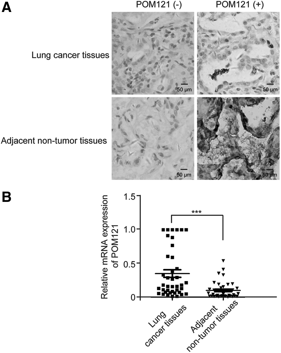
POM121 expression in lung cancer tissues.
POM121 Expression in Paired Lung Cancer Tissues and Adjacent Nontumor Tissues
p < 0.05, p-value is based on the McNemar χ2 test.
POM121, pore membrane protein 121 kDa.
Relationship between POM121 protein expression and clinicopathological features of lung cancer patients
The 75 cases of lung cancer patients for IHC detection were divided into POM121 protein positive expression group and negative expression group. We collected these general and clinicopathological features of lung cancer patients, such as gender, age, smoking, alcohol intake, allergic history, past history, family history of lung cancer, carcinoembryonic antigen, CA125, CA199, pathological classification, tumor diameter, tumor node metastasis (TNM) stage, and lymphatic metastasis. As shown in Table 2 and Supplementary Table S1, POM121 protein expression was significantly correlated with gender, TNM stage, and lymphatic metastasis (p < 0.05). There was no relationship between POM121 protein expression and other features of lung cancer patients (p > 0.05). Multivariate logistic regression analysis showed that POM121 protein expression was related to the gender (OR = 0.333, p < 0.05) and lymphatic metastasis (OR = 0.329, p < 0.05, Table 3).
Association Between the Expression of POM121 and Clinicopathological Features of Lung Cancer Patients
Bold values indicate significance. Pathological classification was according to the latest WHO classification; these cases were all invasive adenocarcinomas.
CEA, carcinoembryonic antigen; TNM, tumor node metastasis.
Multivariable Logistic Regression Analysis of POM121 Expression in Lung Cancer Patients
Bold values indicate significance.
CI, confidence interval; OR, odds ratio.
POM121 protein expression was correlated with the prognosis of lung cancer patients
The median survival time of lung cancer patients in the POM121 protein positive expression group and negative expression group was 46 months (95% confidence interval [95% CI]: 37.18-54.82) and 80 months (95% CI: 48.19-111.81) by Kaplan-Meier method, respectively. After log-rank test, the survival time of lung cancer patients with POM121 protein positive expression was significantly shorter compared with lung cancer patients with POM121 protein negative expression (p < 0.05, Fig. 2), indicating that the positive expression of POM121 protein was associated with poor prognosis of lung cancer patients. After adjusted age of lung cancer patients, gender, TNM stage, metastasis, and POM121 protein expression were included in the COX regression analysis according to inclusion criteria for 0.05 and exclusion criteria for 0.10. However, we did not get statistically significant results (p > 0.05, Table 4), which may be caused by the small number of samples in this study.

The relationship between POM121 expression and the prognosis of lung cancer patients from IHC result. IHC, immunohistochemistry.
COX Regression Analysis for Overall Survival of Lung Cancer Patients After Surgery
Adjusted for age.
Discussion
In this study, POM121 protein expression was measured by IHC method in lung cancer tissues. The result showed that POM121 protein positive expression was more in lung cancer tissues (48.00%) than that in the paired adjacent nontumor tissues (25.33%), and the difference was statistically significant. And then, the clinicopathological features of these lung cancer patients were analyzed to explore the association with POM121 protein expression. Our findings showed that POM121 protein expression was significantly correlated with gender, TNM stage, and lymphatic metastasis, indicating that POM121 may be involved in the proliferation, migration, and invasion of lung cancer. Moreover, the overall survival time of lung cancer patients with POM121 protein positive expression was shorter compared with POM121 protein negative expression showed by Kaplan-Meier survival curves. The above results indicated that POM121 might be involved in the occurrence and development of lung cancer.
POM121 is one kind of nucleopore protein, which is named for its location and relative molecular mass. The protein has a short luminally exposed N-terminal tail, a single transmembrane domain, and a large C-terminal portion adjoining the NPC (Soderqvist and Hallberg, 1994). And a third of the C-terminal contains a pentapeptide XFXFG repeat sequence, which can help identify the nuclear localization signal and facilitate the nuclear entry of biological macromolecules, suggesting that it is related to the transport of incoming nuclear proteins (Gorlich and Mattaj, 1996; Walde and Kehlenbach, 2010). Previous studies in cell biology elaborated the related functions of POM121, such as biological processes as nuclear pore assembly (Rasala et al., 2008), NPC assembly (Doucet et al., 2010; Funakoshi et al., 2011; Talamas and Hetzer, 2011), NE formation (Antonin et al., 2005; Anderson et al., 2009), nucleocytoplasmic transport (Yavuz et al., 2010), and cell apoptosis (Beckman et al., 2004). The versatility of POM121 beyond NE functions has been increasingly studied in accumulated researches in recent years. A study indicated that POM121 had a strong inhibitory effect on macrophage inflammation, which was due to the inhibiting ability of POM121 on phosphorylated p65 nuclear accumulation in NF-κB signal pathway (Ge et al., 2019). The involvement of POM121 in HIV infection was demonstrated in another research (Guo et al., 2018). And the ectopic introduction of PAX5 and POM121 genes resulted in fusion expression of the two proteins and caused acute lymphoblastic leukemia (Denk et al., 2014; Fortschegger et al., 2014).
However, there are few studies on POM121 in cancer, and there is no relevant report on POM121 in lung cancer. In prostate cancer cells, POM121 ectopic introduction could promote cell proliferation and cause tumor drug resistance and tumorigenesis, while POM121 interruption could inhibit tumor proliferation, improve chemotherapy, and suppress tumor formation in vivo (Lim and Wong, 2018). As POM121 disturbance had no effect on the integrity of NPCs, the oncogenic role of POM121 played in cancer might be in its nucleocytoplasmic transport function-dependent way (Stavru et al., 2006). POM121 is also overexpressed in colorectal cancer and oral squamous cell cancer tissues, which was consistent with the results of our study. According to the relevant literature, it is suggested that POM121 may be involved in the occurrence, invasion, and metastasis of lung cancer through regulating DNA replication and repair, its own nucleocytoplasmic transport function, and interaction with other proteins (Rodriguez-Bravo et al., 2018; Ma et al., 2019; Wang et al., 2019).
In this study, we found that both gender and lymphatic metastasis had a significant correlation with POM121 protein expression after multivariate logistic regression analysis. The odds ratios (ORs) suggested that males with lung cancer expressed POM121 protein at a statistically higher rate than females, and lung cancer patients with lymphatic metastasis had a lower probability to express POM121 protein. A previous population-based nonsmall cell lung cancer cohort showed that males had poorer overall survival compared to women following a lung cancer diagnosis (Pitz et al., 2013), which might explain the difference of POM121 protein between males and females with lung cancer. In addition, although TNM stages III and IV had a statistically significant increase in expression of POM121 over stages I and II, there was a really significant expression of POM121 even in these lower stages of lymph node involvement. This was also consistent with the correlation between POM121 protein expression and lymphatic metastasis. However, a larger sample of human tissues and cell biological function assays are further needed to clarify the molecular mechanism of POM121 in lung cancer carcinogenesis and progression in future.
In summary, our study found that POM121 was overexpressed in lung cancer tissues, and POM121 protein expression had an association with lung cancer metastasis and prognosis in lung cancer patients. Our study suggests that POM121 will be a potential prognostic biomarker for lung cancer patients.
Footnotes
Author Disclosure Statement
No competing financial interests exist.
Funding Information
This work was supported by the National Natural Science Foundation of China (81860586, 81860602), Natural Science Foundation of Guangxi Province (2018GXNSFAA281054, 2018GXNSFBA281216), and Key Science and Technology Research and Development Program Project of Guangxi (AB17292074).

References
Supplementary Material
Please find the following supplemental material available below.
For Open Access articles published under a Creative Commons License, all supplemental material carries the same license as the article it is associated with.
For non-Open Access articles published, all supplemental material carries a non-exclusive license, and permission requests for re-use of supplemental material or any part of supplemental material shall be sent directly to the copyright owner as specified in the copyright notice associated with the article.
