




Abstract
Objective:
The study was designed to analyze the expression of CSNK1D in hepatocellular carcinoma (HCC) and to investigate the relationship between the expression of CSNK1D and the prognosis of HCC patients.
Methods:
The CSNK1D and AFP expression levels in patients with hepatocellular carcinoma and their corresponding clinical data were downloaded from TCGA and sorted with a Perl program. CSNK1D and AFP expression differences in normal liver tissue and liver cancer were compared and analyzed, based on the online database HCMDB, the relationships between the expression levels of CSNK1D and AFP and the proliferation and metastases of hepatocellular carcinoma were explored. The immunohistochemical data obtained from the Human Protein Atlas Database further verified the differences in the expression levels of CSNK1D and AFP in liver tissues and liver cancer tissues. Through Kaplan-Meier survival analysis, the effects of CSNK1D and AFP expression levels on the prognosis of patients with hepatocellular carcinoma were investigated, and the influences of and patients' gender, age and grades of cancer cells, tumor size, the status of lymph node metastasis, distant metastasis, and tumor stage on the expression of CSNK1D were analyzed with R language. The influence of differential expressions of CSNK1D on survival time was compared and the prognostic factors influencing the survival of HCC patients were statistically explored by univariate and multivariate analyses. The potential influencing mechanism of CSNK1D on the prognosis of HCC was explored by GSEA enrichment.
Results:
The expression levels of CSNK1D and AFP in cancer foci were significantly higher than that in normal tissues, However, in the same patient, the expression levels of AFP in para-carcinoma tissues and cancer tissues showed no significant differences. The expression levels of CSNK1D in hepatocellular carcinomas with distant metastases were higher than that in those without metastasis, but the expression levels of AFP in metastatic hepatocellular carcinomas were lower than that in those hepatocellular carcinomas without metastases. In immunohistochemical tests, CSNK1D was moderately positive in normal liver tissues, slightly positive in normal bile duct tissues, and highly positive in hepatocellular carcinoma. AFP was slightly positive in normal liver tissues and negative in hepatocellular carcinoma, but it was not detected in normal intrahepatic bile duct tissue. Survival analyses results suggested that higher expression levels of CSNK1D corresponded to the shorter survival times, whereas the expression level of AFP showed no significant influence on survival time. The expression levels of CSNK1D were not correlated with gender, age, lymph node metastasis or distant metastasis The main factors influencing the expression levels of CSNK1D included tumor size, cancer cell grade and tumor stage. The expression levels of CSNK1D in T2 and T3 were higher than that in T1. The expression levels of CSNK1D in G3 and G4 were higher than that in G1. The expression levels of CSNK1D in Stage II and Stage III were higher than that in Stage I. Univariate analysis suggested that tumor size, cell grade, distant metastasis, clinical stage, and CSNK1D expression level were prognostic factors influencing the survival of patients. Multivariate analysis suggested that CSNK1D expression levels are an independent factor influencing the HCC prognosis. GSEA enrichment analysis indicated that CSNK1D mainly influenced the prognosis of HCC through cell cycle, WNT signaling pathway, amino acid degradation metabolism and other pathways.
Conclusion:
CSNK1D is an independent factor influencing HCC prognosis and has the potential to be developed as a potential therapeutic target for HCC, and better than AFP in predicting the prognosis of hepatocellular carcinoma.
Introduction
Liver cancer is one of the most common malignancies in the world, and its mortality rate ranks second in the world (Vallazza et al., 2015). Traditional treatments for liver cancer, including surgery, radiofrequency ablation, and chemotherapy, can reduce the tumor load, but they cannot control metastasis or recurrence (Wang et al., 2019a). Early surgical resection is still the first option for the treatment of liver cancer, but the recurrence rate in the second year after surgery is as high as 50.56% and the 5-year overall survival rate is 26-58% (Chen et al., 2018; Blüthner et al., 2019). High recurrence rate and extensive metastasis are the key factors to reduce the survival rate of patients due to hidden early symptoms, easy metastasis, and recurrence of liver cancer, as well as the lack of effective early markers for judging and assessing the prognosis. Many factors affect the prognosis of hepatocellular carcinoma (HCC) recurrence, so it is difficult to accurately predict the prognosis of patients with liver cancer with traditional predictors. Molecular markers of tumor can show the useful information other than routine clinical pathological characteristics (Bernhardt et al., 2017). Therefore, it is necessary to screen new molecular indicators of the prognosis to further reduce the recurrence rate and mortality and prolong the survival period of liver cancer patients.
CSNK1D (CK1), a member of the CK1 family, was first isolated by Graves and his colleagues in the early 1990s, and its gene is on the long arm of human chromosome 17 (17q25.3) (Xu et al., 2019). In recent years, scholars observed that CSNK1D was closely related to the development of tumors and showed significant differences in the expression level and activity in different cancers (Lin et al., 2020; Liu et al., 2021). High CSNK1D expression was correlated with the shorter survival period of the patients with glioblastoma, lung cancer, and colorectal cancer (Schittek and Sinnberg, 2014; Richter et al., 2016). However, in patients with breast cancer, chronic lymphocytic leukemia, and astrocytoma, it was correlated with the longer survival period (Cheong and Virshup, 2016; Bar et al., 2018). The study aims to investigate the above correlation between the expression level of CSNK1D and the prognosis of HCC.
Materials and Methods
Data downloading and arrangement
After logging to the The Cancer Genome Atlas (TCGA) database, with “Liver and intrahepatic bile ducts,” “TCGA - LIHC”, “Transcriptome Profiling,” and “Clinical” as the keywords, the retrieval time from the establishment day of the library to November 13, 2019, HCC RNA-seq data were downloaded. In total, 50 normal samples and 374 HCC samples as well as clinical data of 377 patients with HCC were obtained. With Perl language, gene ID conversion of the original data as well as script merging was performed to remove the incomplete data (including the data without visiting information or important items for R language analysis).
R language analysis
Through Student's t-test, the expression difference of CSNK1D and alpha-fetoprotein (AFP) in normal liver tissue and liver cancer tissue was analyzed and paired Student's test was performed to analyze the expression difference of CSNK1D and AFP in cancer tissue and adjacent tissues in the same patient. The median expression of CSNK1D and AFP was used as the baseline to divide the data into the low-expression group and high-expression group of CSNK1D and AFP. With Kaplan-Meier method, the survival analysis was performed to compare the survival differences between the two groups. With Wilcoxon test method, the influences of clinicopathological factors on the expression level of CSNK1D were analyzed for the comparison between the two groups. With Kruskal-Wallis test method, the influences of clinicopathological factors on the expression level of CSNK1D were analyzed for the comparison among multiple groups. Multivariate logistic regression model was used to analyze the risk factors for the upregulation of CSNK1D expression in clinicopathological factors. p < 0.05 was considered to be statistically significant.
Online data validation
Based on the online database human cancer metastasis database, the relationships between the expression levels of CSNK1D and AFP and the proliferation and metastases of HCC were evaluated and immunohistochemical data were obtained from the Human Protein Atlas Database to further verify the differences in the expression levels of CSNK1D and AFP in liver tissues and liver cancer tissues.
Gene Set Enrichment Analysis (GSEA)
The expression levels of CSNK1D in liver cancer tissues were sorted in ascending order and divided into phenotype-h group and phenotype-l group according to the median. The gene function was analyzed in Gene Set Enrichment Analysis (GSEA) 4.0.1. The data set was obtained from the Molecular Signatures Database on the GSEA website, and then the enrichment analysis was performed with the default weighted enrichment method. The normalized enrichment score was acquired by the permutation analysis for 1000 times. Gene sets with normal p-value <0.05 and false discovery rate (FDR) <0.25 were considered to be significantly enriched. Six closely related signal pathways were respectively extracted from the phenotype-h group and the phenotype-l group for visual display.
Results
Differential expression
In the comparison of different patients, the statistical analysis showed that the expression level of CSNK1D and AFP in cancer tissues was significantly higher than that in normal tissues (p = 2.017e-23 and p = 2.397e-06). In the same patient, the expression level of CSNK1D in cancer tissues was significantly higher than that in paracancer tissues (p = 1.357e-13), but AFP was not differentially expressed in paracancer and HCC (Fig. 1).
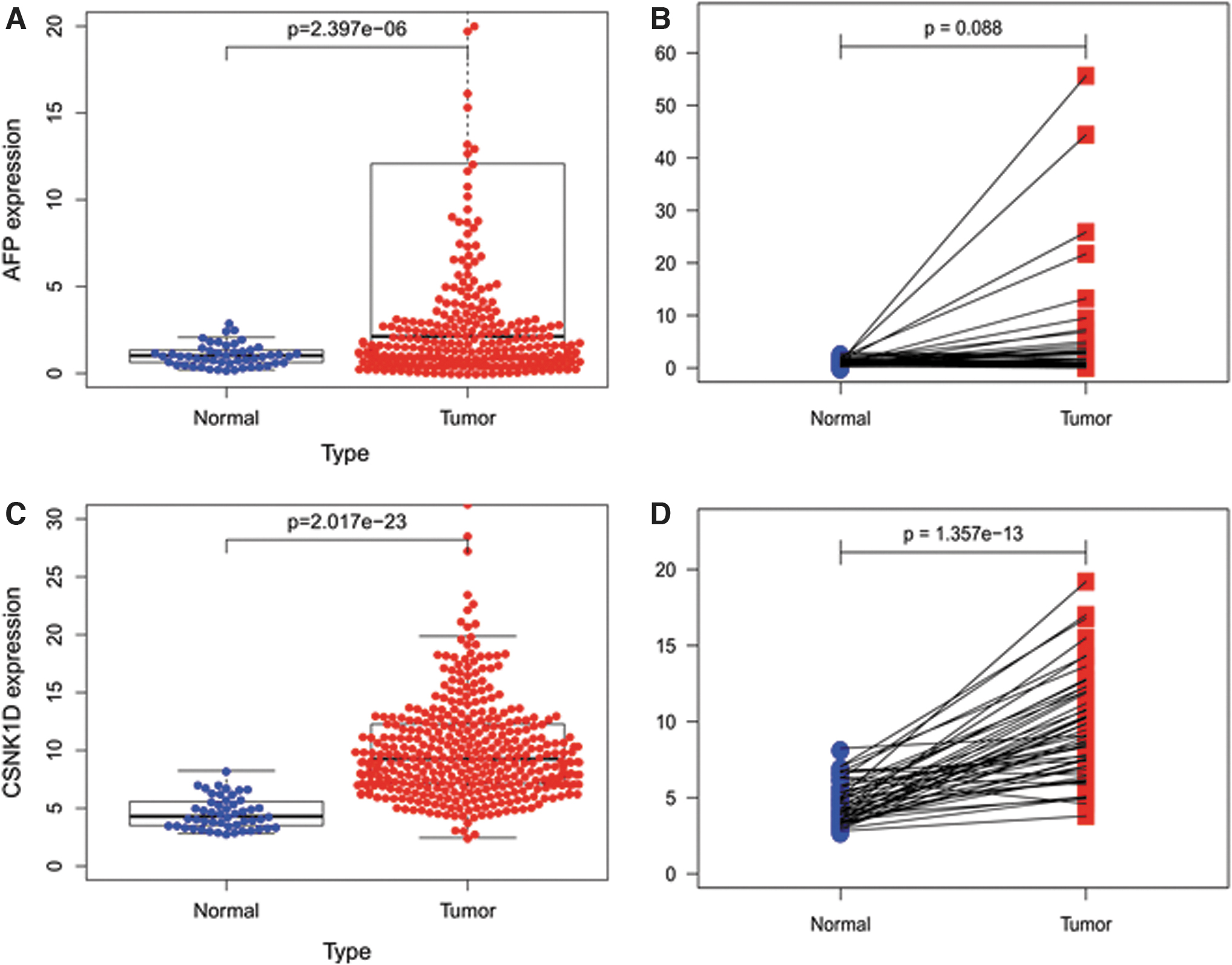
Differential expression analysis of CSNK1D and AFP.
Relationships between the expression levels of CSNK1D and AFP and the proliferation and metastases of HCC cells
The data came from the same HCC data set, Experiment Details for EXP00134. The box plot indicated that the expression level of CSNK1D in patients with distant metastases was significantly higher than that in patients without metastasis, but the expression levels of AFP in patients with distant metastases were significantly lower than that in those without metastases (Fig. 2).

The relationship between CSNK1D and AFP expression and HCC metastasis. Group A was HCC with metastasis, and Group B was HCC without metastasis. HCC, hepatocellular carcinoma. Color images are available online.
Histological differential expressions of CSNK1D and AFP
In normal liver tissues, CSNK1D was moderately positively expressed in liver tissues, weakly positively expressed in intrahepatic bile duct tissues, and highly positively expressed in HCC. AFP was weakly positively expressed in normal liver tissues and negatively expressed in HCC, but it was not expressed in normal intrahepatic bile duct tissues (Fig. 3).

Expression of CSNK1D and AFP in normal and hepatocellular cancerous tissues. Color images are available online.
Survival difference
The survival analysis showed that the total survival period of the low-expression group on CSNK1D was significantly longer than that of the high-expression group. There were 178 cases in the high-expression group and 181 cases in the low-expression group (p = 4.333e-04). The 3-year, 5-year, and 7-year overall survival rates in the high-expression group were, respectively, about 50.6%, 38%, and 25.3%, the median survival time was 0.833 years, and the median mortality was 24%, whereas the 3-year, 5-year, and 7-year overall survival rates in the low-expression group were, respectively, 71.3%, 56%, and 33.9%, the median survival was 1.67 years, and the median mortality was 17.5%. There were 178 cases in the high-expression group of AFP and 180 cases in the low-expression group, and there were no statistically significant differences in overall survival rate and median survival time between the two groups (Fig. 4).
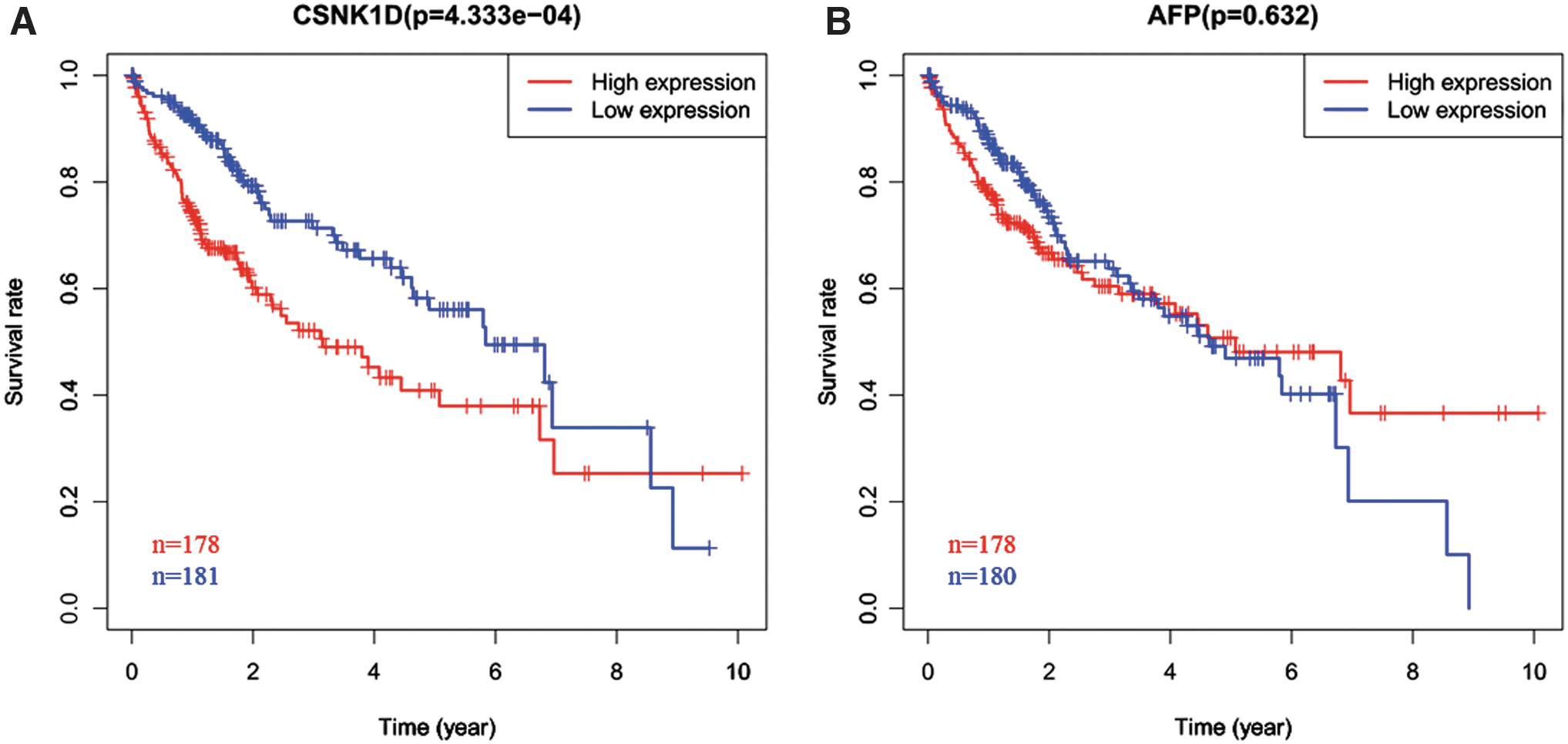
Kaplan-Meier curves.
Correlation between CSNK1D expression and clinical pathological factors
Among clinical pathological factors, the onset age, gender, the status of lymph node metastasis or distant metastasis of the patients did not affect the expression of CSNK1D in liver cancer tissues. The expression of CSNK1D was significantly upregulated when the tumor size was larger (p = 3.272e-4). The expression of CSNK1D was significantly upregulated when the cancer cell grading was higher (p = 0.002). The expression of CSNK1D was higher in the later tumor stages (p = 2.92e-4). Therefore, tumor size, cancer cell grade, and tumor stage were important factors influencing the expression of CSNK1D (Fig. 5). As indicated by the results of multifactor logistic regression analysis, the expression levels of CSNK1D in Stages II and III were higher than that in Stage I and corresponding odds ratio (OR) values were, respectively, 2.05 and 2.01 (p < 0.01). The expressions of CSNK1D in G3 and G4 were higher than that in G1 and corresponding OR values were, respectively, 3.02 and 3.79 (p < 0.05). The expressions of CSNK1D in T2, T3, and T4 were higher than that in T1 and corresponding OR values were, respectively, 2.28, 1.90, and 3.33 (p < 0.05), indicating statistically significant differences. The logistic regression analysis results indicated that tumor size, cancer cell grade, and tumor stage were risk factors for the upregulation of CSNK1D expression (Table 1).
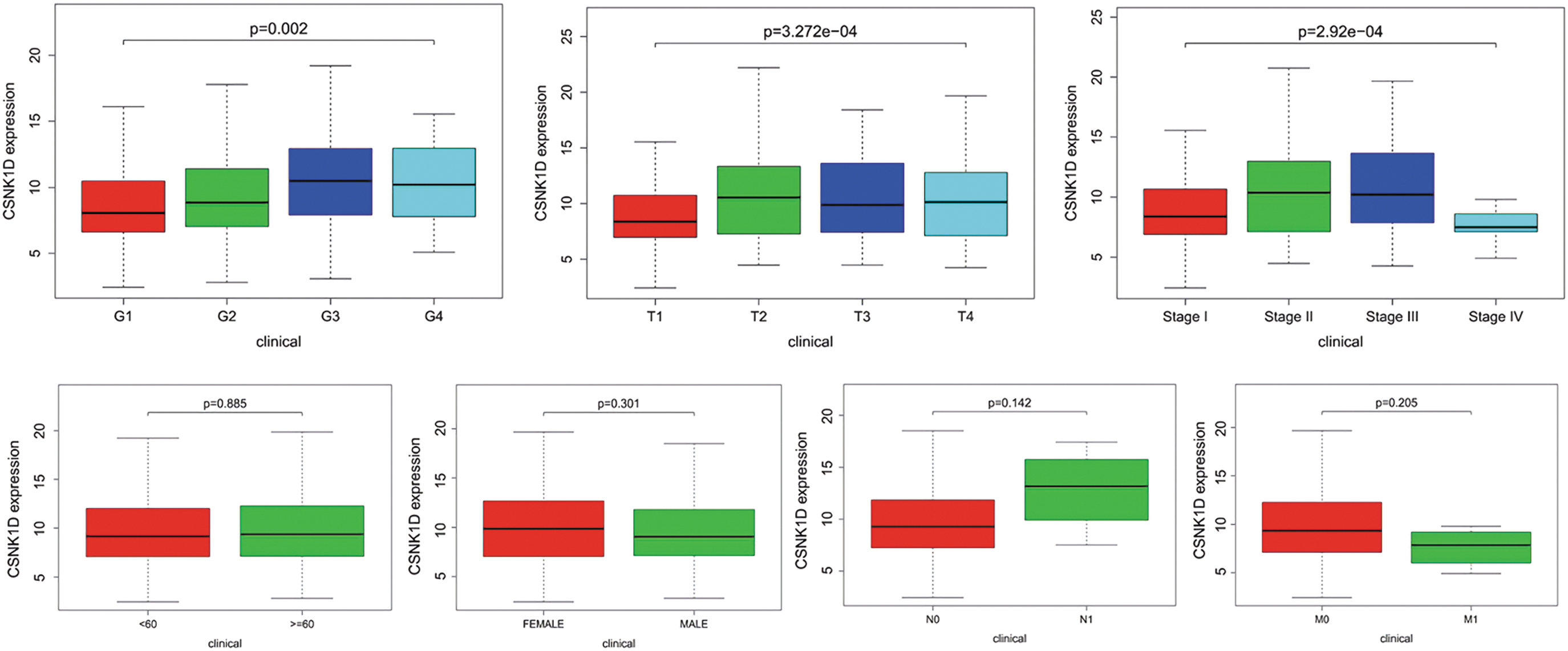
Impact of clinicopathologic parameters on CSNK1D expression. G, tumor cell classification; M, distant metastasis; N, regional lymph node metastasis; T, tumor size. Color images are available online.
Logistic Regression Analysis of the Correlation Between Clinicopathologic Parameters and CSNK1D Expression
OR, odds ratio.
COX analysis of prognostic factors
Univariate analysis results showed that age, gender, grade of cancer cells or the status of lymph node metastasis of the patients did not affect the survival period of the patients, and the difference was not statistically significant. Influencing factors of the survival period of HCC patients mainly included tumor stage, tumor size, distant metastasis status, and CSNK1D expression level. Among the above factors, distant metastasis status, and CSNK1D expression level had the most significant influences on the survival period of patients and corresponding HR values were, respectively, 3.85 and 2.33. However, multifactor analysis indicated that only the expression level of CSNK1D could have an independent influence on the survival period of patients and corresponding HR was 2.09. Therefore, the expression level of CSNK1D was an independent factor affecting the prognosis of HCC patients (Table 2) (Fig. 6).
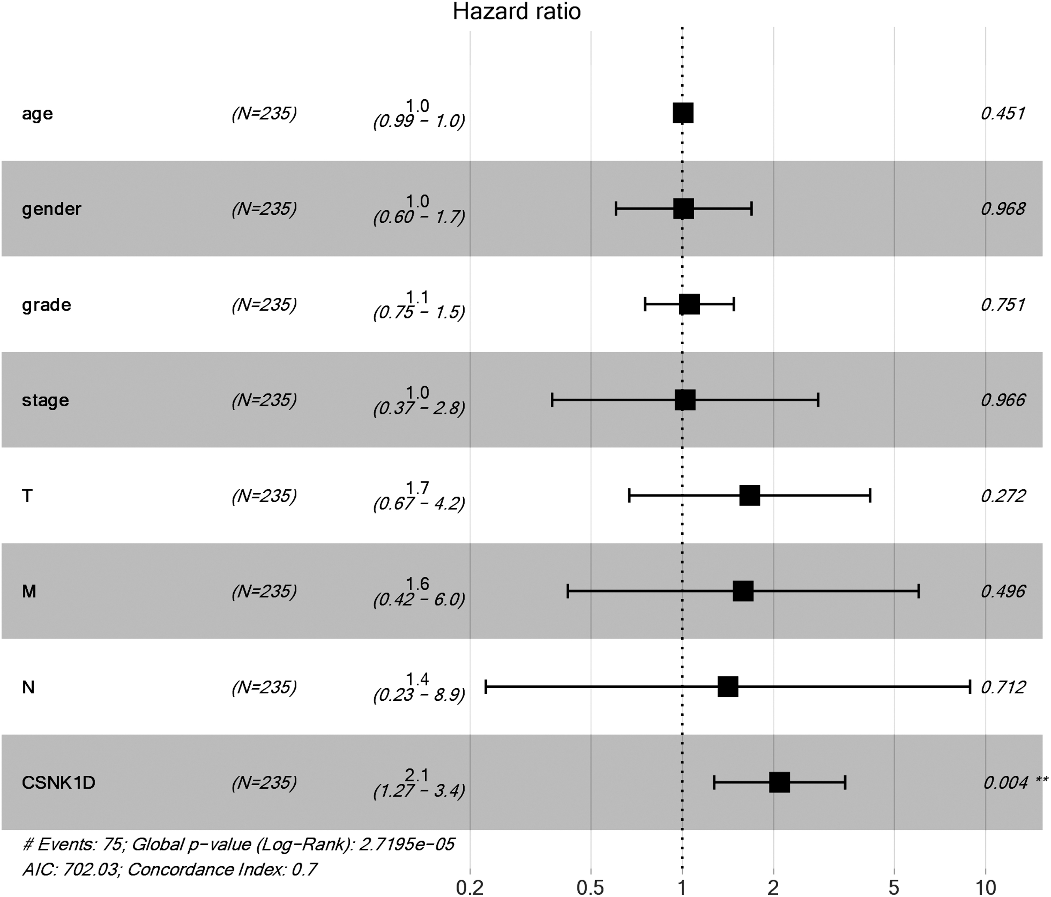
Forest plots of Cox regression analysis of prognostic factor.
Cox Regression Analysis of Prognostic Factors
HR, hazard ratio.
GSEA enrichment analysis
In the phenotype-h group, 95 gene sets with normal p-value <0.05 and 118 gene sets with FDR <0.25 were obtained and the expression of CSNK1D was mainly concentrated in cell cycle, apoptosis, WNT, and other signal pathways. In phenotypic-I group, 13 gene sets with normal p-value <0.05 and 18 gene sets with FDR <0.25 were obtained, and the expression of CSNK1D was mainly enriched in the decomposition pathways of valine, leucine, isoleucine, tryptophan, and alanine (Table 3) (Figs. 7 and 8).
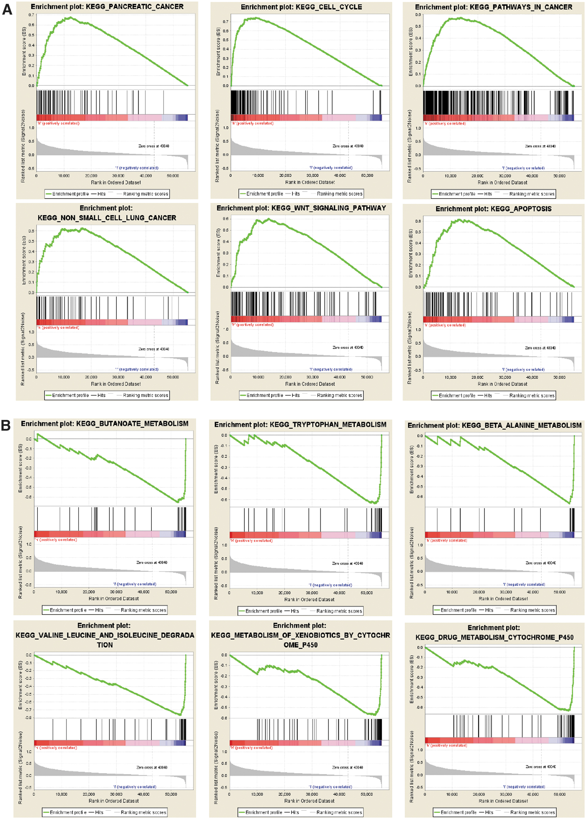
GSEA enrichment was adopted to explore the mechanism of CSNK1D affecting HCC.
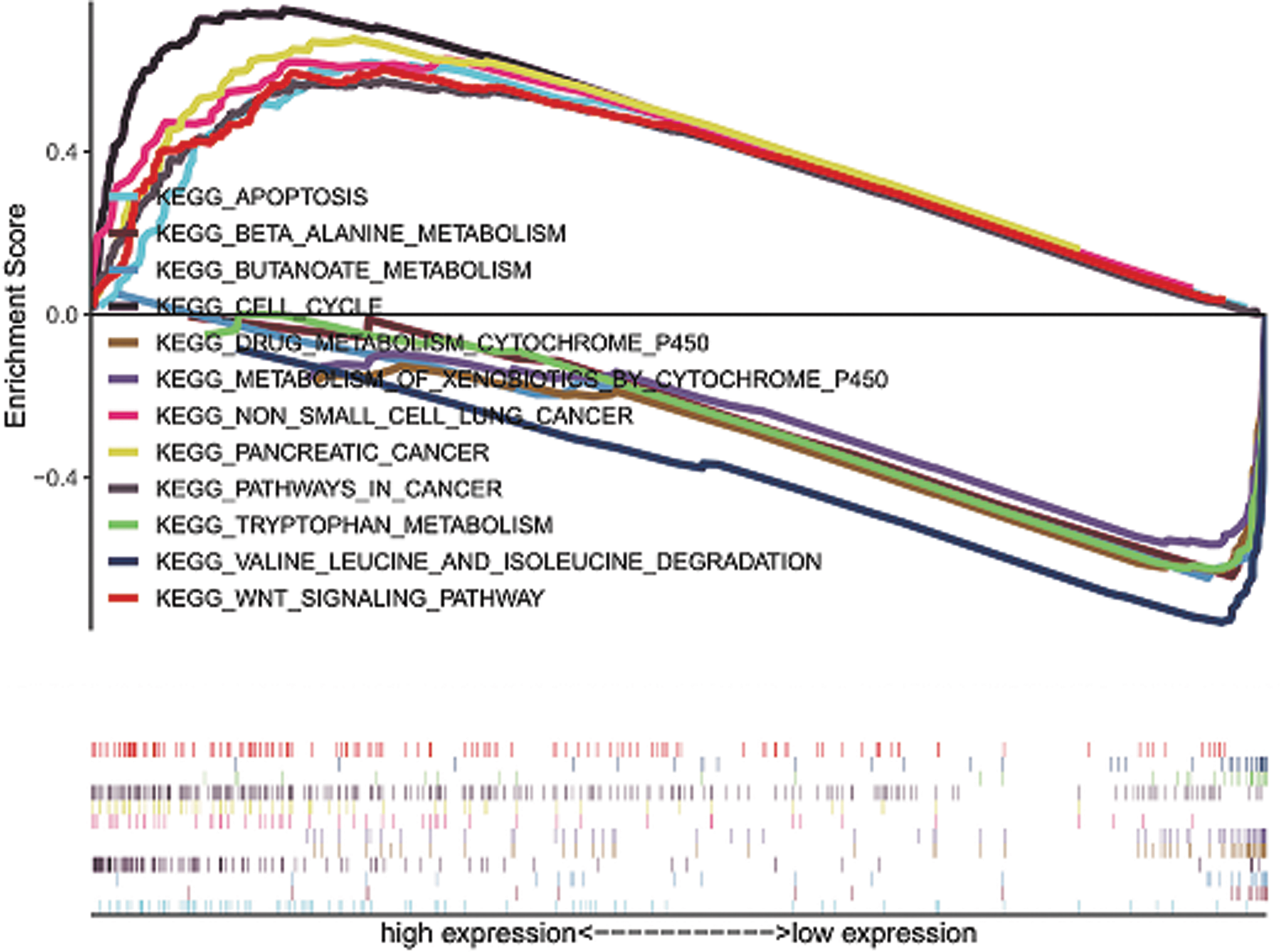
GSEA enrichment was adopted to explore the pathway and correlation of CSNK1D affecting HCC. Color images are available online.
Gene Set Enrichment Analysis Enrichment Was Adopted to Explore the Pathway of CSNK1D Affecting Hepatocellular Carcinoma
ES, enrichment score; FDR, false discovery rate; NES, normalized enrichment score; NOM, nominal.
Discussion
The poor long-term efficacy of HCC has been widely concerned worldwide. At present, the scientific evaluation system for its prognosis has not been established. The study on the factors influencing the prognosis and recurrence of HCC as well as the pathogenesis and progression of HCC may provide the basis for exploring diagnostic and prognostic markers and developing potential drug targets for HCC patients. There are many factors influencing the prognosis of HCC. In general, they are related to the following four aspects: the disease status of patients, the clinicopathological characteristics, the effect of surgery, and the sensitivity of the patient to the comprehensive postoperative treatment (Hung et al., 2016; Shimoda et al., 2016; Sun et al., 2016; Garnelo et al., 2017). This research mainly explored the influences of clinical pathological factors and the expression level of CSNK1D on the prognosis of HCC. At present, it is believed that the biological characteristics of tumors that affect the recurrence and metastasis of liver cancer include tumor size, origin, number, hepatic vascular invasion, tumor thrombus, and immune microenvironment. This study suggested that tumor size, tumor stage, and distant metastasis are the prognostic factors of HCC. It is widely believed that tumor size affects the prognosis of liver cancer. Tumors with the diameter of 3 cm or less are the critical point of biological characteristics of liver cancer since the incidence of microvascular tumor thrombus in liver cancer tumors with the diameter larger than 3 cm (23.5%) was higher than that in tumors with the diameter of <3 cm (6.9%) (Lu et al., 2011; Garnelo et al., 2017). In addition, liver cancer tumors with the diameter of 3 cm or less showed the higher degree of differentiation, more complete capsule, and the less invasion in surrounding tissues (Lu et al., 2011; Li et al., 2019). There are several liver cancer staging systems in the world (Liu et al., 2016). Although the prediction values of various staging systems for the prognosis of patients are different, each staging system has a certain prediction value for the survival period and prognosis of most liver cancer patients in various countries (Shao et al., 2016). All staging systems reflect that the later the stage is the shorter the survival period is (Shao et al., 2016). The distant metastasis of cancer indicates the later stage, which is one of the characteristics of advanced liver cancer (Chen et al., 2014; Wang et al., 2019b). Patients with distant metastasis undoubtedly have a poor prognosis (Greer et al., 2017). Our research suggested that the expression level of CSNK1D was closely related to the size, grade, and stage of tumors, but it was different from the traditional HCC marker, AFP. CSNK1D was differentially expressed in normal and cancerous tissues of different patients or in paracarcinoma tissues and cancer tissues in the same patients. The positive rate of CSNK1D in liver cancer tissues was higher. The expression level of CSNK1D significantly affected the proliferation and metastases of HCC and could better predict the survival period of patients with HCC.
This study suggested that tumor size, tumor stage, and distant metastasis status could affect the prognosis of HCC, but multivariate COX risk regression analysis showed that the above factors were not the independent prognostic factor for HCC survival. CSNK1D was the only independent prognostic factor for HCC survival. Moreover, Kaplan-Meier survival analysis showed that CSNK1D expression level was the influencing factor for the survival of HCC patients. The higher the expression level of CSNK1D was the shorter the survival period of patients was. Therefore, it was speculated that CSNK1D might be a new marker for the diagnosis of HCC and the prognosis evaluation. However, the influencing mechanism of CSNK1D on the features of HCC cells, cell proliferation ability, and antiapoptotic ability should be further explored.
Through GSEA enrichment analysis, we found that CSNK1D could affect the occurrence and development of HCC through cell cycle, WNT signaling pathway, amino acid metabolism, and other pathways. CSNK1D played an important role in cell cycle and genomic stability. Greer et al. (2017) found that silencing CK1DNA decreased ChK1 and CDC2/CDK1, and inevitably affected cell differentiation, which was an important feature to maintain the undifferentiated state of stem cells. The WNT signaling pathway is one of the important pathways involved in the development, regeneration, cell proliferation, and dynamic balance of tissues (Basu et al., 2016; Chai et al. 2016; Duchartre et al., 2016; and Pohl et al., 2017). Changes or mutations in the WNT pathway have been observed in various cancers, and CSNK1D can positively or negatively affect the WNT pathway through phosphorylation. CSNK1D can undergo posttranslational modifications through intramolecular autophosphorylation or site-specific phosphorylation of upstream cell kinases, reversibly regulate and fine-tune kinase activity, and generate new sequences through autophosphorylation (Amit et al., 2002; Zeng et al., 2005). The motif is pSer/pthr-X-X-Y, where X represents any amino acid and Y represents any amino acid other than threonine or serine. These motifs can act as pseudosubstrates to compete the catalytic centers of kinases (Rivers et al., 1998). CK1 kinase activity is reduced by phosphorylation (c-terminal), thus affecting the biological behaviors of cancer cells.
In summary, the overexpression of CSNK1D is closely related to the progression of HCC, and CSNK1D is expected to become a biomarker and a therapeutic target for HCC prognosis evaluation.
Footnotes
Authors' Contributions
H.Z. and X.L. designed the study. C.Q., H.Z., T.Z., and W.L. analyzed the data. All authors have given approval to the final version of the article.
Author Disclosure Statement
No competing financial interests exist.
Funding Information
This work was supported by Guangdong provincial health and family planning commission (no. B2021088).