




Abstract
Aim:
The aim of this study was to explore the possible associations between single nucleotide polymorphisms (SNPs) and DNA methylation levels of seven genes in the inflammatory response pathway with susceptibility to chronic periodontitis (CP) among the Uighur population of the Xinjiang Autonomous Region of China.
Methods:
A total of 444 eligible subjects (279 CP patients and 165 healthy controls) were enrolled in the study. Genomic DNA was obtained from gingival tissue for genotyping eight SNPs and performing methylation measurements of seven genes.
Results:
SNP rs2070745 in the formyl peptide receptor 1 (FPR1) gene achieved statistical significance in a standard allelic association analysis for CP (p = 0.02). The frequency of the rs2070745 minor allele G was higher in the cases than in controls (0.367 vs. 0.291). Additionally, rs2070745 was significantly associated with CP under the dominant genetic model (p = 0.03). Using logistic regression analysis, rs2070745 was found to be consistently associated with CP under the additive dominant model, and this association remained significant after covariates were taken into account [odds ratio (OR) = 1.49 (1.09-2.05), p = 0.014; OR = 1.58 (1.04-2.40), p = 0.031, respectively]. No significant gene-gene interactions were identified. Although we did not find a polymorphism in interleukin 6 (IL6) associated with CP in our study, the methylation level of a CpG island region located within the promoter region of IL6 was significantly less in CP patients compared with controls (p < 0.05).
Conclusions:
The genetic polymorphism rs2070745 in FPR1 and the methylation level of the promoter region of IL6 might be associated with CP in the Uighur population of China.
Introduction
Chronic periodontitis (CP) is a chronic infectious disease of the periodontal supporting tissue caused by a plethora of factors, including microbial compositions as well as both the innate and adaptive immune response mechanisms leading to inflammation and destruction of periodontal supporting tissues (gingiva, periodontal membrane, cementum, and alveolar bone) (Nanci and Bosshardt, 2006). Its clinical manifestations include inflammation of the gingiva, formation of periodontal pockets, and absorption of alveolar bone, which eventually can lead to loosening and loss of teeth (Socransky and Haffajee, 1997). The results of epidemiological investigations show that CP has become the primary cause of tooth loss among the Chinese population (Zhao et al., 2015). It not only affects the oral health of patients but is also closely related to multiple systemic diseases, including diabetes (Movva et al., 2014), metabolic syndrome (Morita et al., 2010), pneumonia (Iwasaki et al., 2018), and most importantly, vascular diseases leading to myocardial infarction and stroke (Kane, 2017). Thus, early diagnosis and effective treatments are paramount. Currently, the etiology of CP is not fully understood, but it is likely that in addition to infectious agents, the host's genetics plays a role.
Individual susceptibility to CP may be influenced by systemic risk factors, including genetic factors. Early epidemiological studies in the Netherlands have shown that CP in adults has familial clustering (Petit et al., 1994). In a study of twins, 38-82% of the differences between the twins' teeth and clinical indicators were also found to be due to genetic factors (Michalowicz et al., 1991).
The relationship between genetic factors and CP has been a topic of interest. CP is not a single-gene disease, but rather results from the contribution of susceptibility alleles from multiple genes. Accumulating evidence suggests that the host immune response contributes both protective and/or destructive effects toward the development of periodontal disease (Cekici et al., 2014; Meyle et al., 2017). Genetic factors that play a role in regulating the periodontal disease immune response pathways include, but are not necessarily limited to, interleukin 1 beta (IL1B) (Ribeiro et al., 2016; Majumder et al., 2019), C-C motif chemokine receptor 2 (CCR2) (Gunpinar et al., 2017), C-C motif chemokine receptor 5 (CCR5) (Shih et al., 2014; Cavalla et al., 2018), C-X-C motif chemokine ligand 8 (IL8), interleukin 6 (IL6) (Zhao and Li, 2018), STAT1 (Wei et al., 2019), and formyl peptide receptor 1 (FPR1) (Gunji et al., 2007). It has been shown that there are extensive differences in the occurrence of different mutations and polymorphisms in these genes among patients from different ethnic populations (Folwaczny et al., 2003; Gunji et al., 2007; Shao et al., 2009; Zhu et al., 2010). Since genetic factors cannot fully explain the susceptibility and disease progression of CP, epigenetic regulation has also been posited to play an important role in the pathogenesis of periodontitis. In the gingival tissue of CP patients, aberrant promoter methylation has been described for several genes involved in the immune activation pathways, including TLR2, PTGS2, IFNG, IL6, IL8, and TNF (Jurdziński et al., 2020). In the case of CP risk in the Uighur population in Xinjiang, China, this association remains unclear. In our research, to determine the association between immune response pathway genes and their potential role in CP, we selected CP patients and periodontally healthy persons from the Uighur population in Xinjiang as the research subjects and performed comparative genotypic and methylation analyses.
Methods
Ethics statement
This study was approved by the Ethics Committee of the First Affiliated Hospital of Xinjiang Medical University (Urumqi, China). All participants agreed to provide sufficient gingival tissue samples and filled out questionnaire information. Written informed consent from all participants was obtained.
Study participants
The subjects were recruited from the First Affiliated Hospital of Xinjiang Medical University from April 2014 to November 2016. A total of 444 individuals, including 279 patients and 165 healthy controls, were recruited for this study. All subjects lived in Moyu, Hetian, or their surrounding areas and are ethnically Uighurs. According to the new classification standard proposed by the 1999 working conference of the American society of periodontal diseases, all subjects had to meet the diagnostic standard of extensive CP. Our sample inclusion criteria were as follows: in the periodontitis group, patients had a probing depth (PD) of >7 mm and a clinical attachment loss (CAL) of >5 mm. Subjects in the healthy control group had no history of periodontal abscess, no gingival swelling or spontaneous bleeding, and no bleeding on probing (BOP), with PD at all sites of less than 3 mm. The exclusion criteria were systemic diseases affecting the progression of periodontal disease; female breastfeeding and pregnancy; history of oral local radiotherapy; and long-term use of anti-inflammatory drugs and antitumor drugs. A detailed workflow of the sample enrollment is shown in Figure 1.
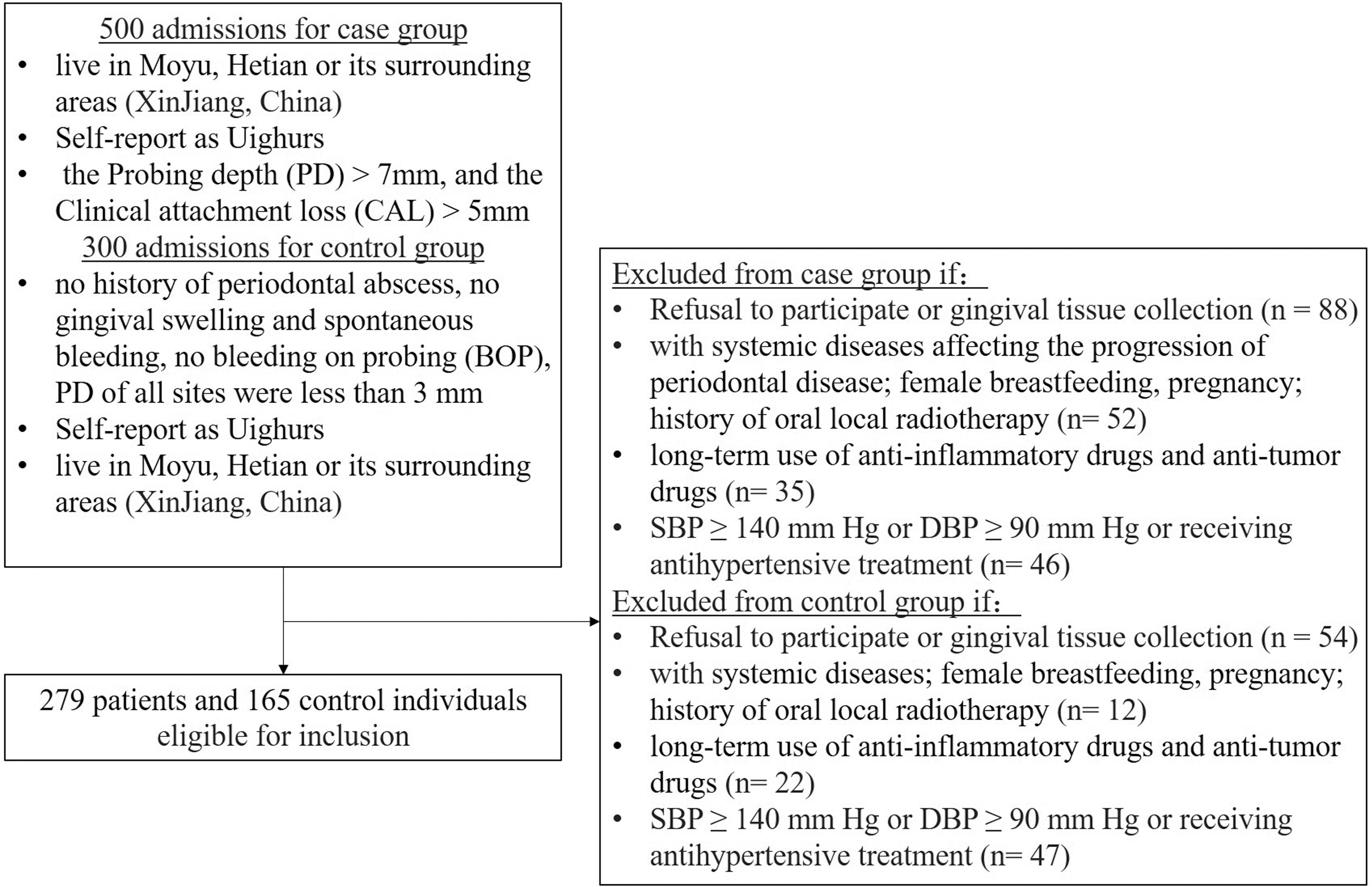
Workflow of sample enrollment of this study.
Single nucleotide polymorphism selection and genotyping
Referring to the existing literature in regard to single nucleotide polymorphisms (SNPs) affecting CP, we selected rs1143634 in IL1B (da Silva et al., 2018), rs1799864 in CCR2 (Damrongrungruang et al., 2014), rs333 in CCR5 (Folwaczny et al., 2003), rs4073 in IL8 (Andia et al., 2011), rs1800795 and rs2069837 in IL6 (Farhat et al., 2017), rs3771300 in STAT1 (Saraiva et al., 2013), and rs2070745 in FPR1 (Gunji et al., 2007), which had a minor allele frequency of >1% in the Chinese subgroups from the 1000 Genomes Project (Han Chinese in Beijing and Han Chinese in the south). Gingival tissue samples were collected from all participants. DNA was extracted from tissue samples using a commercial DNA purification kit (GoldMag, China) and quantified by spectrometry (DU530 UV/VIS spectrophotometer, Beckman Instruments, Fullerton, CA). Polymorphisms were genotyped using an improved, multiple ligase, detection reaction method (Thomas et al., 2004), with technical support from the Shanghai Genesky Biotechnology Company. Detailed information regarding the protocol and quality control has been described previously (Zhang et al., 2014). The primer list can be found in Supplementary Table S1.
DNA methylation analysis
Genomic DNA samples from gingival tissues of 42 patients and 27 age- and sex-matched healthy controls were used for the DNA methylation analysis. The DNA methylation level was measured using the MethylTarget™ method (Genesky Biotechnologies, Inc., Shanghai, China) based on multiplex PCR amplification and was followed by next-generation sequencing. Detailed information on this method has been previously reported (Idriss et al., 2018; Sun et al., 2018; Zhu et al., 2019). Specifically, CpG islands in the promoter were defined according to the following criteria for the genes of interest: (1) >200 bp; (2) >50% GC content; and (3) ratio of observed/expected CpG >0.6. The EZ DNA Methylation™-GOLD Kit (Zymo Research) was used for sodium bisulfite conversion of DNA, following the manufacturer's protocols. Primers were designed for multiplex amplification of CpG island regions of the genes of interest and the amplification products were used for library construction. Libraries were further sequenced on an Illumina HiSeq benchtop sequencer using the 2 × 150 paired-end sequencing protocol according to the manufacturer's guidelines. The methylation level at each CpG site was calculated as the percentage of methylated cytosines over the total tested cytosines. The average methylation level of all measured CpG sites within the amplified region or the gene was calculated and used for identifying differentially methylated amplicons and genes.
Statistical analyses
The categorical variables are presented as percentages (%), and continuous variables are expressed as mean ± standard deviation. Differences between the case and control groups were measured using the χ2 test for categorical variables or t tests for continuous variables. Logistic regression models were used to investigate the association between polymorphisms and CP. The Mann-Whitney U test was used to compare methylation levels between CP patients and healthy controls. All statistical analyses were performed using SPSS 18.0 software (SPSS, Chicago, IL). PLINK software was used for the genetic association analysis. A generalized multifactor dimensionality reduction (GMDR) model was used to screen for the best gene-gene interaction combinations (Xu et al., 2016). A two-tailed p value of <0.05 was considered statistically significant.
Results
Baseline characteristics
A total of 444 eligible subjects (279 patients and 165 healthy controls) were included in this study. Baseline characteristics of the subjects are presented in Table 1. We found that there were significant differences between the two groups in the number of males and number of females (p = 0.01). No statistical differences were found for age and body-mass index. As predicted, the values of the clinical parameters, PD and CAL, were higher in the CP group than in healthy controls (p < 0.001).
General Characteristics of Chronic Periodontitis Patients and Healthy Controls
BMI, body-mass index; BOP, bleeding on probing; CAL, clinical attachment loss; PD, probing depth; SD, standard deviation.
Association between SNPs and CP risk
Eight SNPs were successfully genotyped. The basic information of SNPs and genotype distributions in the case and control cohorts is listed in Table 2. Polymorphism rs333 did not satisfy the Hardy-Weinberg equilibrium (HWE) and was removed from further analyses (p = 0.02). A standard allelic association analysis for all seven remaining SNPs found that rs2070745 in FPR1 was significantly associated with CP (p = 0.02). The frequency of the rs2070745 minor allele G was higher in cases than in controls (0.367 vs. 0.291). Additionally, rs2070745 was significantly associated with CP under the dominant genetic model (p = 0.03) (Table 3). For logistic regression analysis assuming three common genetic models (additive, dominant, and recessive inheritance), the rs2070745 SNP was consistently associated with CP under the additive and dominant models; the association remained significant after covariates were adjusted for [Table 4; odds ratio (OR) = 1.49 (1.09-2.05), p = 0.014; OR = 1.58 (1.04-2.40), p = 0.031]. No evidence was found for the association between the other SNPs and CP, regardless of whether the covariates were adjusted or not. We used GMDR to look for gene-gene combinations for CP; however, we did not identify any significant gene-gene interaction combinations (Table 5).
Basic Information of Single Nucleotide Polymorphisms Examined in the Related Genes
AGTCAGTATCAATTCTGGAAGAATTTCCAGACA; MAF: minor allele frequency.
CCR2, C-C motif chemokine receptor 2; CCR5, C-C motif chemokine receptor 5; FPR1, formyl peptide receptor 1; IL1B, interleukin 1 beta; IL6, interleukin 6; SNP, single nucleotide polymorphism.
The Hardy-Weinberg Equilibrium and Chi-Square Test Results in Allele and Genotype Models of Single Nucleotide Polymorphisms
Significant results (p < 0.05) are shown in bold.
HWE, Hardy-Weinberg equilibrium; CI, confidence interval; OR, odds ratio.
Logistic Analyses Under Different Genetic Models
Significant results (p < 0.05) are shown in bold italic.
Values in bold indicate that the locus is statistically significant.
CP, chronic periodontitis.
Generalized Multifactor Dimensionality Reduction Analysis for Gene-Gene Interaction
CV, cross-validation.
Differentially methylated sites, amplicons, and genes
Methylation levels of these genes in the gingival tissues of the case and control individuals were further measured. No CpG islands were identified in the promoter regions of FPR1, IL1B, CCR2, and CCR5. We only identified one CpG region in each of the promoters of IL8 and STAT1 and two CpG regions in the promoter of IL6; these three regions were individually amplified and sequenced following bisulfite conversion (the chromosome regions of the amplicons and a list of primers can be found in Supplementary Table S2). A mean coverage of >800 X was observed and the bisulfite conversion efficiency for all samples was above 98.66%. There was no statistically significant difference in the bisulfite conversion efficiency between the case and control groups (Supplementary Figure S1). Results showed that three of the 70 CpG sites located within the first of the two IL6 gene region amplicons were differentially methylated in CP patients when compared with controls (all p < 0.05) (Fig. 2). Furthermore, the group difference of the methylation level was >0.01. The first amplicon in the IL6 promoter region was also differentially methylated in CP patients compared with controls (p = 0.009, Table 5). We did not find any significant differences in DNA methylation levels of the IL8, IL6, and STAT1 genes between the CP patients and controls (all p > 0.05, Table 6).
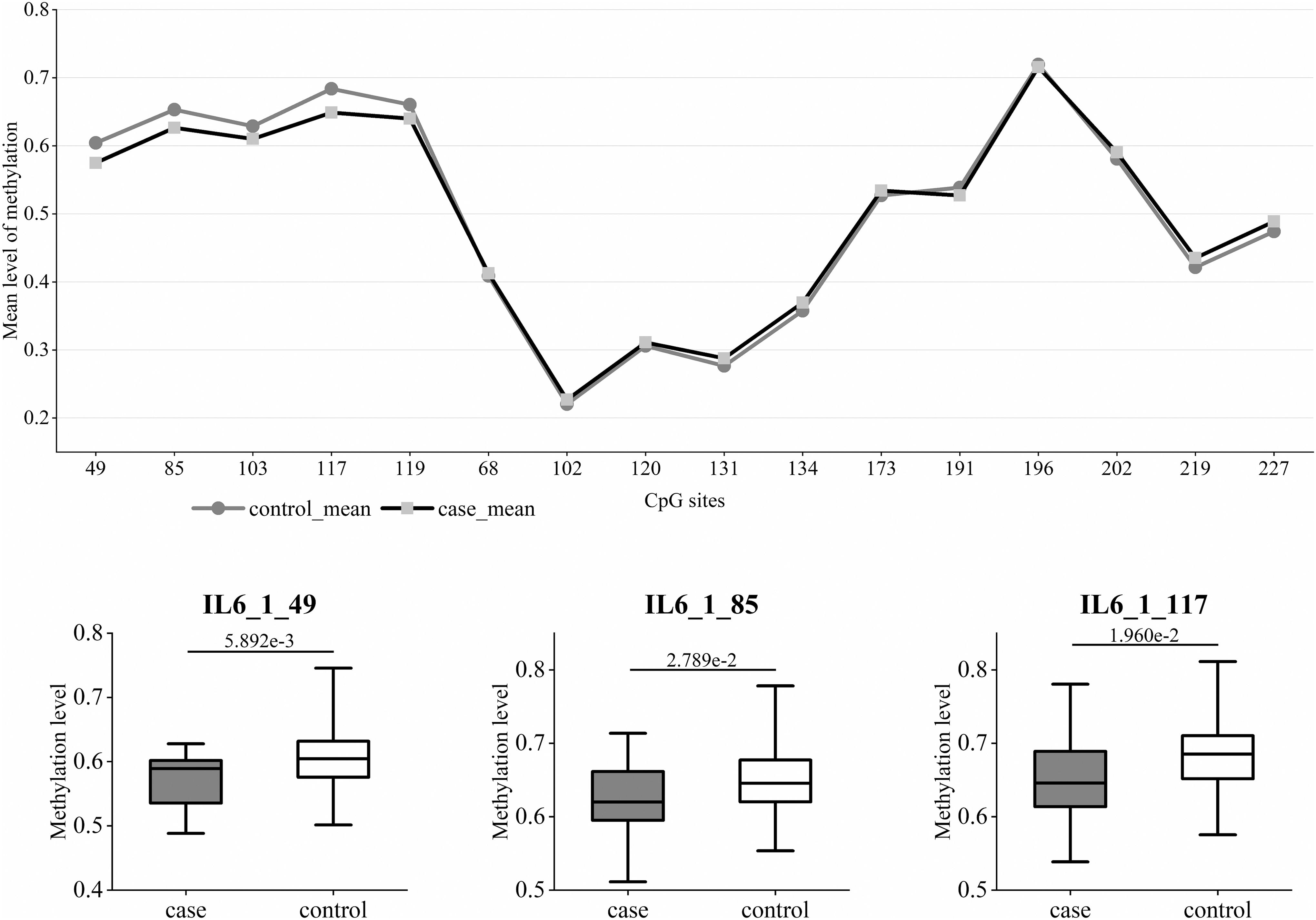
Methylation level analysis of CpG sites in the promoter region of IL6. IL6, interleukin 6. Top: Methylation levels of all CpG sites in the promoter region of IL6. Bottom: Box plots showing three CpG sites with significantly different methylation levels between CP tissues and normal gingival tissues. CP, chronic periodontitis.
Differences in Methylation Levels (%) Between Chronic Periodontitis Patients and Controls
Significant results (p < 0.05) are shown in bold.
Mean in case, average methylation degree of the CP group; Mean in control, average methylation degree of the control group; MethylDiff, average methylation degree of the CP group minus average methylation degree of the control group; p (U-test): the U-test model is used to calculate the p value.
IL8, C-X-C motif chemokine ligand 8.
Discussion
It is well known that genetic polymorphisms and DNA methylation can have an effect on the regulation of gene expression, which leads to differences in susceptibility and severity of disease among individuals (Shang et al., 2020; Smith et al., 2020). To provide new ideas for the treatment and prevention of CP, it is necessary to explore contributions of the immune response pathway to the susceptibility of CP. In this study, we successfully genotyped seven SNPs located in genes that are part of the immune response pathway. We found that rs2070745 in the FPR1 gene was associated with CP risk. Furthermore, compared with the control individuals, the methylation level of one CpG island region in the IL6 promoter was significantly lower in the gingival tissues of CP patients.
Polymorphonuclear leukocytes (PMNs) play an important role in the first line of immune defense against bacterial and fungal infections (Sandborg and Smolen, 1988). FPR1, which belongs to the seven-transmembrane domain G-protein-coupled family, is expressed on the surface of PMNs and is associated with multiple leukocyte functions (Le et al., 2002). Diminution in the number of high-affinity FPR1s on PMNs in CP patients has been reported (Perez et al., 1991), and a role for FPR1 SNPs in CP and aggressive periodontitis progression has been suggested (Gunji et al., 2007; Maney et al., 2009). The polymorphism rs2070745 located in exon 2 of the FPR1 gene (301 C > G) involves an amino acid change in the third transmembrane domain (p.V101L). In 2013, Zhou et al. (2013) found that variants of FPR1 carrying a single amino acid substitution of leucine for valine at position 101 (p.Leu101) displayed significantly improved receptor affinity. In our study, individuals who carry the G allele are at a significantly higher risk for developing CP. Given these results, low receptor affinity of FPR1 associated with the G allele of rs2070745 might contribute to the pathogenesis of CP. Furthermore, the MAF value for p.V101L (0.291) found in the control samples of the present study is similar to that derived from the African population in the 1000 Genomes Project (0.26), but it is significantly lower than those found in European (0.38) and Asian (0.47) populations, respectively. This result might indicate novel genetic characteristics of rs2070745 in the Chinese Uighurs.
IL6 is well known as a proinflammatory cytokine associated with the development of periodontal disease (Zhu and Guo, 2016). Increased expression of IL6 has been found in peripheral blood mononuclear cells (Moreira et al., 2007), saliva (Costa et al., 2010), and gingival tissue (Prabhu et al., 1996) and is associated with periodontal disease severity. Although no polymorphism in IL6 was found to be associated with CP in our study, the methylation level of a CpG island region located on the promoter region of IL6 was significantly lower in CP patients compared with controls. In 2003, Stefani et al. (2013) explored the mRNA expression, polymorphisms, and methylation levels of IL6 in periodontal tissues and concluded that the expression of IL6 was higher in the CP group. Similar to our study, no statistical differences in the distribution of genotypes or the overall level of DNA methylation were observed (Stefani et al., 2013). It is worth noting that although the average methylation levels of all CpG sites in IL6 were consistent between case and control individuals, we detected significantly lower methylation levels of one CpG region in the promoter region of IL6 in the CP tissues than in normal gingival tissues (Fig. 2). It may be that methylation differences in this region affect transcriptional regulation and result in the higher expression of IL6 in CP tissues.
Our research is not without limitations: (1) the small size of study cohorts somewhat limited the statistical power and thus validation analyses with larger sample sizes should be performed. (2) CP is a multifactorial disease; the relationship between host genetics and disease susceptibility cannot be considered alone. The true relationship between genetic susceptibility and CP should be analyzed in combination with multiple environmental risk factors. (3) Since RNA samples of the gingival tissue were not quantitated for mRNA expression, the evidence is not conclusive as to whether polymorphisms in FRP1 or the DNA methylation levels of IL6 have an effect on the mRNA expression of corresponding genes.
Conclusion
Our study preliminarily looked at the association between polymorphisms and DNA methylation of seven genes in the immune response pathway and risk of CP in the Xinjiang Uighur population. For the first time, our study demonstrates that the genetic polymorphism rs2070745 in FPR1 and the methylation level of the promoter region of IL6 might be associated with susceptibility of CP in the Uighur population of China.
Footnotes
Authors' Contributions
A.A. conceived and designed the experiments, J.L. performed the experiments, T.M. analyzed the data, and A.A. and J.Z. wrote the manuscript.
Author Disclosure Statement
No competing financial interests exist.
Funding Information
This study was supported by the National Natural Science Foundation of China (Grant No. 81760194).

References
Supplementary Material
Please find the following supplemental material available below.
For Open Access articles published under a Creative Commons License, all supplemental material carries the same license as the article it is associated with.
For non-Open Access articles published, all supplemental material carries a non-exclusive license, and permission requests for re-use of supplemental material or any part of supplemental material shall be sent directly to the copyright owner as specified in the copyright notice associated with the article.
