




Abstract
Background:
Colorectal cancer (CRC) is a common malignancy of the digestive system, but its specific mechanisms of occurrence and development remain incompletely understood. F-Box and leucine-rich repeat protein 7 (FBXL7) is a subunit of the Skp-cullin-F-box ubiquitin ligase, involved in cell cycle regulation, endothelial cell damage, and inflammatory immunological responses. However, the role of FBXL7 in CRC remains unknown. In this study, we investigated the clinical significance and potential mechanism of FBXL7 expression in CRC progression.
Methods:
We utilized data from The Cancer Genome Atlas (TCGA) and the University of California Santa Cruz Xena (UCSC Xena) database for bioinformatic analyses. Clinical CRC samples were used to confirm FBXL7 expression. Gene set enrichment analysis (GSEA) and various databases, such as TCGA, UCSC Xena, cBioPortal, University of ALabama at Birmingham CANcer data analysis portal, MethSurv, Tumor Immune Estimation Resource (TIMER), TIMER2.0, Tumor-Immune System Interaction Database, and Tumor Immune Dysfunction and Exclusion Database (TIDB), were used to investigate the role of FBXL7 in CRC. Statistical analysis was performed using R (v.3.6.3) or GraphPad Prism 8.0.
Results:
Our findings revealed the predictive significance of FBXL7 in CRC patients. FBXL7 expression was associated with tumor stage, lymph node stage, pathological stage, perineural invasion, and lymphatic invasion. GSEA analysis identified associations between FBXL7 and extracellular matrix organization, as well as immune-related pathways. Immunological analysis revealed a correlation between high FBXL7 expression and the development of an immunosuppressive microenvironment.
Conclusion:
Identifying FBXL7 as a novel biomarker for CRC could shed light on the promotion of CRC development by the immune environment.
Introduction
Colorectal cancer (CRC) has the second-highest incidence and the fifth-highest mortality rate among all cancers in China, as well as the third-highest incidence and the second-highest mortality rate worldwide (Sung et al., 2021). Adenocarcinomas account for most cases of CRC, with squamous cell carcinoma, adenosquamous carcinoma, spindle cell carcinoma, and undifferentiated carcinoma accounting for the other subtypes (Fleming et al., 2012). Various treatment modalities, including surgical resection, chemotherapy, radiation, and a combination of methods, are used for treating CRC. Recent advancements in CRC treatment have led to the approval of targeted therapy and immunotherapy, significantly improving the prognosis of patients.
Numerous recent articles highlight the efficacy of these treatments, underscoring their impact on patient outcomes (Zhao et al., 2022). The immune system plays a complex role in cancer development and significantly impacts CRC progression (Markman and Shiao, 2015). The discovery of novel therapeutic approaches and an improvement in the prognosis of patients with advanced CRC could both be aided by further analysis of immune regulatory processes in advanced CRC.
F-Box and leucine-rich repeat protein 7 (FBXL7) is a crucial component of the E3 ubiquitin ligase Skp-cullin-F-box, involved in substrate recognition for ubiquitination. It participates in various biological activities, including cell cycle progression, cell death pathways, and inflammatory immunological responses (Coon et al., 2012; Keskin et al., 2019; Ni et al., 2021). FBXL7's association with immunity regulation was discovered in a comprehensive study on hepatocellular carcinoma (HCC) prognosis (Zhang et al., 2023), suggesting the immunomodulatory effects of FBXL7. However, the specific immunomodulatory effects of FBXL7 in CRC remain unknown. Investigating FBXL7's role in immune regulation within the context of CRC holds promising avenues for further research.
Herein, we conducted bioinformatic analysis and verified the expression of FBXL7 in CRC tissues. We investigated the correlation between FBXL7 expression levels and clinical characteristics and prognosis of patients. In addition, we aimed to explore the clinical significance and functional mechanisms of FBXL7 in CRC through comprehensive analysis.
Materials and Methods
Data and sample collection
We retrieved pan-cancer sequencing data and ribonucleic acid (RNA) sequencing (RNA-Seq) data for FBXL7 from The Cancer Genome Atlas (TCGA) database (https://portal.gdc.cancer.gov/). In addition, we obtained the messenger RNA (mRNA) expression data of FBXL7 in CRC tissues and relevant clinicopathological features, including tumor stage (T stage), lymph node stage (N stage), metastasis stage (M stage), pathological stage, primary outcome, age, residual tumor, perineural invasion, lymphatic invasion, overall survival (OS), disease-specific survival (DSS), progression-free interval (PFI), and disease-free survival (DFS) from the University of California Santa Cruz (UCSC) Xena database (https://xenabrowser.net/datapages/).
CRC tissues and paired paracancerous tissues were collected from a tumor tissue specimen database consisting of samples from 26 patients at the First Affiliated Hospital of Chongqing Medical University (Chongqing, China). All tissue specimens were identified and stored at −80°C. The study was conducted in accordance with the guidelines and approved by the Ethics Committee of the First Affiliated Hospital of Chongqing Medical University (approval number: 2022-K414).
RNA extraction
TRIzol® reagent (Life Technologies, Carlsbad, CA) was utilized to extract total RNA from both CRC tissues. The extracted RNA was quantified using a NanoDrop 2000 spectrophotometer (Thermo Fisher Scientific, Rockford, IL), and subsequently stored at −80°C for further analysis.
Real-time quantitative reverse transcription polymerase chain reaction
Quantification of FBXL7 expression was carried out using quantitative reverse transcription polymerase chain reaction (qRT-PCR) in CRC tissues. Total RNA (1 μg) was reverse transcribed into complementary DNA using a reverse transcription system (Promega, Madison, WI), resulting in a final volume of 20 μL. qRT-PCR was performed on an ABI 7500 Real-Time PCR system (Applied Biosystems, Foster City, CA) using SYBR® Green PCR Master Mix (Thermo Fisher Scientific, Hong Kong, China). The amplification was conducted with β-actin serving as the internal control. The primer sequences utilized are provided in Table 1.
The Primer Used in This Study
FBXL7, F-Box and leucine-rich repeat protein 7.
Comparison of the FBXL7 expression level
The expression of FBXL7 was assessed in a total of 647 CRC tissues and 51 normal colorectal tissues, as well as in 50 CRC tissues along with their paired adjacent normal colorectal tissues, across 33 different human cancer types using data from TCGA. To evaluate its diagnostic potential, a receiver operating characteristic (ROC) curve was constructed using R programs. Furthermore, qRT-PCR data were utilized to analyze the expression of FBXL7 in comparison to paired adjacent normal tissues and at different pathological stages of CRC.
Correlation analysis of FBXL7 and clinicopathological characteristics
The TCGA colorectal adenocarcinoma (COADREAD) datasets obtained from the UCSC Xena database were analyzed using R software. Kaplan-Meier plots and log-rank tests were performed using the survival package to assess the survival outcomes.
Genetic alteration in patients with CRC
Two different COADREAD datasets were utilized in this study: COADREAD (Dana-Farber Cancer Institute [DFCI], Cell Reports 2016) and COADREAD (TCGA, Firehose legacy). While both datasets focus on COADREAD, they have distinct origins and may differ in terms of patient cohorts, data collection, and analysis methods. Kaplan-Meier plots and log-rank tests were used to determine the significance of the discrepancy in the survival curves.
DNA methylation details of FBXL7
The University of ALabama at Birmingham CANcer data analysis portal (UALCAN) (http://ualcan.path.uab.edu/index.html) was used to evaluate the FBXL7 promoter DNA methylation level in CRC (Chandrashekar et al., 2017). In the subsequent analysis, we utilized the MethSurv database (https://biit.cs.ut.ee/methsurv/) to evaluate the DNA methylation status of FBXL7 in the TCGA dataset (Modhukur et al., 2018). Specifically, we assessed the association between DNA methylation at specific loci in FBXL7 and clinical outcomes, including survival differences between samples with and without the methylated loci. This analysis aimed to investigate the potential predictive significance of CpG methylation in FBXL7.
Gene set enrichment analysis and Tumor Immune Estimation Resource
Gene set enrichment analysis (GSEA) was performed to explore the potential signaling pathways associated with FBXL7 in CRC. This analysis utilized normalized RNA-Seq data from TCGA, and differential gene expression analysis was conducted using DESeq2 (Yu et al., 2012). The significance of various functional collections of groups with different FBXL7 expression levels in CRC was assessed using predefined gene sets from the Molecular Signatures Database (https://www.gsea-msigdb.org/gsea/msigdb/index.jsp).
Tumor Immune Estimation Resource (TIMER) (https://cistrome.shinyapps.io/timer/) was utilized to evaluate immune cell infiltration, including B cells, CD4+ T cells, CD8+ T cells, macrophages, neutrophils, and dendritic cells (DCs). Its purpose was to examine the association between tumor purity and FBXL7 expression in CRC (Li et al., 2017). The correlation between FBXL7 expression and immune cell infiltration was analyzed using Spearman's correlation analysis. The TIMER2.0 server (http://timer.cistrome.org/) was also used to investigate the correlation between FBXL7 expression and immune infiltration of three immunosuppressive cells, namely cancer-associated fibroblasts (CAFs), tumor-associated macrophages (TAMs), and regulatory T (Treg) cells in CRC (Li et al., 2020).
Tumor-Immune System Interaction Database and Tumor Immune Dysfunction and Exclusion
Tumor-Immune System Interaction Database (TISIDB) (http://cis.hku.hk/TISIDB/index.php) is an open web-integrated repository portal that serves as a comprehensive resource for tumor immune system interactions (Ru et al., 2019). The relationship between FBXL7 and the immune system in CRC (including expression, methylation, copy number variation [CNV], and mutation) was evaluated using TISIDB. Tumor Immune Dysfunction and Exclusion (TIDE) analysis (http://tide.dfci.harvard.edu/query/) was performed to determine the survival benefit of cytotoxic T-lymphocyte (CTLs) infiltration in CRC patients with high or low FBXL7 expression (Fu et al., 2020; Jiang et al., 2018).
Validation of immunosuppressive gene correlation
Data from the TCGA COADREAD dataset available on the UCSC Xena database were utilized to investigate the correlation between FBXL7 expression and immunosuppressive genes. The statistical analysis and correlation calculations were performed by the website itself, while the R ggplot2 package was used for data visualization purposes.
Statistical analysis
For the statistical analysis and visualization, R software (version 3.6.3) or GraphPad Prism 8.0 was used. Specific R packages were utilized for different analyses. The pROC package (version 1.17.0.1) was used for ROC curve analysis, while the ggplot2 package (version 3.3.3) was used for visualization purposes. The TCGA colorectal adenocarcinoma (COADREAD) datasets obtained from the UCSC Xena database were performed by the website itself. Kaplan-Meier plots and log-rank tests were constructed using the survminer package (version 0.4.9) for visualization and the survival package (version 3.2-10) for statistical analysis of survival data.
To compare differences across groups, the Wilcoxon rank-sum test, Student's t-test, or analysis of variance (ANOVA) were used, depending on the appropriateness of the data. Pearson's or Spearman's correlation tests were used to establish correlations. The statistical significance was denoted as follows: p > 0.05 (not significant), *p < 0.05, **p < 0.01, and ***p < 0.001. The threshold for statistical significance was set at p < 0.05.
Results
FBXL7 is abnormally expressed in various cancers, including CRC
FBXL7 expression was initially analyzed in 33 types of tumor samples and normal samples using pan-cancer sequencing data retrieved from TCGA to explore the possible roles of FBXL7. The mRNA FBXL7 levels increased in cholangiocarcinoma, kidney renal clear cell carcinoma, and liver HCC and decreased in bladder urothelial carcinoma, breast invasive carcinoma, cervical squamous cell carcinoma, endocervical adenocarcinoma, colon adenocarcinoma (COAD), kidney chromophobe, lung adenocarcinoma, lung squamous cell carcinoma, pheochromocytoma and paraganglioma, prostate adenocarcinoma, rectal adenocarcinoma (READ), thyroid carcinoma, and uterine corpus endometrial carcinoma compared with their corresponding normal samples (Fig. 1A).

The FBXL7 expression level in various human cancer types.
Moreover, we observed that FBXL7 expression in all these CRC tissues was lower than that in the normal colorectal tissue in the unpaired groups (p < 0.001) (Fig. 1B), and the result remained consistent in the paired groups (p < 0.001) (Fig. 1C). Subsequently, 26 pairs of CRC tissues and corresponding paracancer tissues were collected to verify the FBXL7 mRNA expression level using qRT-PCR. The results suggested that the FBXL7 mRNA expression level also decreased in these CRC tissues compared with the corresponding paracancer tissues (p = 0.0143) (Fig. 1D). Further analysis revealed that the FBXL7 mRNA expression level was higher in advanced-stage COAD (Fig. 1E), whereas the FBXL7 mRNA expression level was the lowest in stage II and relatively higher in the other stages in READ (Fig. 1F).
These findings demonstrate that FBXL7 expression is downregulated in CRC. However, its expression is higher in advanced CRC tissues compared to early CRC tissues, indicating that FBXL7 expression is regulated in a complex manner as CRC progresses.
Relationship between FBXL7 expression and clinical characteristics
The correlation between FBXL7 expression levels and various clinicopathological characteristics of CRC patients was examined. Despite the overall decrease in FBXL7 mRNA expression levels in CRC, higher expression levels were observed in advanced T stages, N stages (p < 0.01) (Fig. 2A, B), and pathological stages (p < 0.05) (Fig. 2D). FBXL7 expression was also higher in CRC patients with perineural invasion (Fig. 2H, p < 0.05) or lymphatic invasion metastases (Fig. 2I, p < 0.01). In addition, FBXL7 expression was higher in READ compared to COAD (p < 0.05) (Fig. 2J). No significant differences were found in FBXL7 expression levels concerning the M stage (Fig. 2C), primary outcome (Fig. 2E), age (Fig. 2F), and residual tumor size (Fig. 2G). Overall, patients with advanced-stage CRC exhibited higher levels of FBXL7 expression.
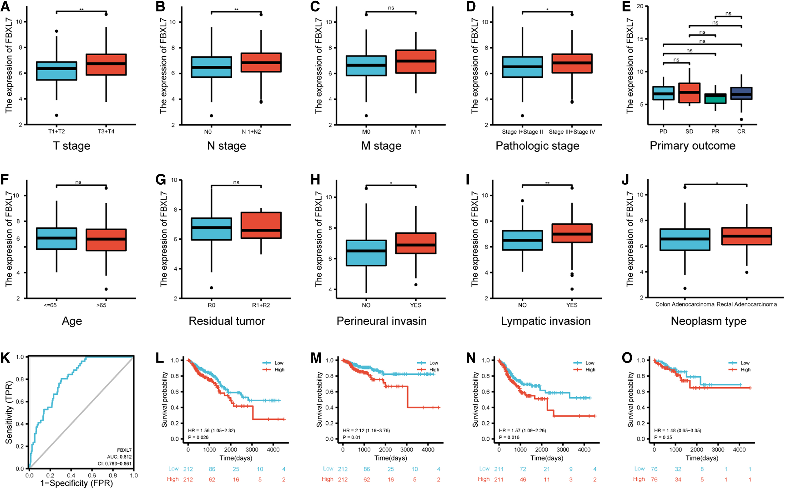
The clinical value of FBXL7 expression in CRC.
The ROC curve analysis for diagnosis indicated that FBXL7 expression could help distinguish between tumors and normal tissue (Youden index = 49.8%, sensitivity = 80.4%, specificity = 69.4%, and area under the curve = 0.812) (Fig. 2K). Kaplan-Meier survival curves demonstrated that CRC cases with high FBXL7 expression exhibited poor OS (hazard ratio [HR] = 1.56, p = 0.026) (Fig. 2L), DSS (HR = 2.12, p = 0.01) (Fig. 2M), and PFI (HR = 1.57, p = 0.016) (Fig. 2N). However, no significant difference was observed in DFS (HR = 1.48, p = 0.35) (Fig. 2O). These preliminary results suggest the potential utility of FBXL7 as a candidate diagnostic and prognostic marker for CRC.
FBXL7 genetic alteration and methylation in patients with CRC
A total of 1259 CRC patients from two datasets, cBioPortal—TCGA Firehose Legacy and DFCI Cell Reports 2016, were analyzed. The percentage of FBXL7 genetic alterations in CRC was found to be 4% (Fig. 3A), with the alteration rate ranging from 2.1% to 5.01% (6/377) (Fig. 3B). Kaplan-Meier plots and log-rank tests indicated significant differences in OS (p = 0.0378) (Fig. 3C), suggesting that patients with FBXL7 alterations had a poor prognosis in terms of OS. However, no significant difference in DFS (p = 0.728) was observed between patients with and without FBXL7 alterations (Fig. 3D).
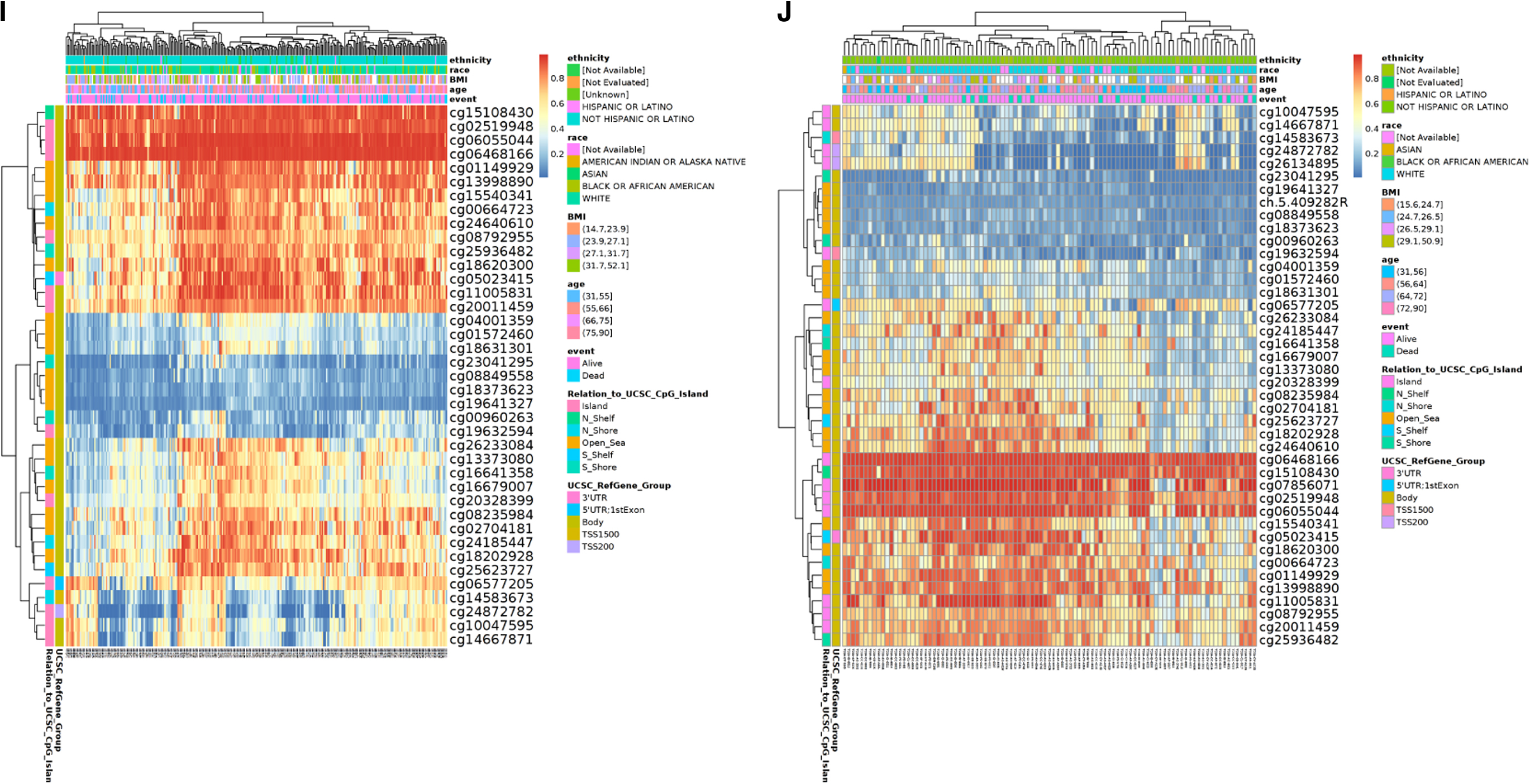
FBXL7 genetic alteration and methylation in CRC.
UALCAN data indicated that FBXL7 DNA methylation levels were higher in CRC cancer tissues compared with normal samples (p < 0.001) (Fig. 3E, F). Furthermore, the methylation status of FBXL7 was markedly higher in early-stage COAD (Fig. 3G), while it was higher in the middle stage and lower in the other stages of READ (Fig. 3H).
Using MethSurv, the prognostic value of each CpG was assessed, and high methylation levels were observed in CRC for cg15108430, cg06468166, and cg06055044. However, these methylation levels did not demonstrate statistical significance in terms of prognosis (Fig. 3I, J; Table 2).
The Relationship Between the DNA Methylation Sites of F-Box and Leucine-Rich Repeat Protein 7 and Prognosis
CI, confidence interval; COAD, colon adenocarcinoma; HR, hazard ratio; LR, likelihood ratio; READ, rectal adenocarcinoma.
GSEA investigation of FBXL7 and the correlation between FBXL7 expression and immune cell infiltration
GSEA results revealed a significant association between FBXL7 and extracellular matrix (ECM) organization and immune-related pathways (Fig. 4A). This indicates that FBXL7 is involved in modulating the tumor immune microenvironment.
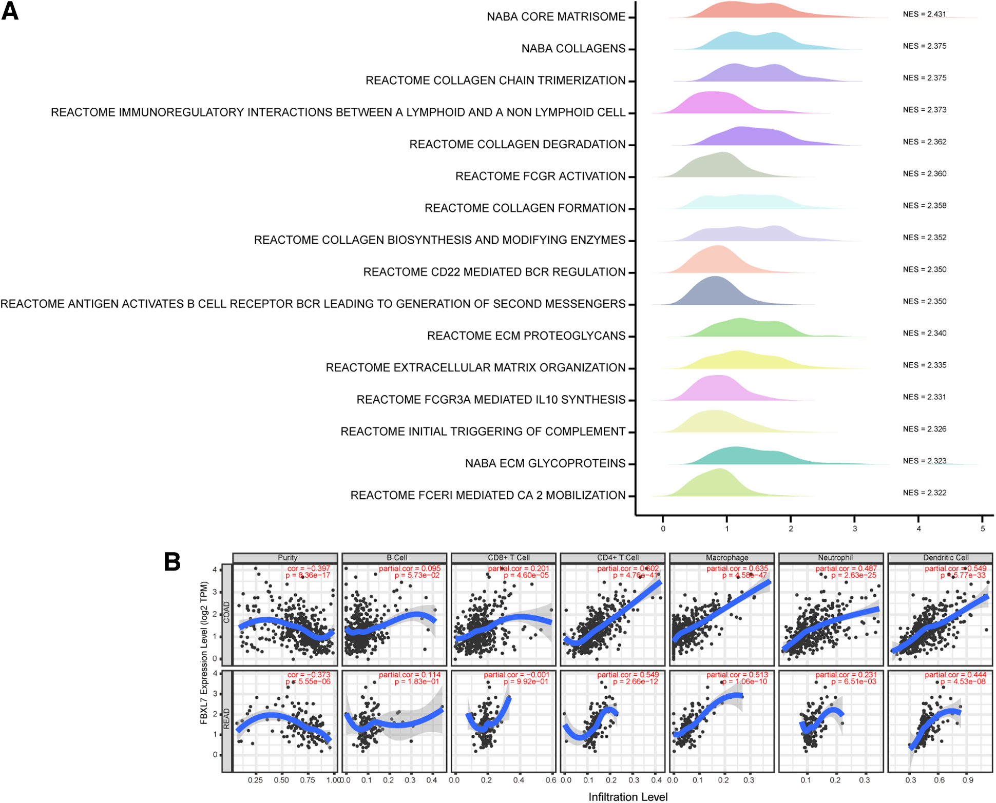
Correlation between FBXL7 expression and the involved pathway or immune infiltrates.
The relationship between FBXL7 expression and immune cell infiltration, adjusted by purity, was evaluated using TIMER. The results demonstrated a positive correlation between FBXL7 expression and infiltrating levels of B cells (r = 0.095, p = 8.36e−17), CD8+ T cells (r = 0.201, p = 4.60e−05), CD4+ T cells (r = 0.602, p = 4.76e−41), macrophages (r = 0.635, p = 4.58e−47), neutrophils (r = 0.487, p = 2.63e−25), and DCs (r = 0.549, p = 5.77e−33), as well as a negative correlation with tumor purity (r = −0.397, p = 8.36e−17) in COAD.
In READ, a positive correlation was observed between FBXL7 expression and infiltrating levels of B cells (r = 0.114, p = 1.83e−01), CD8+ T cells (r = −0.001, p = 9.92e−01), CD4+ T cells (r = 0.549, p = 2.66e−12), macrophages (r = 0.513, p = 1.06e−10), neutrophils (r = 0.231, p = 6.51e−03), and DCs (r = 0.444, p = 4.53e−08), along with a negative correlation with tumor purity (r = −0.373, p = 5.55e−06) (Fig. 4B). These findings suggest that FBXL7 may play a crucial role in CRC progression by regulating the immune microenvironment.
Relationship between FBXL7 expression and CRC immune regulation
The TISIDB database was used to establish relationships between FBXL7 (including expression, methylation, CNV, and mutation) and immunosuppressive genes, immune-activating genes, major histocompatibility complex (MHC) genes, chemokine genes, and chemokine receptor genes. The results showed that FBXL7 expression was positively correlated with most markers of immunosuppressive genes, immune-activating genes, MHC II genes, chemokine genes, and chemokine receptor genes in CRC. On the contrary, FBXL7 methylation was negatively correlated with most markers of immunosuppressive genes, immune-activating genes, MHC II genes, chemokine genes, and chemokine receptor genes in COAD. CNV and mutation did not significantly contribute to the effect on these markers (Figs. 5A-E).
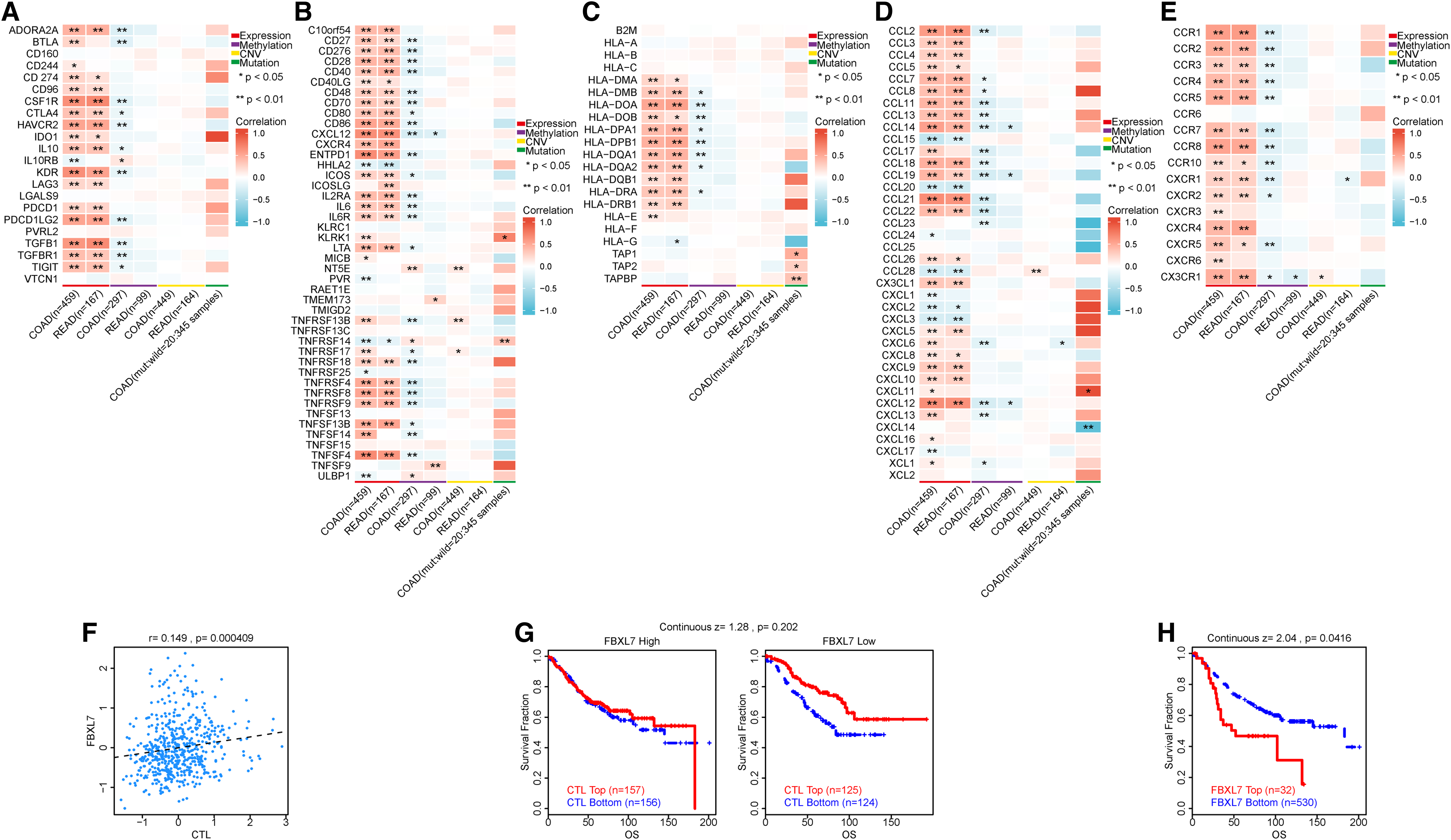
The correlation between FBXL7 and immunoregulation-related genes or prognosis.
Dysfunctional T cell phenotype analysis using the TIDE revealed an association between FBXL7 expression and CTLs (r = 0.149, p = 0.000409) (Fig. 5F). Although not statistically significant (p = 0.202), the CTL top group in the FBXL7 low-expression group showed a trend of longer survival compared to the FBXL7 high-expression group (Fig. 5G). This suggests that CTL function may be reduced with high FBXL7 expression, but further evidence is required to support this finding. In addition, high FBXL7 expression was associated with shorter survival durations in CRC cohorts (continuous z = 2.04, p = 0.0416) (Fig. 5H). Thus, a correlation has been observed between FBXL7 expression and immune system regulation, indicating a potential association with a less favorable prognosis in CRC cohorts.
Analysis of immunosuppressive mechanisms
CAFs, TAMs, and regulatory T cells (Tregs) are important immune cell components in the tumor microenvironment (Braster et al., 2017; Chen et al., 2014; Takeuchi and Nishikawa, 2016). We used TIMER2.0 to examine the relationship between FBXL7 and CAFs, TAMs, and Tregs in CRC. Our observations revealed that FBXL7 expression was positively correlated with CAF, TAM, and Treg infiltration in CRC. Among them, CAFs showed the strongest positive correlation, and M0 and M2 macrophages showed stronger positive correlations compared to M1 macrophages (Fig. 6A). Previous studies have reported that macrophages are attracted to the tumor microenvironment through chemokines such as CCL2, CCL3, CCL4, CCL5, CCR5, and CXCL12 (Chen et al., 2018), and they are polarized into the M2 type immune suppressor through the action of CCL5, CCR5, and hypoxia-inducible factor 1-alpha (HIF1A) (González-Martín et al., 2011; Halama et al., 2016; Lewis and Murdoch, 2005).

Correlation between FBXL7 expression and immune-related cells or gene markers.
CXCR3 stimulates CD8+ T cell infiltration into the tumor microenvironment (Chow et al., 2019). TIM3 (HAVCR2) inhibits the function of CD8+ T cells (Kang et al., 2015), and LAG3 inhibits the function of CD4+ T cells (Huard et al., 1994). We analyzed the correlation between FBXL7 and the above-mentioned genes to further support the association between FBXL7 and immunosuppression in CRC. The results showed a positive correlation between FBXL7 and all the mentioned genes (Figs. 6B-6K). Particularly, CCL2 (r = 0.727, p < 0.001) and CXCL12 (r = 0.802, p < 0.001) exhibited the strongest correlation with FBXL7 expression. Thus, a close association was observed between FBXL7 and macrophage recruitment. Furthermore, compared with CXCR3, which demonstrated a slightly positive correlation, HAVCR2 and LAG3 demonstrated significant positive associations. These findings consistently suggest that high FBXL7 expression in CRC may have immunosuppressive effects.
Discussion
FBXL7 exhibits abnormal expression patterns in various cancers. Interestingly, we observed higher levels of FBXL7 in advanced CRC tissues compared to early-stage tissues, while it exhibited a marked downregulation in early-stage CRC. Consistent with a previous study (Wang et al., 2022), our research also demonstrated that high FBXL7 expression was associated with a poor prognosis in CRC patients. As CRC mortality rates vary depending on its stage, FBXL7 has the potential to serve as a prognostic biomarker for this disease.
The high mutational load has been linked to longer DSS in CRC (Giannakis et al., 2016). However, tumor cells can undergo evolutionary pressure, leading to a reduction in antigenicity through various mechanisms. These mechanisms include impaired antigen presentation, immunosuppressive molecules suppressing immunological function, and immune checkpoint molecules suppressing immune activity of T cells (Kather et al., 2018). These factors may contribute to the poor prognosis observed in patients with FBXL7 variations, as revealed by our genetic changes and CNV analysis.
Considering the impact of evolutionary pressure on tumor cells and the potential immunological implications, the findings from our genetic changes and CNV analysis further support the notion that FBXL7 variations may modulate the clinical course of CRC.
In line with our results, a previous study demonstrated that miR-152-5p can regulate FBXL7 expression, resulting in its downregulation in gliomas. However, as the stage of glioma progresses, the expression of miR-152-5p decreases, leading to an upregulation of FBXL7 and promoting glioma progression (Kong et al., 2020). Similarly, our data analysis revealed a high level of promoter methylation associated with FBXL7 downregulation in CRC. Although the specific methylation sites impacting CRC prognosis were not identified in our study, further investigations are necessary to identify key methylation sites associated with FBXL7 and elucidate the underlying regulatory mechanisms.
Our GSEA results uncovered associations between FBXL7 expression and stromal remodeling as well as immune-related pathways, suggesting a potential role for FBXL7 in modulating the immunoregulatory microenvironment and contributing to CRC development. Consistently, our immune infiltration analysis revealed a significant correlation between FBXL7 expression and the level of immune cell infiltration, particularly CD4+ T cells and macrophages. These findings suggest that FBXL7 may exert its influence on CRC by impacting specific immune cell populations, which play crucial roles in the immune response and tumor microenvironment. The observed correlations between FBXL7 expression and immune cell infiltration, along with the GSEA results, support the notion of FBXL7's involvement in the immune modulation of CRC.
These findings provide insights into the potential immunomodulatory role of FBXL7 and its contribution to CRC progression. Further investigations are warranted to elucidate the underlying mechanisms and functional implications of the observed correlations between FBXL7 and specific immune cell subtypes, shedding light on potential therapeutic strategies targeting FBXL7 and the immune microenvironment in CRC.
According to the tumor immune interaction analysis using TISIDB, FBXL7 showed positive correlations with most immunostimulators and immunoinhibitors. Notably, FBXL7 exhibited a strong positive correlation with human leukocyte antigen-D-related isotype (HLA-DR), the predominant MHC class II molecule produced in CRC. In addition, FBXL7 expression was not significantly correlated with MHC class I molecules, suggesting that it was not directly associated with CD8+ T cells. The presence of MHC II molecules in paracancerous CRC tissues, increasing as CRC progresses, may explain the relationship between high FBXL7 expression, HLA-DR expression, and CD4+ T cell infiltration in advanced CRC.
Moreover, our TIDB analysis revealed a positive correlation between CTLs and FBXL7 expression. Interestingly, the CTL top cohort in the low FBXL7 expression group had a better prognosis compared to the CTL bottom cohort. However, this difference was less prominent in the high FBXL7 expression group. These findings suggest that FBXL7 potentially limit CTL function. This observation aligns with previous research demonstrating that the immunosuppressive microenvironment can inhibit CD8+ T cell function, even in the presence of normal CD8+ T cell functioning (Weng et al., 2022).
Furthermore, FBXL7 expression positively correlated with CAFs, TAMs, and Tregs, indicating its potential role in regulating immunosuppression within the CRC microenvironment. The interactions between FBXL7 and various molecules, such as CCL2, CXCL12, CCR5, HIF1A, CXCR3, HAVCR2, and LAG3, further validate FBXL7's immunosuppressive function. Moreover, the higher FBXL7 expression in CRC may contribute to ECM disorganization, which in turn hampers the efficacy of immunotherapy by impeding drug and immune cell delivery to the tumor. ECM irregularities also lead to hypoxia, upregulation of immunomodulatory molecules, enhanced angiogenic signals, and ultimately immune escape.
Furthermore, ECM irregularities promote tumor-related angiogenesis and inflammation, rendering the tumor microenvironment resistant to immunotherapy (Chen et al., 2021; Lu et al., 2012). In line with the consensus molecular subtype (CMS) classification (Guinney et al., 2015), higher FBXL7 expression in CRC is more likely to be associated with CMS type 4, known as the immune tolerant subtype, highlighting the potential for antiangiogenic treatment regimens.
However, it is essential to validate the relationship between FBXL7 and immunoregulation in CRC using cell and animal models to further substantiate our findings. In addition, comprehensive studies are needed to explore the functional implications of FBXL7-mediated immunosuppression and its potential impact on immunotherapeutic strategies.
Conclusions
In summary, our study provides valuable insights into the complex role of FBXL7 in CRC. The observed variations in FBXL7 expression, methylation, and their associations with immune-related pathways and cell populations highlight the potential clinical significance of FBXL7 in CRC prognosis and immunoregulation. Further investigations are warranted to unravel the underlying mechanisms and functional implications of FBXL7 in CRC development and its potential as a therapeutic target.
Footnotes
Acknowledgments
We acknowledge Xiantao online tools for providing platforms and contributors for uploading meaningful datasets.
Authors' Contributions
Conceptualization, S.W. and T.L.; data curation, S.W., X.Z., S.Z., and J.X.; formal analysis, S.W., X.Z., S.Z., and J.X.; funding acquisition, T.L.; investigation, S.W. and S.Z.; methodology, S.W.; project administration, T.L.; resources, X.Z.; software, S.W.; validation, S.W.; visualization, S.W.; writing—original draft, S.W. and S.Z.; writing—review and editing, T.L.
Author Disclosure Statement
No competing financial interests exist.
Funding Information
No funding was received for this article.