




Abstract
Background:
In Dayao County, Chuxiong Yi Autonomous Prefecture, Yunnan Province, Southwest China, 5% of the surface is scattered with blue asbestos, which has a high incidence of pleural mesothelioma (PMe). Simian virus 40 (SV40) is a small circular double-stranded DNA polyomavirus that can cause malignant transformation of normal cells of various human and animal tissue types and promote tumor growth. In this study, we investigate whether oncogenic SV40 is associated with the occurrence of PMe in the crocidolite-contaminated area of Dayao County, Yunnan Province, Southwest China.
Methods:
Tumor tissues from 51 patients with PMe (40 of whom had a history of asbestos exposure) and pleural tissues from 12 non-PMe patients (including diseases such as pulmonary maculopathy and pulmonary tuberculosis) were collected. Three pairs of low-contamination risk primers (SVINT, SVfor2, and SVTA1) were used to detect the gene fragment of SV40 large T antigen (T-Ag) by polymerase chain reaction (PCR). The presence of SV40 T-Ag in PMe tumor tissues and PMe cell lines was detected by Western blotting and immunohistochemical staining with SV40-related antibodies (PAb 101 and PAb 416).
Results:
PCR, Western blotting, and immunohistochemical staining results showed that the Met5A cell line was positive for SV40 and contained the SV40 T-Ag gene and protein. In contrast, the various PMe cell lines NCI-H28, NCI-H2052, and NCI-H2452 were negative for SV40. PCR was negative for all three sets of low-contamination risk primers in 12 non-PMe tissues and 51 PMe tissues. SV40 T-Ag was not detected in 12 non-PMe tissues or 51 PMe tissues by immunohistochemical staining.
Conclusion:
Our data suggest that the occurrence of PMe in the crocidolite-contaminated area of Yunnan Province may not be related to SV40 infection and that crocidolite exposure may be the main cause of PMe.
The Clinical Trial Registration number: 2020-YXLL20.
Introduction
Malignant mesothelioma (MM) is a rare aggressive tumor that mostly originates in the mesothelial cells of the pleura and peritoneum and less commonly in the pericardium and tunica vaginalis testis. Pleural mesothelioma (PMe) is the most common entity, accounting for 81% of MM (Viscardi et al., 2020). It has long been believed that a history of occupational or environmental asbestos exposure can cause chronic inflammation and induce the release of oxygen-free radicals, activating various pathways that promote cell growth and leading to 80% of PMe cases (Wadowski et al., 2020). The incidence of PMe is still rising worldwide, and it is estimated that peak incidence will occur within the next few years (Sinn et al., 2021). Due to a latency period of more than 40 years between asbestos fiber exposure and the development of disease, most cases are usually identified at advanced stages and have a very poor prognosis. Treatment options for this tumor are very limited. Only early-stage cases and less symptomatic patients may benefit from aggressive surgical management and multimodality treatments. In advanced cases of PMe, the combination of pemetrexed and platinum remains the only established treatment to improve overall survival (Viscardi et al., 2020). Despite the latest advancements in chemotherapy, molecular targeted therapy, and immune checkpoint inhibitor therapy, long-term survival remains an unmet goal (Zhao et al., 2023). The median survival is 12-18 months with a 5-year survival rate <10%, and cured cases are rare (Nadal et al., 2021; Süß and Kölbl, 2018). Therefore, it is urgent to identify novel molecular biomarkers and immune targets for early screening and diagnosis, treatment, and prognostic monitoring in the hope of developing individualized precision treatment strategies and effectively improving patient survival and quality of life.
However, PMe may result from other nonasbestos factors, including genetics, erionite, radiation, and simian virus 40 (SV40), which may work alone or in combination (Yang et al., 2008). The polyomavirus SV40 is a potentially oncogenic virus of rhesus monkeys’ origin, which is built of small circular double-stranded DNA polyomavirus of approximately 5243 base pairs. SV40 is a potent DNA tumor virus that is known to induce cancer in laboratory animals (Carbone et al., 2020). SV40 can cause malignant transformation of human and animal cells in tissue culture and induce the formation of various tumors in hamsters and transgenic mice. The large tumor antigen (T-Ag) is the main gene product involved in SV40 replication and mediates cellular transformation. It is also known as one of the most potent transforming proteins and can transform different types of cells in the absence of other viral genes (Butel and Lednicky, 1999; Pepper et al., 1996). T-Ags induce DNA synthesis in host cells and prolong the onset of S-phase through the inhibition of the tumor suppressor proteins p53 and Rb (Butel and Lednicky, 1999). These interactions may contribute to the development of PMe by rendering mesothelial cells more susceptible to other carcinogens. Recently, there has been a growing concern among the identification of SV40 in human tumors, and SV40 has been implicated in the development of several cancers, including PMe, breast cancer, prostate cancer, glioma, and lung cancer (Limam et al., 2020; Sato et al., 2020; Unterberger et al., 2022; Yin et al., 2021). However, a definitive role of the virus in human mesothelioma has not been unequivocally demonstrated but has been rigorously debated (Carbone et al., 2020).
Crocidolite is scattered on 5% of the land in Dayao County, Yunnan Province, Southwest China (Jia et al., 2016). In the early 1980s, crocidolite was mined and used indiscriminately by local residents, causing serious environmental pollution. In addition, the most recent annual statistics indicate that the incidence of MM in the area reached 8.5/100,000 (1977-1983) and 17.75/100,000 (1987-1995), which is 10-fold higher than that in the general population (0.1-0.6/100,000) (Wang et al., 2023). Although the use of asbestos has been regulated and restricted in Western countries, the increasing use, import, and export of asbestos in many non-Western developing countries render PMe a continuing global health problem (Baas et al., 2015). In addition, SV40 may act synergistically with crocidolite asbestos to transform mesothelial cells (Cleaver et al., 2014). However, the role of SV40 in PMe and SV40 has not yet been studied in a Chinese population, especially in the crocidolite-contaminated area of Dayao County, Yunnan Province. The purpose of this study is to identify the correlation between the incidence of PMe and SV40 in the population of crocidolite-contaminated areas in Yunnan Province by polymerase chain reaction (PCR), Western blotting, and immunohistochemical staining. In conclusion, no association was found between SV40 and the occurrence of PMe in the crocidolite-contaminated area in Dayao County, Yunnan Province, Southwest China.
Materials and Methods
Patients and tissue collection
Tumor and pleural samples were obtained from patients with pathologically confirmed PMe at the Department of Thoracic Surgery of the Fourth Affiliated Hospital of Dali University (the First People’s Hospital of Chuxiong Yi Autonomous Prefecture) and the First People’s Hospital of Dayao County between 2014 and 2019. Of these, 51 PMe cases and 12 non-PMe cases, including pulmonary maculopathy and tuberculosis patients, were selected. All cases were obtained from PMe patients in the crocidolite-contaminated area of Dayao County. None of the PMe patients received radiotherapy, chemotherapy, hormone therapy, or other related antitumor treatments before surgery. Written informed consent was obtained from each patient before entering this study. Samples were collected upon the signing of informed consent and immediately frozen and stored in the institutional biobank. All studies were conducted after approval by the Medical Ethics Committee of the Dali University Hospital Consortium (approval number: 2020-YXLL20).
Cell culture
The human embryonic kidney cell lines HEK239, HEK293T, and HEK293JCT, the PMe cell lines NCI-H28 (epithelial type), NCI-H2052 (sarcomatous histology type), and NCI-H2452 (biphasic mixed type), and the human pleural mesothelial cell lines LP9 and Met5A (SV40-transformed pleural mesothelial cells) were purchased from the American Type Culture Collection. All cell lines were identified by short tandem repeat analysis. All cell types were routinely checked for mycoplasma contamination using the TransDetect Luciferase Mycoplasma Assay Kit (Servicebio, Wuhan, China). Cell lines were cultured in RPMI-1640 (Gibco, Thermo Fisher Scientific, USA) supplemented with 10% fetal bovine serum (FBS, Gibco, Thermo Fisher), 100 U/mL penicillin, and 0.1 mg/mL streptomycin (Beyotime, Shanghai, China) at 37°C and 5% carbon dioxide. The medium was changed every 2-3 days, and trypsin was added for digestion and passage when the cells were 80% confluent.
Hematoxylin-eosin staining
After the collection of PMe and non-PMe tissues, the histological structure was evaluated by hematoxylin and eosin (HE) staining analysis using a HE staining kit (Haoxin Biotech, China). Briefly, tissues were fixed in formaldehyde, dehydrated using gradient alcohol, and cleared using xylene (Sinopharm Chemical Reagent Co., Ltd., China). Next, the tissues were embedded in paraffin. Three-micrometer-thick sections were stained using an HE staining kit for 3-8 min. After dehydration using graded tap water, the samples were stained with eosin to visualize the cytoplasm for 1-3 min. The samples were washed with tap water, dehydrated, and fixed. The sections were mounted in neutral gum and observed by light microscopy.
Genomic DNA extraction and identification
An Animal Genomic DNA Extraction Kit (TIANGEN, Beijing, China) was used to extract genomic DNA (gDNA) from PMe tissues and cell line samples. The proteins were extracted strictly according to the kit instructions, and then a 1.5% agarose gel was used for electrophoresis detection and identification. Specifically, the OD260 and OD280 of genomic DNA were measured using an ultra-micro ultraviolet spectrophotometer (Thermo Scientific, USA), and high-purity DNA (OD260/OD280 = 1.6-1.8) was selected for subsequent experiments.
PCR
We performed PCR amplification of SV40 using specific primers with low-contamination risk reported in the literature (López-Ríos et al., 2004) and selected the β-actin gene as the reference gene. The primer sequences (Table 1) and the positions of the SV40 T-Ag primers (Fig. 1) are shown later. PCR amplification was carried out in a 25 µL reaction containing 12.5 µL of 2× Taq PCR Master Mix, 9.5 µL of double-distilled water, 1.0 µL of each primer (10 µmol/L), and 1 µL of high-purity genomic DNA extracted from fresh tissues or cell lines. After a predenaturing step of 3-10 min at 94-95°C, 50 amplification cycles were performed. Each cycle consisted of 1 min at 94°C, 30 s at 58°C, and 1 min at 72°C. The specific PCR conditions are shown in Table 2. Finally, the DNA fragments were visualized by 1.5% agarose gel electrophoresis with Nucleic Acid Dye (TIANGEN) staining under a Gel Imaging System.
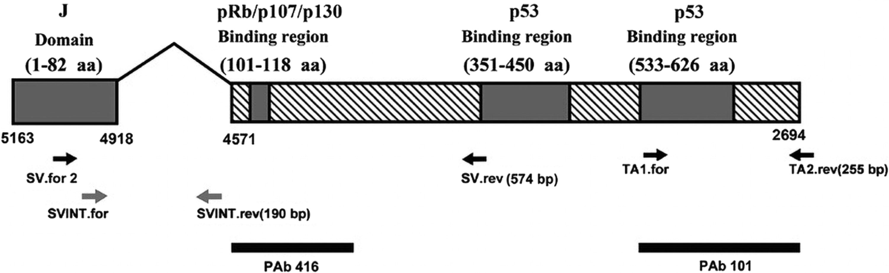
Location of SV40 primer sets and antibodies. The black boxes correspond to the DNA of the four regions of SV40 T-Ag. The numbering direction of the SV40 genome is opposite to that of T-Ag transcription. The 3 pairs of specific primers with low contamination risk used in this study are marked with arrows. SV40, simian virus 40; T-Ag, tumor antigen.
Primer Sequences
PCR, polymerase chain reaction.
PCR Conditions
PCR, polymerase chain reaction.
Western blotting analysis
Total protein from pleural mesothelial cells and PMe cells was obtained using Western/IP cell lysis buffer (Beyotime, Shanghai, China) for further assays, and the proteins were quantified by a Bicinchoninic Acid (BCA) Protein Quantification Kit (Beyotime, Shanghai, China). The proteins were denatured with sodium dodecyl sulfate (SDS) loading buffer and immediately subjected to standard Western blotting. Western blotting was performed using 10% sodium dodecyl sulfate-polyacrylamide gel electrophoresis (SDS-PAGE) gels for protein separation and nitrocellulose membranes (Millipore, USA) for protein blotting. The membranes were blocked with 5% nonfat milk (Beyotime, Shanghai, China) in Tris-buffered saline and incubated at 4°C with primary antibodies diluted in primary antibody dilution buffer (Beyotime, Shanghai, China) (1:1000). Bound antibodies were detected using the Chemiluminescence Imaging System (Bio-Rad, USA) and fluorochrome-labeled secondary antibodies. Quantitative analysis was performed with ImageJ software. The following antibodies were used: Pab101 (MA5-12458, Invitrogen, USA) and Pab416 (ab16879, Abcam, USA).
Immunohistochemical staining
Immunohistochemical staining was performed by the streptavidin perosidase (SP) method. Briefly, the non-PMe pleural tissues and PMe tissues were adhered to slides at a thickness of 4 µm. The tissue sections were fixed in 10% neutral formalin, paraffin embedded, and then dewaxed in water. After deparaffinization with xylene, the sections were rehydrated in graded ethanol, and heat-mediated antigen retrieval was performed in citric acid. Tissue sections were incubated with monoclonal antibodies against Pab101 (MA5-12458, Invitrogen) and Pab416 (ab16879, Abcam) at 4°C overnight and labeled with horseradish peroxidase (HRP) (rabbit) secondary antibody (ab8227, Abcam) at room temperature for 60 min. Finally, sections were developed in diaminobenzidine (DAB) solution under microscopic observation. The scoring of SV40 expression was performed based on both the ratio and the intensity of positive-stained cells (Jenniskens et al., 2021). Ten high-power fields of positive cells enriched in each section were randomly selected for observation. These scores were performed by two experienced pathologists in a blinded manner independently, and averaged percentage values were taken.
Statistical analysis
GraphPad Prism 9.0 and SPSS 23.0 software were used for statistical analysis and graphing. The χ2 test or Fisher’s exact probability test was used for statistical analysis. Differences were considered statistically significant at p<0.05.
Results
Clinical characteristics of the participants
The study included 12 non-PMe patients and 51 PMe patients. In the non-PMe patient group, 6 patients (50.00%) were male, 6 patients (50.00%) were female, and 8 patients (66.67%) were aged ≥50 years. In the PMe patient group, there were 38 (74.51%) males and 13 (25.49%) females. The majority of patients developed PMe when they were aged 50 or older (45 cases, 88.24%). The occupational asbestos exposure history was available for 40 (78.43%) patients. Among them, 22 patients (43.14%) were smokers. The clinicopathological data are summarized in Table 3.
General Data and Clinicopathological Data of Participants
PMe, pleural mesothelioma.
HE staining morphology of PMe tissue and non-PMe pleural mesothelial tissue
HE sections showed that the surface of non-PMe pleural mesothelial tissue was covered by benign mesothelial cells and fibrous, adipose, and vascular tissues underneath (Fig. 2A). Among the 51 PMe patients, 86.27% (44/51) of the specimens were the epithelioid type. Tumor cells were mainly composed of two types of cells, one with moderate amounts of eosinophilic cytoplasm and the other with eosinophilic or basophilic cytoplasm. These two types of cells could be dominated by one or mixed. Due to the different degrees of differentiation, there were significant differences in tissue structure, which could manifest as tubular, papillary, tubular papillary, cleft-like, microcystic, solid-nesting, and patchy structures, and the gaps were filled with abundant edematous mucinous stroma (Fig. 2B). There were 5.88% (3/51) cases of the sarcomatoid type. Tumor cells were spindle-shaped, rich in cells, often interwoven into bundles, and focal whirlpool-like, spoked, and hemangiopericytoma-like structures could also be seen. The nuclei were atypical, with deep staining and mitotic figures common (Fig. 2C). The biphasic mixed type accounted for 7.84% (4/51). This type of mesothelioma had both epithelioid and sarcomatoid mesothelioma components mixed in different proportions (Fig. 2D).
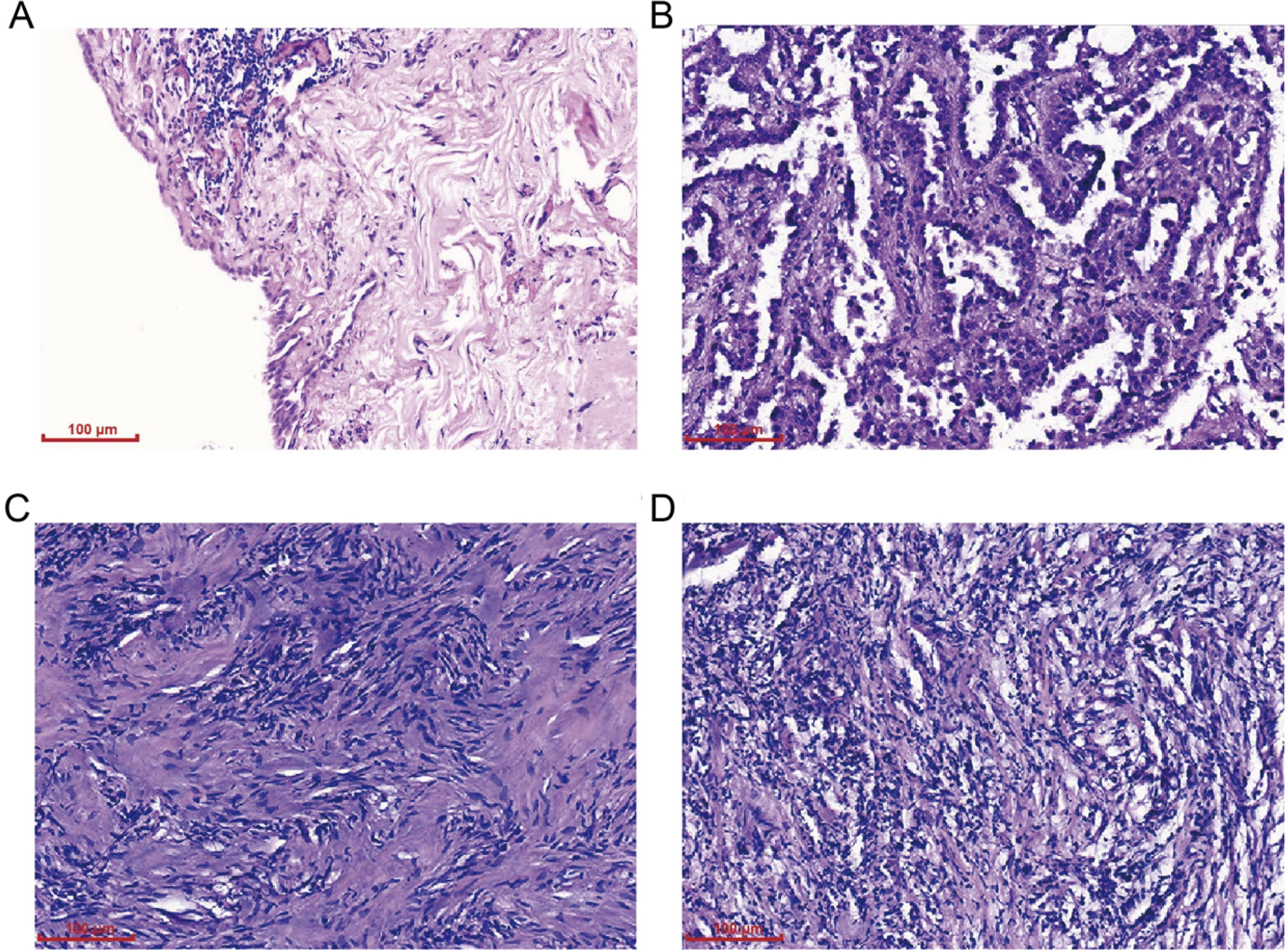
HE staining results of PMe tissues and non-PMe pleural mesothelial tissues (200×).
Validation of amplification specificity of PCR primers
The 293T cells are human embryonic kidney 293 cells that have been genetically modified with viral genes to become stable immortalized cells that can express SV40. The PCR product visualization on a 1.5% agarose gel electrophoresis with Nucleic Acid Dye staining showed that appropriately sized segments were detected in the 293T cells, whereas the 293 cells were negative (Fig. 3). The PCR results indicate that the three pairs of low-contamination risk primers used in this experiment were reasonably designed with strong specificity, and the amplified fragment size was completely consistent with the expected fragment size.

Verification of the specificity for three pairs of low-contamination risk primers. The appropriately sized segments were detected in the 293T cells, whereas the 293 cells were negative.
SV40 DNA was negative in PMe tissues and PMe cell lines
The presence of SV40 T-Ag was investigated in all samples using the PCR method through specific primers. The PCR results of 12 non-PMe pleural mesothelial tissues and 51 PMe tissues were negative (Fig. 4). The PCR results of normal mesothelial cell lines and PMe cell lines showed that SV40 was negative in the normal mesothelial cell line LP9, whereas the SV40-transformed normal mesothelial cell line Met5A was positive for SV40. In addition, SV40 T-Ag-specific fragments were negative in PMe cell lines of different pathological types (Table 4). These findings demonstrate that neither PMe tissues nor PMe cell lines failed to show SV40 DNA amplification.

PCR detection of SV40 expression in PMe tissues. The PCR results of the experimental groups (containing 12 non-PMe tissues and 51 PMe tissues) (1-10) were negative. Negative control group without DNA (11) and the positive control group of 293T cell (12) all showing corresponding correct results. PCR, polymerase chain reaction; PMe, pleural mesothelioma; SV40, simian virus 40.
Results of PCR and Immunohistochemistry Assays
PCR, polymerase chain reaction; SV40, simian virus 40; T-Ag, tumor antigen.
PMe cell lines negative for SV40 T-Ag protein
Western blotting failed to detect SV40 T-Ag protein in the normal pleural mesothelial cell line LP9, whereas SV40 T-Ag was detected in the SV40-transformed Met5A cell line. In addition, no sequences corresponding to SV40 T-Ag protein were detectable in any case of the different PMe cell lines NCI-H28 (epithelioid type), NCI-H2052 (sarcomatoid type), and NCI-H2452 (biphasic mixed type) (Fig. 5).
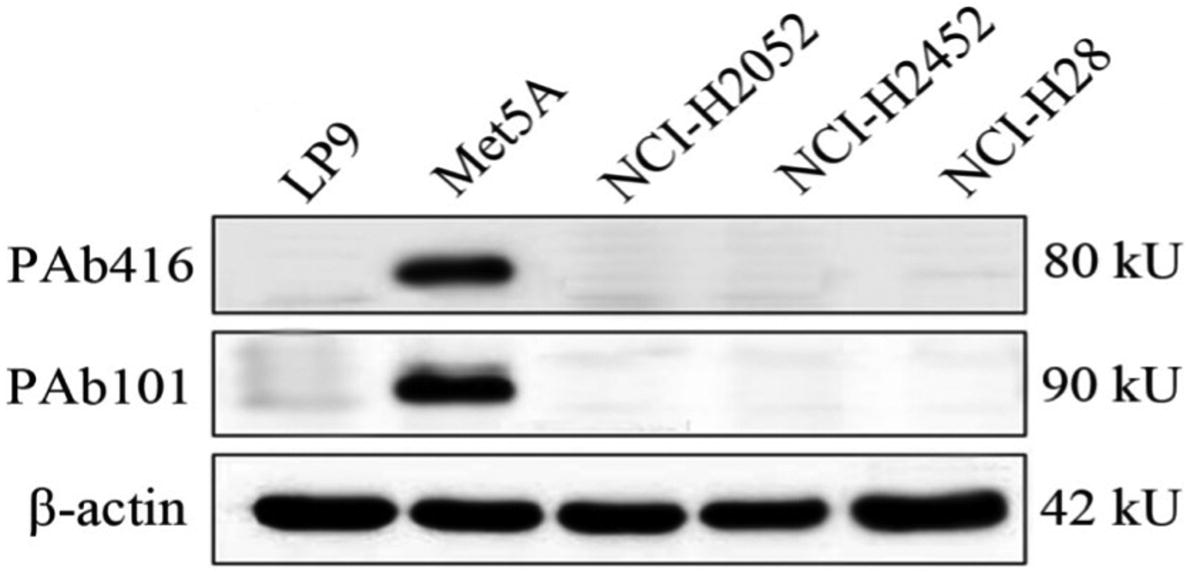
Detection of SV40 T-Ag protein expression in normal pleural mesothelial cell lines and PMe cell lines by Western blotting. Western blotting failed to detect SV40 T-Ag protein in the normal pleural mesothelial cell line LP9 and the different PMe cell lines NCI-H28, NCI-H2052, and NCI-H2452, whereas SV40 T-Ag was detected in the SV40-transformed Met5A cell line. PMe, pleural mesothelioma; SV40, simian virus 40; T-Ag, tumor antigen.
SV40 T-Ag expression in PMe tissues and PMe cell lines
Immunohistochemical staining results showed that the normal pleural mesothelial cell line LP9 was negative for SV40 T-Ag protein. The positive control cell line Met5A was strongly positive, showing obvious yellow or brownish yellow staining, and was mainly expressed in the nucleus. Our results confirmed the sensitivity and specificity of the PAb101 antibody for SV40 T-Ag antigen detection. However, none of the 12 non-PMe pleural mesothelial tissues and 51 PMe tissues studied showed positive immunohistochemical staining for SV40, with no staining observed in the cytoplasm or nucleus (Fig. 6). In addition, no evidence of SV40 T-Ag was found in the various PMe cell lines examined (Table 4). Overall, immunohistochemical staining analysis failed to show SV40 T-Ag protein in PMe tissues and PMe cell lines.
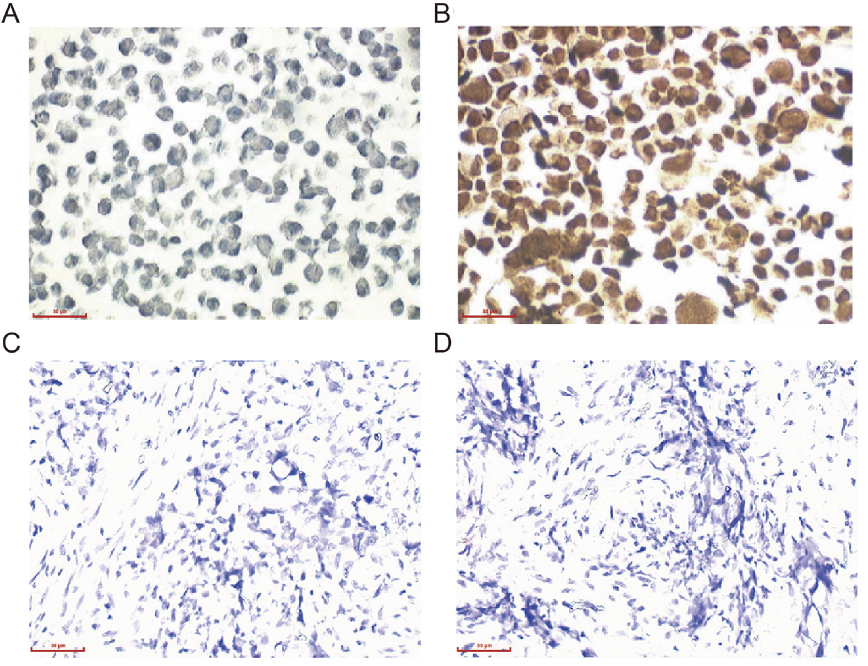
Immunohistochemical detection of SV40 T-Ag expression in normal pleural mesothelial cells and PMe tissues. The normal pleural mesothelial cell line LP9
Discussion
The possible association between SV40 and PMe has inspired much interest, especially after Carbone et al. reported the presence of SV40 in approximately 60% of mesotheliomas (Carbone et al., 1994). Several subsequent studies have investigated the relationship between PMe and SV40, but the results have varied (Thanh et al., 2016). One study that investigated the presence of SV40 DNA sequences in 60% (29/48) of mesothelioma cases suggested a relationship between SV40 and human MM. In a case-control study, PCR analysis revealed the presence of SV40 DNA in 8 of 19 PMe cases exposed to asbestos, suggesting that SV40 may increase the risk of PMe among asbestos-exposed individuals (Cristaudo et al., 2005). Many studies in European countries and the United States have confirmed this finding, suggesting that these countries may have received a polio vaccine contaminated with SV40 between 1955 and 1967 (Attanoos et al., 2018; Pandolfi et al., 2018).
However, many recent investigators have been unable to detect SV40 in human mesothelioma tissues, such as studies in Turkey, Finland, Japan, and Germany, which may suggest that their vaccines were not contaminated (Aoe et al., 2006; Us, 2013). In a study by Eom et al. of the 36 human PMe tissues screened, none contained detectable SV40 T-Ag sequences, arguing against the role of SV40 in the development of human PMe (Eom et al., 2013). These differences are perhaps related to the significant geographic variation of SV40 infection in contaminated poliovirus vaccines (Aoe et al., 2006). In a study by Alchami et al., no role was identified in a sensitive RNA T-Ag in situ hybridization technology on potentially affected birth cohorts (Alchami et al., 2020). Immunohistochemistry also showed that none of the PMe samples expressed SV40 T-Ag protein, suggesting that the PCR positivity may be due to plasmid contamination. In addition, smaller amplification products may lead to higher positivity rates.
In this study, no evidence of SV40 T-Ag was found in any of the PMe tissues and cell lines examined, either by high-sensitivity PCR molecular diagnosis, Western blotting, or immunohistochemistry. The present results showed that there was no association between SV40 and PMe development in the crocidolite-contaminated area of Dayao County, Yunnan Province, China. Our study is the first to propose a correlation between SV40 and PMe in the crocidolite-contaminated area of Dayao County, Yunnan Province, China, suggesting that SV40 may not be involved in the pathogenic mechanism of PMe in this region. In addition, these results are identical to a large amount of evidence indicating that many recent investigators have been unable to detect SV40 DNA sequences in human mesothelioma tissues (Mohammad-Taheri et al., 2013). Overall, combined with the substantial evidence of SV40 T-Ag negativity in PMe tissues reported worldwide, our findings also do not support the oncogenic role of SV40 in human PMe.
It is worth noting that the role of SV40 in human PMe remains controversial. Based on the enzyme-linked immunosorbent assay of SV40 T-Ag, Mazzoni et al. suggested that SV40 transformation activity was due to T-Ag expression. SV40 T-Ag may bind and inactivate the tumor suppressor genes pRB and TP53 (Mazzoni et al., 2022). Therefore, patients with SV40 infection are at greater risk of developing PMe after long-term exposure to asbestos. However, antibody-based experimental methods may be interfered with by autoantibodies and heterophilic antibodies, resulting in false positives. Moreover, this study only performed protein-level detection and did not verify SV40 T-Ag at the gene level. Therefore, the conclusion of whether SV40 can cause p53 inactivation still lacks clear and direct experimental support. Serological studies suggest that many reports of SV40 seropositivity may be due to cross-reactivity with the closely related polyomaviruses John Cunningham virus (JCV) and BK virus (BKV) (Manfredi et al., 2005). Some scholars also suggested that positive PCR results of SV40 may be due to laboratory contamination, as the two have identical sequence segments (López-Ríos et al., 2004). This study suggests that PCR detection results may have some limitations. Therefore, this evidence failed to convincingly suggest the correlation between SV40 and the occurrence of most human PMe. Furthermore, our results differ from those of Mazzoni et al. in Italy (Mazzoni et al., 2012), Jin et al. in Japan (Jin et al., 2004), Thanh et al. in Vietnam (Thanh et al., 2016), and Hübner et al. in Belgium (Hübner and Van Marck, 2002). Whether the differences in the correlation between PMe and SV40 between China, Italy, Japan, Vietnam, Belgium, and other countries are due to geography still requires further research.
In our opinion, the differences in the detection of SV40 T-Ag in PMe may be due to the following reasons. (1) These differences are perhaps related to the distribution of SV40 in contaminated poliovirus vaccines, so there are some significant geographic differences. However, epidemiological investigations failed to show that exposure to contaminated poliovirus vaccine populations results in an increased risk of developing PMe (Rotondo et al., 2019). (2) Some investigators consider that the high detection rate of SV40 T-Ag in PMe and other human tumors may be caused by plasmid contamination containing SV40 T-Ag DNA sequences widely used in laboratories (Manfredi et al., 2005). As DNA samples with genomic presence or occasional plasmid contamination cannot be detected, DNA-negative controls should be rigorously set to prevent potential false-positive results. (3) Previous studies have shown that different primer sets for SV40 T-Ag may lead to different proportions of positive rates in PCR detection (López-Ríos et al., 2004). The commonly used primers SV. for 3/SV. rev can span the DNA sequence of small T-Ag to amplify the pRb-binding coding region on SV40 T-Ag. The T-Ag gene fragment is commonly found in various commercial expression vectors and reporter vectors, thus increasing the possibility of detecting the 105 bp SV40 DNA sequence at the nucleotide between 4468 and 4372 bp (Saribas and Safak, 2020). In the present study, we designed and used three pairs of low-contamination risk primers spanning the intronic sequence of the SV40 T-Ag gene to further avoid false positives. In addition, due to the PAb101 monoclonal antibody used in this study, that reacts specifically with SV40 T-Ag, and not cross-reacting with T-Ag expressed by BKV and JCV, false-positive results caused by the widespread presence of the latter two viruses and the expression of T-Ag in the normal population can be excluded. Although our results failed to reflect all cases of PMe, combined with the large amount of negative evidence provided for SV40 detection worldwide, considering that 3 pairs of low contamination risk primers were used for genetic detection of SV40 T-Ag in this study, as well as the high sensitivity and specificity of antibodies for SV40 T-Ag protein detection, we have reason to believe that SV40 infection is not the main pathogenic factor for PMe in the crocidolite-contaminated area of Dayao County, Yunnan Province.
Conclusion
In conclusion, our study suggests that SV40 is not closely related to PMe in the crocidolite-contaminated area of Yunnan Province. Therefore, clinicians should continue to consider environmental and occupational asbestos exposure as the most likely and definitive risk factors for PMe.
Footnotes
Acknowledgments
The authors express their gratitude to the Pathology Department of the First People’s Hospital of Chuxiong Prefecture for providing PMe samples.
Authors’ Contributions
R.-A.L and W.X. conceptualized and designed the study and assessed the outcomes of the study. Tissue samples were collected by B.-Y.W., W.M., and Y.-P.Z. X.C. and Y.-Q.P. analyzed the collected data. R.-A.L. drafted the article. J.-J.Z. and L.Q. participated in the modification and revision of the article. R.-A.L. and W.X. confirm the authenticity of all original raw data. All authors read and approved the final version of the article.
Author Disclosure Statement
No competing financial interests exist.
Funding Information
This research was funded by the National Natural Science Foundation of China (82160516).