
Abstract
Abstract
Introduction
Case
A 47 year-old-woman, para 3, presented to the outpatient department with a complaint of a mass protruding outside her vulva for last 6 months. The mass was reducible. She had a history of burning during micturition. On pelvic examination, her anterior vaginal wall was found to be protruding outside the vulva. First-degree descent of the cervix was present and there was no associated rectocoel. She was provisionally diagnosed as having a large cystocoel (Fig. 1). Considering her age, she was prepared for a vaginal hysterectomy with cystocele repair and perineorrhaphy. Preoperatively, when the patient was examined by the senior consultant, surprisingly, the mass turned out to be a sessile cystic growth, 12 cm X 6 cm X 4 cm, arising from the anterior vaginal wall. With this new diagnosis of a vaginal-wall cyst, the patient was taken up for excision of the cyst. A wide excision of the tumor was done. There were no intraoperative complications and her estimated blood loss was ∼250 mL. The pathologic evaluation revealed spindle-shaped fibroblasts and myofibroblasts lacking significant nuclear pleomorphism or mitotic figures in a loose collagenous myxoid background, with numerous medium-sized vessels characteristic of aggressive angiomyxoma (Fig. 2). The surgical borders were free of tumor.
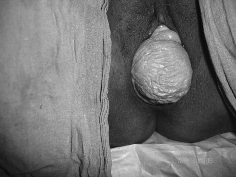
Aggressive angiomyxoma appearing as a cystocoel.

Histopathologic view of tumor (H.E. 100×).
Results
Close follow-up of the patient is still going on without recurrence, 24 months after surgery. A recent computerized tomography (CT) scan of her abdomen and the pelvis was normal.
Discussion
Angiomyxoma is considered to be aggressive because of its propensity to cause local recurrences. It has been found in the pelvic soft tissue, retroperitoneum, urinary bladder, vagina, vulva, perineum, scrotum (in men), and buttocks, and manifests as a polypoid or cystlike lesion or as an ill-defined mass in the pelvic region. AAM characteristically grows slowly and insidiously, has no capsule, and is locally infiltrative. 2 Histologically, mitosis is not commonly seen, but occasional cases may show mild atypia. Despite an innocuous looking histologic pattern, AAM recurs in 70% of patients who have it. 3
Imaging studies, such as CT scan and magnetic resonance imaging, help in preoperative evaluation and postoperative follow-up as this tumor is ill-defined clinically.4,5 Other tests, such as a barium enema, intravenous pyelography, or ultrasonography, might be used, based on clinical judgment. Given that the preoperative diagnosis is different in most cases as was shown in the current case, the results of these imaging studies are commonly overestimated. Although AAM is a rare lesion, it must be considered as a differential diagnosis for any mass in the perineum or soft tissue of the pelvis in females. 6
Gungor et al. reported a case of a patient needing repeat surgery that could have been avoided by an adequate preoperative evaluation. 1 Wide local excision is the therapy of choice. 6 However, resection of these tumors can be technically difficult and the value of extensive surgery to obtain clear margins has been questioned. 2 As a result of the cells' low mitotic activity, it is unlikely that radiation therapy or chemotherapy would be a useful adjunct to primary surgical treatment. Long-term follow-up is necessary. 7 Because the tumor grows slowly and is often symptomatic only when it is large, radiographic follow-up is the best option. 8
Conclusions
As a result of AAM's rare occurrence and locally invasive nature, early detection of this kind of tumor requires a high index of suspicion. The case in this report highlights the importance of meticulous examination and evaluation of similar cases preoperatively by a senior consultant. This case also demonstrates that, when vaginal-wall descent is disproportionately large, compared to the descent of the cervix, other differential diagnoses should always be kept in mind.
Footnotes
Disclosure Statement
No competing financial conflicts exist.