
Abstract
Abstract
Introduction
There are several techniques of creating a neovagina: using olive-shaped plastic forms combined with traction devices (Vecchietti technique) 3 ; Hegar dilators or specially constructed bicycle seats for passive dilatation; bowel transplants; labial, thigh, or subcutaneous abdominal flaps; and insertion of an inlay split-thickness skin graft, amnion, or peritoneal graft. The goal of any method is to create a vaginal canal of adequate diameter and length to accommodate sexual intercourse. However, there is a lack of consensus regarding the best therapeutic approach. We share our experience using oxidized regenerated cellulose as a substitute for skin grafts or amnion for construction of a new vagina.
Materials and Methods
A prospective, longitudinal study was designed at a tertiary care setting. Over a period of 2 years, oxidized regenerated cellulose (Surgicel®, Johnson & Johnson, India) was used in 10 women undergoing vaginoplasty for MRKH syndrome. The diagnosis was made after a complete workup, including sex steroid hormone levels, chromosomal analysis, pelvic ultrasonography, intravenous pyelography, x-ray of the spine, and magnetic resonance imaging if required. All patients gave written informed consent, and received appropriate counseling prior to surgery. Ethical clearance for the study was obtained from the Institutional Review Board. The procedure was performed with patients under either spinal or epidural anesthesia. Patients were positioned in the lithotomy position, and after ensuring strict asepsis, an indwelling Foley catheter was placed to continuously drain the bladder. Vaginal dimples were identified. The rudimentary müllerian ducts were dilated along the pelvic axis using Hegar dilators (up to size 14) in a stepwise fashion. This created two cavities on both sides of the median raphe, which was subsequently divided (Fig. 1). With further blunt dissection with fingers or sponge on holder, the space was dissected until the peritoneum was reached. An attempt was made to create a neovagina that would easily accommodate the length of two fingers. At the time of dissection, placing an assistant's finger in the rectum helped in guiding the dilators to prevent rectal injury. Subsequently, a vaginal mold made from tightly rolled sterile foam, placed in a condom, and further covered with oxidized cellulose mesh (Surgicel) was inserted in the neovagina. The Surgicel was wrapped over the mold like a sleeve and sutured into place using delayed absorbable Vicryl™ sutures (2–0 polyglactin 910, Ethicon, Johnson & Johnson Ltd., Mumbai, India) (Fig. 2). The mold was tailored to the size of the neovagina, and was kept in place by suturing the labia together. Postoperatively, the urinary catheter was left in situ, and all patients received antibiotics (ciprofloxacin and metronidazole) for 8–10 days to prevent infection. Analgesics (diclofenac sodium) were administered on demand. Perineal hygiene was maintained. The foam mold and urinary catheter were removed 8–10 days later with the patient under intravenous sedation. The neovagina was irrigated with saline and povidone iodine solution, and a glass mold was inserted at the same sitting. Patients were taught how to insert the glass mold prior to discharge from the hospital, and were instructed to use it day and night for the first 3 months; then at night-time only, until regular intercourse was possible.
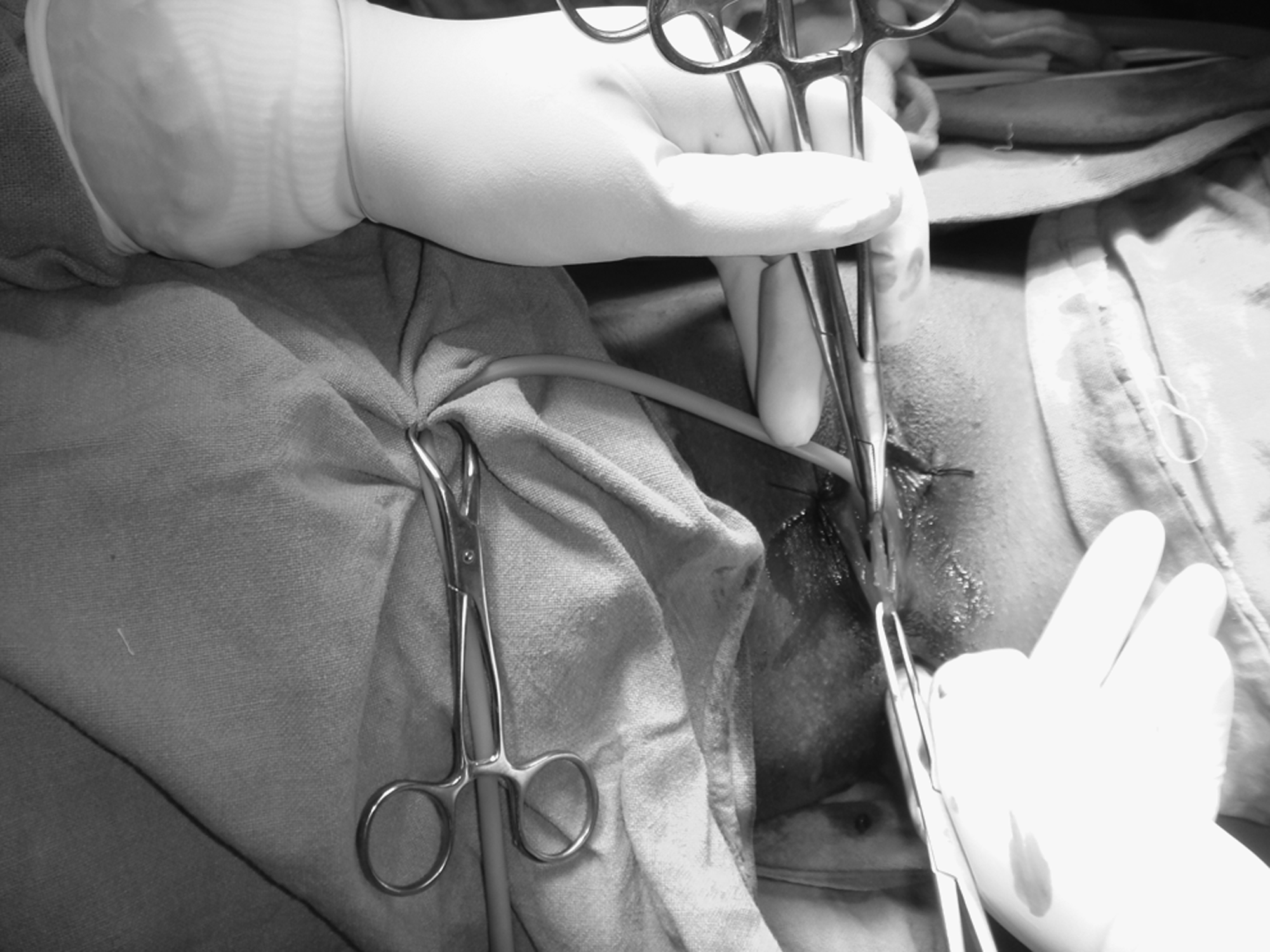
Median raphe created following blunt dissection along müllerian ducts.
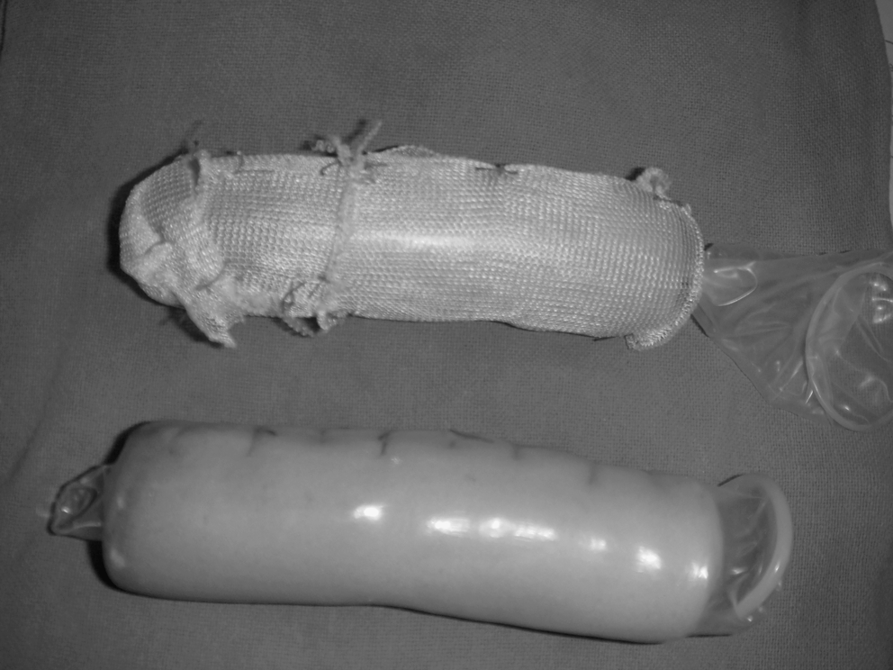
Foam mold wrapped in condom, and also covered in Surgicel® prior to insertion.
Results
The mean age of patients was 20.4 years (range: 17–25 years). The secondary sexual characters were well developed in all women. Sex hormones and karyotype were normal in all. Two (2) patients had spina bifida and another 2 were diagnosed to have renal anomalies: absent right kidney and malrotated ectopic kidney with duplication of collection system, respectively. On imaging, the uterus was rudimentary in all. Four (4) women were married and 6 were engaged to be married.
There were no intraoperative complications. Average operating time was 15 minutes (range: 10–20 minutes). The procedure was performed by a single operator (V.D.) as the primary surgeon to maintain uniformity. Adequate vaginal length (7–10 cm) was achieved in all cases on foam mold removal. On follow-up at 6 months, the vagina was well epithelialized in all, and in a majority of patients (9 of 10) at least two fingers in width and 7–10 cm in depth. Partial vaginal stenosis was encountered in 1 patient, who had not used the glass mold postoperatively as directed. She has been advised to use dilators and is under follow-up. One (1) patient had extensive granulation tissue, with resultant dyspareunia, persisting for 8 weeks postoperatively. The vagina healed well with the topical application of silver sulfadiazine cream. None of the remaining 9 patients reported dyspareunia, need for vaginal lubrication, or any coital difficulty.
Discussion
Most women with MRKH syndrome are diagnosed, in our setting, when they present with primary amenorrhea at 16–18 years of age, or with inability to have successful sexual intercourse after marriage. Absence of vagina and uterus can impose immense psychologic stress on a woman's well-being, and recognition of this emotional aspect, with adequate counseling, is as imperative as a successful surgical procedure. In addition, choosing the right procedure is individualized, depending largely on patients' preferences and willingness for postoperative care, along with the surgeon's experience and preference.
It is a standard practice in our unit to use a modified Sheares technique: creation of neovagina using Hegar dilators to dilate rudimentary müllerian bulbs followed by insertion of a foam mold put in a condom, then covered with amnion for epithelialization of neovagina, with satisfactory results over several years. 4 Due to unavailability of amnion at times, and concern regarding risk of transmission of infection, we have now started using oxidized cellulose (Surgicel) for epithelialization of neovagina and have presented our experience. This method of creating neovagina takes less time and causes less patient discomfort compared to other techniques of creation of neovagina, requiring skin graft or bowel vaginoplasty. The average time taken per procedure was just 15 minutes.
Most surgical procedures aim at creating a potential space by dissection of fibroareolar tissue between bladder and rectum. The difference lies in the substance used to line the neovagina to help epithelialization. Wharton recommended placing a condom-covered mold in the neovagina in order to allow vaginal granulation tissue to epithelialize. 5 This has a problem of prolonged discharge from granulation tissue 6 and led to modification: use of skin grafts, a technique originally described by Abbe 7 and later modified by McIndoe. 8 However, this leaves a permanent scar at the site where graft was obtained. The same is true for bowel and peritoneal vaginoplasties, which require laparotomy and increase morbidity, operating time, and possible postoperative vaginal stenosis. 9
Use of human amnion for lining neovagina was an important step forward, as it avoids scarring and laparotomy. 6 Amnion has been shown to act as a biological dressing, permitting and possibly promoting vaginal epithelialization. 10 Use of amnion raises logistic problems and theoretical concern for viral transmission. Surgicel, on the other hand, is easily available, biodegradable, and does not carry risk of infection. It has been used in pelvic surgery; Surgicel rapidly forms a gelatinous mass that acts as a protective coating on raw surfaces, allowing re-epithelialization to occur. 11 For epithelialization of neovagina, oxidized regenerated cellulose has been used successfully, but in a small number of patients (Table 1). Jackson and Rosenblatt first described use in 4 patients with satisfactory results. 12 Complete epithelialization occurred by 3–6 months, no strictures occurred, and vaginal depth in these women ranged from 6 to 12 cm. Motoyama et al. reported successful use in a second series of 10 patients. 13 They reported satisfactory epithelialization of neovagina 1–4 months after surgery and all patients were satisfied with the procedure. The authors further reported that the neovagina created with this procedure was not much different from the normal adult vagina with respect to histology and physiologic conditions. Sharma et al. also used Surgicel in 10 cases of vaginoplasty, and of 9 patients followed up, 8 had satisfactory intercourse, while 1 had mild dyspareunia due to partial stenosis. 14
UTI, urinary tract infection.
Conclusions
Based on our study and review of three other studies, we conclude that oxidized cellulose is an effective alternative to skin grafts and amnion for epithelialization of neovagina. It is easily available, safe, and biodegradable. It decreases the duration of surgery and complications compared to use of skin grafts and bowel vaginoplasty.
Footnotes
Disclosure Statement
No competing financial interests exist.