
Abstract
Abstract
Introduction
Although an accurate preoperative diagnosis may be impossible to achieve in some cases, other cases may provide an opportunity to gain the correct diagnosis either through additional diagnostic tests or through reconsideration of clues that point away from gynecologic and toward gastrointestinal primary cancers.4,5 Every effort should be expended to achieve an accurate diagnosis at the onset of treatment planning because, whereas the distinction between gynecologic and gastrointestinal tumors can be subtle, the implications for all aspects of the patient's care can be quite profound. For example, most gastrointestinal cancers metastatic to the ovary carry an extremely poor prognosis, with a median survival of 17 months, ranging from 1 to 86 months.6,7 Patients with primary ovarian malignancies, even when giant, survive significantly longer and can sometimes even be cured.8,9
Establishing the correct diagnosis prior to initial surgery is beneficial not only to the patient but also to the oncologic team caring for the patient. For the patient, the potential to avoid added procedures and anesthetics as well as the process of informed consent is facilitated. For the medical oncologist, the decision to initiate neo-adjuvant therapy or consider the patient for enrollment in a clinical protocol is possible. For the surgeon, operative planning is facilitated by having the necessary personnel and equipment available to provide definitive surgical care at the time of initial operation, and measures can be taken to minimize the need for diverting or permanent colostomy. 10 In cases of disseminated mucinous tumors or pseudomyxoma peritoneii, consideration for debulking with heated intraperitoneal chemotherapy is also possible.11–13 If such measures cannot be undertaken locally, then transfer of the patient to a tertiary center remains an option at that point in time when the patient is likely to receive the greatest benefit from expert care, before chemotherapy or surgical therapy is initiated.
The purpose of this study was to critically analyze cases in a single institution where large pelvic tumors, initially of uncertain or incorrect diagnosis, were ultimately determined to be of colorectal origin. The study sought to determine whether a complete preoperative workup was simply misinterpreted, not pursued, or assigned minimal significance. The study further sought to determine the implications of the incorrect diagnosis on subsequent surgical procedures. Finally, in performing this analysis, it was hoped that recommendations going forward to avoid missed diagnoses and maximize the opportunity for optimal treatment planning and consent, could be established.
Methods
Patients who had undergone laparotomy for pelvic tumor of unknown origin with a discharge diagnosis of colorectal cancer between 2002 and 2009 were identified from the hospital tumor registry and discharge databases after approval as an exempt study was obtained from the University and Medical Center Institutional Review Board. Patients were further identified by the specialty of the admitting physician at the time of presentation or by the operating surgeon of record during the initial surgical resection. If the admitting physician or operating surgeon of a patient with a discharge diagnosis of colorectal cancer was a gynecologic surgeon, the chart was reviewed to determine whether the presenting diagnosis and final diagnosis were discordant.
In cases for which the admitting and discharge diagnoses were discordant, patient records were further analyzed to determine whether the data necessary to confirm the ultimate diagnosis had been available at the time of initial surgery, or if the data available including presenting signs or symptoms warranted further diagnostic studies to achieve a more accurate preoperative diagnosis. In short, the study sought to determine whether the opportunity to establish the correct diagnosis had been present before the initial surgical procedure.
The following signs and symptoms specific to colorectal cancer, or at least warranting further colorectal cancer-directed workup were sought in the preoperative records:
• History of present illness/Review of Systems—Abdominal pain, change in bowel habits, hematochezia, melena, dyschezia, weight loss, nausea/vomiting • Physical examination—Palpable abdominal mass, palpable rectal mass, heme- positive stool • Past medical/surgical/familial history—Personal or family history positive for colorectal cancer, prior surgical and endoscopic findings/pathologic reports • Diagnostic/laboratory studies—Gastrointestinal specific tumor markers including carcinoembryonic antigen (CEA), CA19-9; radiologic findings consistent with colonic obstruction, gastrointestinal tumor growth, and normal ovaries and uterus; and endoscopic findings.
Occasionally, the gross specimen was the only clue to the diagnosis of colorectal cancer and this could only be appreciated at the time of initial surgery. Intraoperative gross findings consistent with an endoscopically identifiable lesion (mucosal defect in colon, rectum, or appendiceal orifice) were regarded as a missed opportunity for preoperative diagnosis in patients who did not have preoperative colonoscopy. Mid-body appendiceal tumors were not considered “discoverable” unless visible on preoperative CT scan. The number of opportunities for obtaining the diagnosis preoperatively or justifying additional workup that would establish the diagnosis was determined for each patient.
Pathology was reviewed for all cases and confirmed to be nongynecologic in origin. Ovarian pathology for the resected specimens was sought and reported with the colonic pathology. Presenting signs and symptoms, radiographic findings, and surgical procedures performed were also catalogued and reported.
Results
Demographics
In total, over 96 months, 10 patients were diagnosed with colorectal cancer after exploration by a gynecologic oncologist for pelvic tumor of unknown origin, each ultimately undergoing colonic resection. Average and median age of these patients was 49 with a range of 40–62. Seven patients were postmenopausal, 2 were premenopausal and 1 had had a hysterectomy. Of these 10 patients, 9 had an unplanned intraoperative consultation to a surgical oncologist, general surgeon, or colorectal surgeon; 1 had a planned consultation because of a high level of suspicion of colonic involvement preoperatively.
Presenting signs and symptoms
The most common clues to a nongynecologic primary cancer were in the presentation and history of the present illness. Table 1 depicts the presenting signs and symptoms of the 10 patients identified in this review. Almost all patients presented with abdominal pain. Fifty percent of the women presented with at least one sign or symptom suggestive of gastrointestinal malignancy, characterized by either a change in bowel habits or dyschezia. Forty percent presented with symptoms consistent with either gastrointestinal or gynecologic malignancy (abdominal pain, ascites, increased abdominal girth, and weight loss) and 1 patient presented with gynecologic-specific symptoms (postmenopausal bleeding only). Eighty percent of patients had more than one presenting complaint, most commonly pain and increased girth or pain and change in bowel habits.
History and physical and tumor markers
Family history was not documented in 6 patients, was nonspecific in 3, and was positive for colorectal cancer in a first degree relative in 1. Past medical and surgical history supported a recurrence of previously diagnosed colorectal cancer in 1 patient. One patient presented with a stage IV colon cancer including obstruction and omental caking within 1 month of total abdominal hysterectomy. Two patients had preoperative tumor marker assays for CEA and CA-125, and none had results for CA 19-9. Both patients whose CEA levels were checked had elevations >20, which prompted CT and colonoscopy.
CT findings
Table 2 summarizes CT findings on these 10 patients as interpreted by the reading radiologist. Seven patients had a preoperative CT scan. All of these had radiologic findings suspicious for colorectal cancer, some more specific than others. Forty-three percent of them had specific radiologic findings consistent with colorectal malignancy including 2 with colonic thickening, 4 with pelvic masses involving the colon, and 2 with a colonic filling defect after taking oral contrast. Many of these patients had more than one CT finding. All patients with a preoperative CT scan had gross findings on the resected colon specimen that suggested a colonoscopy before surgery should have been revealing.
Radiologic finding suggestive of colorectal malignancy
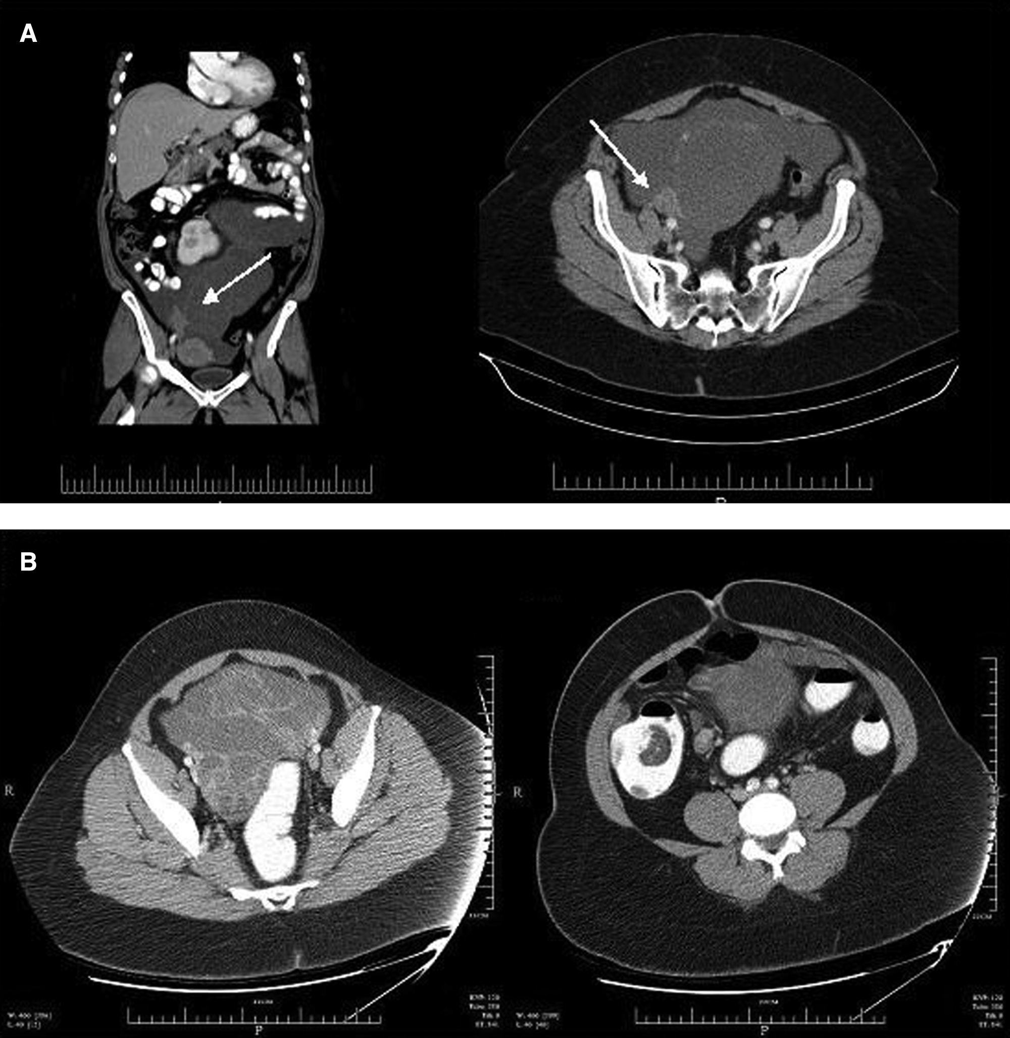
Occasionally, CT findings that showed only pelvic pathology directed patients toward gynecologic oncology. Figure 1a depicts a representative scan of a patient with a 2-cm mid-appendiceal mucinous adenocarcinoma that was not visible on CT scan and a giant right adnexal mass surrounded by a relatively normal-appearing ovary. In the absence of any identifiable gastrointestinal primary cancer, this patient was assumed to have a primary ovarian process arising from the right ovary. Figure 1B represents a patient with a cecal filling defect (right panel) and a complex cystic mass involving the right ovary interpreted by the radiologist as a possible ovarian neoplasm. Despite a recommendation for barium enema or colonoscopy by the reading radiologist, this patient was operated upon without having been given these tests
The gross specimen
Ninety percent of patients had a gross specimen that confirmed a colorectal primary cancer. Excluding the 3 patients who had not undergone endoscopy preoperatively, gross findings were highly suggestive that a colorectal cancer diagnosis could have been obtained had preoperative endoscopy been performed. Endoscopy was, by far, the least used diagnostic study with the highest potential for establishing the correct diagnosis. Only those who had undergone endoscopy had documented rectal examinations. None of the documented rectal exams indicated a that guaiac assay had been performed.
Pathology
The tumor of origin could be confirmed pathologically in all but 1 patient. That patient had no identifiable colorectal cancer lesion and metastatic serosal lesions of the ovaries and tubes. All ovarian pathology were either drop metastases or a local extension of a gastrointestinal primary cancer that could not be distinguished from the normal ovary. None of the patients had normal ovaries and tubes, but 2 patients had benign lesions of one ovary unrelated to the pelvic pathology for which they were explored. Final pathologic diagnosis revealed the origin of the pelvic tumor to be 40% appendiceal, 50% colonic, and 10% undifferentiated adenocarcinoma.
Subsequent procedures
Subsequent laparotomy for additional surgical therapy was indicated in 50% of these patients once the definitive pathologic diagnosis was established. Each of these patients underwent tumor debulking within 4 weeks of initial surgery and 3 had cytoreductive surgery with intraperitoneal heated chemotherapy (2 mitomycin, 1 cis-platinum) for appendiceal mucinous carcinomatosis. Details of these procedures are outlined in Table 3. Two patients had a third procedure performed, 1 underwent further cytoreduction within 90 days of her initial operation, and the other had a complex abdominal wall reconstruction for a hernia resulting from one of the previous two procedures.
TAH, total abdominal hysterectomy; BSO, bilateral salpingo-oophorectomy; RSO/LSO, right/left salpingo-oophorectomy; LOA, lysis of adhesions; HIPEC, heated intraperitoneal chemotherapy.
Missed opportunities for correct diagnosis
Table 4 outlines each patient meeting criteria for inclusion in this analysis and the clues indicating a missed opportunity to establish the correct diagnosis. Three patients (shaded) did have a preoperative endoscopic examination but were still operated upon for presumed gynecologic malignancy. One patient had what was thought to be a diverticular stricture of the sigmoid colon and a pelvic mass that was ultimately determined to be a sigmoid cancer extending into the pelvis. Another patient had a 0.5-cm mid-appendiceal mucinous tumor with a giant (20 cm) drop metastasis that could not be visualized on preoperative endoscopy. The third patient had a cecal mass that was endoscopically biopsied and revealed to be a poorly differentiated adenocarcinoma with signet ring cell features, but was deemed more likely to be a metastasis from an ovarian primary cancer.
ND, not done; NS, nonspecific; N/A, not applicable.
The following findings supported colonoscopy in the 7 cases for which it was not completed preoperatively: gross specimen indicating that endoscopy would have been diagnostic in 6 patients, prior gastrointestinal cancer diagnosis in 1, computed tomography with specific findings consistent with colorectal mass in 3, CT with nonspecific findings consistent with colorectal mass in 4, colorectal cancer in first-degree relatives in 1, and presenting symptoms associated with colorectal cancer including dyschezia, obstruction, or hematochezia in 3.
Discussion
It is sometimes difficult to make a definitive preoperative diagnosis with large pelvic tumors, even if all diagnostic avenues are explored. Because no diagnostic modality is 100% sensitive or specific, misdiagnosis can occur even if the patient undergoes endoscopy. If preoperative diagnosis had been accurate or even if the differential diagnosis prior to laparotomy had been broadened to include gastrointestinal malignancy, informed consent, operation, adjuvant therapy, and prognosis would certainly have been different for at least some of these patients.
In this study, a number of patient charts were reviewed following intraoperative consultations during laparotomy for pelvic tumor of unknown origin, presumed to be gynecologic malignancy, for gross and pathologic findings consistent with gastrointestinal malignancy. Initial clinical presentation explains why all of the women in this study were presumed to manifest with gynecologic tumors. Radiologic presentation was, for the most part, nonspecific, although there were a few findings that should have raised suspicion for gastrointestinal malignancy and prompted preoperative endoscopic examination. An argument can be made that the radiologic findings suggestive of, but nonspecific for, gynecologic or gastrointestinal malignancy such as ascites, carcinomatosis, and complex pelvic masses, should have prompted an endoscopic evaluation. The differential diagnoses for these findings include both types of malignancy without priority for either one.
The serum tumor markers CEA and CA-125 may help differentiate between primary ovarian and primary gastrointestinal tract cancer, specifically if both are evaluated and the CA 125/CEA ratio is taken into consideration. 14 This study was unable to determine the significance of tumor markers in the study because most were not checked. Two of the subjects had a CEA >20, indicating a high likelihood of metastatic disease of gastrointestinal origin. 15 One of the 2 had a CA-125 >35, significant for extensive disease of gynecologic organs. 15 Based on these findings, it would be advisable to check a CEA level in all patients in whom the CA 125 is normal, the diagnosis is in doubt, or other portions of the workup are consistent with a gastrointestinal primary cancer.
Tumors of appendiceal origin seemed to be the most perplexing, even when endoscopy was performed. Two of the 3 patients who had endoscopic evaluation were ultimately diagnosed with primary appendiceal malignancies. One had no abnormal findings on colonoscopy because the tumor was mid-appendix. The other had a large mass in the cecum extending to the hepatic flexure, found intraoperatively and pathologically to have originated from the appendix. Endoscopic biopsy resulted in undifferentiated adenocarcinoma, but the preoperative diagnosis remained gynecologic.
Fifty percent of these patients required a total of 7 additional operations to definitively address their ultimate diagnosis; 3 of whom underwent intraperitoneal heated chemotherapy. Given the expanding body of evidence supporting heated intraperitoneal chemotherapy in peritoneal surface malignancy and select cases of disseminated peritoneal mucinous adenocarcinomatosis, this is not an insignificant observation. Had these individuals been identified a priori as potential patients who could benefit from such surgery, preparation to provide such treatment could have been made at the initial exploration. The authors of this study advocate an approach to such patients that alerts the treating physician to adopt a high level of suspicion and an approach of preparedness for whatever procedures may be necessary to treat such condition at the time of initial exploration.
Conclusions
In order to maximize the chances of achieving these ends, the authors of this study have established the following guidelines at the institution where this study was done, for gastrointestinal workup for a patient with pelvic tumor of unknown origin:
1. Taking a thorough history, including focused gastrointestinal review of symptoms, specifically inquiring about change in bowel habits, dyschezia, melena/hematochezia, nausea and vomiting, and personal and family history results from prior gastrointestinal surgery including appendectomy 2. Performing a physical examination including rectal examination with guaiac test 3. Evaluation for occult anemia 4. CT of abdomen and pelvis 5. Colonoscopy within 3 months of presentation with abnormal symptoms 6. Identifying tumor markers including CEA, CA 125, and CA 19-9.
Any positive findings from these recommended guidelines should raise the level of suspicion and preparedness for something other than a gynecologic primary cancer despite compelling evidence to the contrary.
Footnotes
Acknowledgments
K.M.L. collected, analyzed, and interpreted data; drafted the article; performed critical revisions; and provided final approval of the version to be published.
M.A.W., S.C.A., and S.M. collected and interpreted data, performed critical revisions, and provided final approval of the version to be published. M.S.H. was responsible for the conception and design, performed critical revisions, and provided final approval of the version to be published. E.E. Zervos was responsible for conception and design; collected, analyzed, and interpreted data; drafted the article; performed critical revisions; and provided final approval of the version to be published.
Disclosure Statement
No competing financial conflicts exist.