
Abstract
Abstract
Introduction
Case
A 26-year-old prima gravida presented, without an appointment, to the rural health center associated with this hospital for an antenatal checkup at 18 weeks of gestation. There was no history of drug intake, consanguinity, or family history of congenital malformation. She was referred to the hospital for an ultrasound (US) examination. The US revealed olighydramnios, and a male fetus with a gestational age of 18 weeks. The scan of the fetus revealed a defect in the anterior abdominal wall, with herniation of the abdominal contents outside the abdominal cavity. Absence of the right lower limb was also noted.
Results
The mother was informed of the poor prognosis and after counseling, the pregnancy was terminated. A stillborn male fetus was aborted along with the placenta and membranes.
A fetal autopsy was performed. The fetus weighed 280 g, with a crown–rump length of 145 mm. The right lower limb was missing. A huge abdominal defect extending from the distal sternum to the suprapubic region, involving the right side, was seen, with evisceration of the liver, gallbladder, intestines, and spleen (Fig. 1). The diaphragm was present. Heart and lungs were normal. Thymus was present, anal atresia was observed, and the colon ended in a meconium-filled pouch. Kidneys and urinary bladder were normal. Male external genitalia were identifiable and intra-abdominal testes were noted.
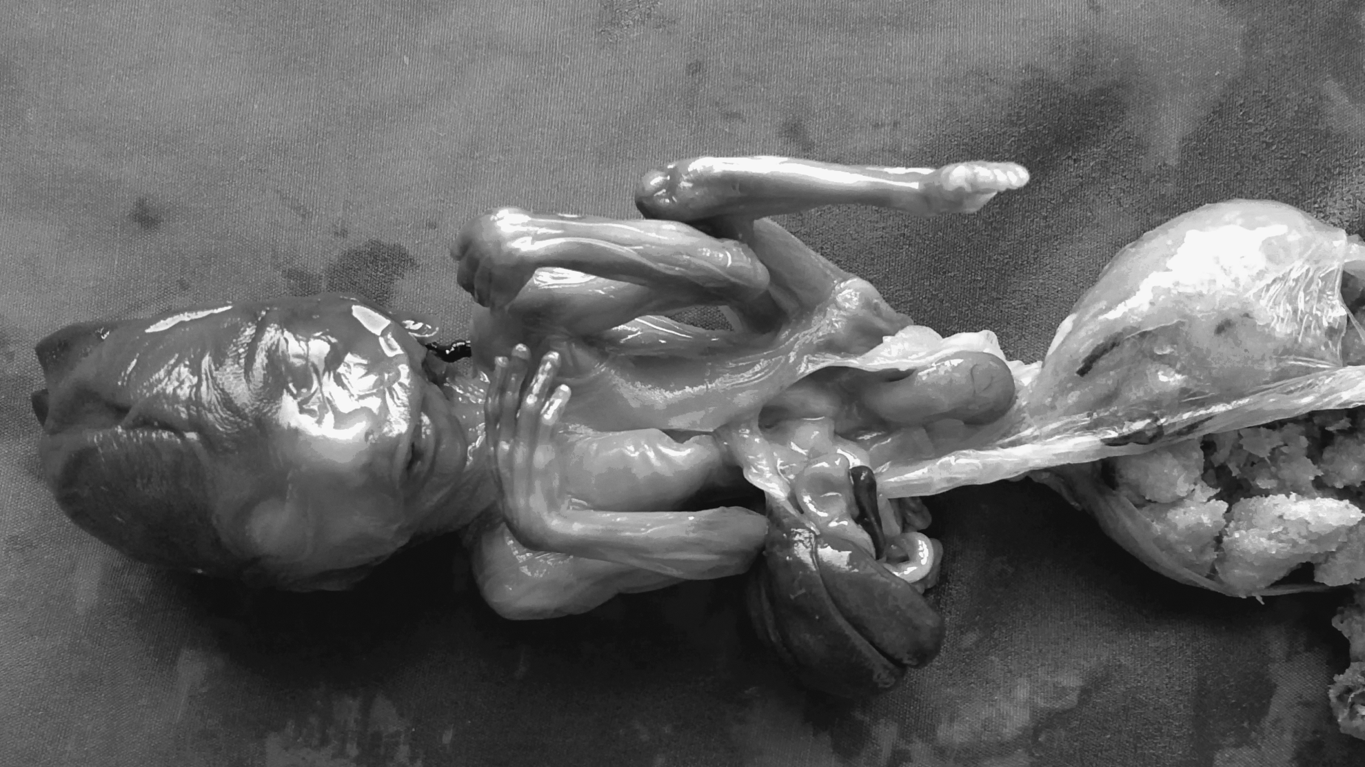
Fetus of 18 weeks' gestation with gastroschisis and evisceration of the liver, gallbladder, intestines, and spleen. Absence of the right lower limb was also noted.
The placenta and the membranes were unremarkable. No amniotic bands were identified. The umbilical cord was extremely short and measured 1.5 cm in length. A cut section showed the presence of single umbilical artery.
Discussion
LBWC is also known as the amniotic deformity, adhesion, and mutilation complex. 1 Its pathogenesis is probably heterogeneous. Three pathogenetic mechanisms have been proposed; namely, the early amnion rupture theory, 4 vascular disruption theory,1,2 and embryonic dysgenesis. 3 The early amniotic rupture theory states that failure of the amnion to fuse with the chorionic plate causes these membranes to dehisce, forming strips floating within the amniotic cavity, where they entangle the fetus, resulting in mechanical or vascular disruption of normally developing structures. According to the vascular disruption theory, amniotic bands not causally related to amniotic rupture are secondary to disruption of fetal blood flow.1,2 The developmental theory was first proposed by Streeter in 1930. 5 His hypothesis was that these defects were the result of abnormal folding of the embryo, which caused maldevelopment of the amniotic cavity and the germ disc. 6
Van Allen, 1 in a study of a series of fetuses with LBWC, found that the major structural defects included limb defects and internal organ malformations (95%), marked scoliosis (77%) and craniofacial defects (56%). The major internal organ malformations included cardiac anomalies, absent diaphragm, anal atresia, absent kidney, renal dysplasia, and exostrophy of the urinary bladder.
Russo et al. 7 have proposed that there are two clearly distinguishable phenotypes: the placenta-cranial and the placenta-abdominal adhesion phenotype. The first phenotype shows craniofacial defects and amniotic bands and/or adhesion, whereas the second is without craniofacial defects and presents with urogenital anomalies, anal atresia, abdominal placental attachment, and persistence of an extra embryonic celom. This case belongs to the placental abdominal adhesion phenotype.
Kalousek et al. divide the amniotic wall defects into LBWC and the amniotic rupture sequence (ARS). It is likely that in LBWC, the initiating event occurs before the fusion of the chorion with the amnion, and in ARS, the insult occurs later when the body wall and the neural tubes have closed. The primary defect in LBWC is thought to be related to abnormal body stalk development, 8 and is an anomaly, therefore, of early embryonic organization and development rather than of postembryonic disruption, as has been proposed for ARS. 9
Distinguishing features are that clefts in ARS do not follow anatomical lines of closure, lesions are not symmetrical, and umbilical cord constriction may be seen.10–12
Conclusions
The malformations caused by ARS must be distinguished from those that may be found in a syndromic setting such as in what is known as “ADAM” (amniotic deformity, adhesions, mutilations) complex, as ARS is sporadic and does not recur except in rare cases associated with collagen defects. The dismal prognosis of LBWC necessitates termination of pregnancy.
Footnotes
Disclosure Statement
No competing financial interests exist.