
Abstract
Abstract
Introduction
C
Case
A 26-year-old woman, gravida 2, para 1, with a previous cesarean section 2 years ago for a breech presentation, presented with persistent vaginal spotting for a duration of 2 months. She was diagnosed with a missed abortion at a local hospital and dilatation and evacuation (D&E) was performed. Her spotting continued and a repeat D&E was performed with the idea that incomplete evacuation had resulted from a failed procedure. However, after the repeat D&E, her complaint of vaginal bleeding persisted and she was referred to the current authors' center for further management.
This patient had no pain in her abdomen. She was hemodynamically stable. There was no palpable mass or tenderness felt on abdominal examination. On speculum examination, the cervical os was noted to be closed and minimal bleeding was present. On vaginal examination, her uterus was noted to be bulky and a soft 4×4–cm bulge was felt in the lower part of the uterus anteriorly. No adnexal masses were felt. There was no cervical tenderness. Baseline investigations, such as a liver-function test and a renal-function test produced results that were within normal limits. Her initial serum ß–human chorionic gonadotropin level (ß-hCG) was 1980 mIU/mL.
A subsequent ultrasound showed a gravid uterus with a gestational sac in the lower uterine segment abutting the urinary bladder (Fig. 1). The clinical presentation, history of a previous lower-segment cesarean section (LSCS), and the initial ultrasound examination raised the suspicion of a CSP with possible isthmic–cervical involvement or a miscarriage in progress. Magnetic resonance imaging (MRI) confirmed an intrauterine pregnancy bulging through the myometrium of the lower uterine segment with a resultant mass effect on the urinary bladder without direct invasion of the urinary-bladder wall. Both the endometrial and endocervical cavities were pushed posteriorly. Enhancing products were seen in the center of the lesion. The findings on MRI were diagnostic of CSP (Fig. 2).

Transvaginal ultrasound shows a hypoechoic mass anterior to the uterus.
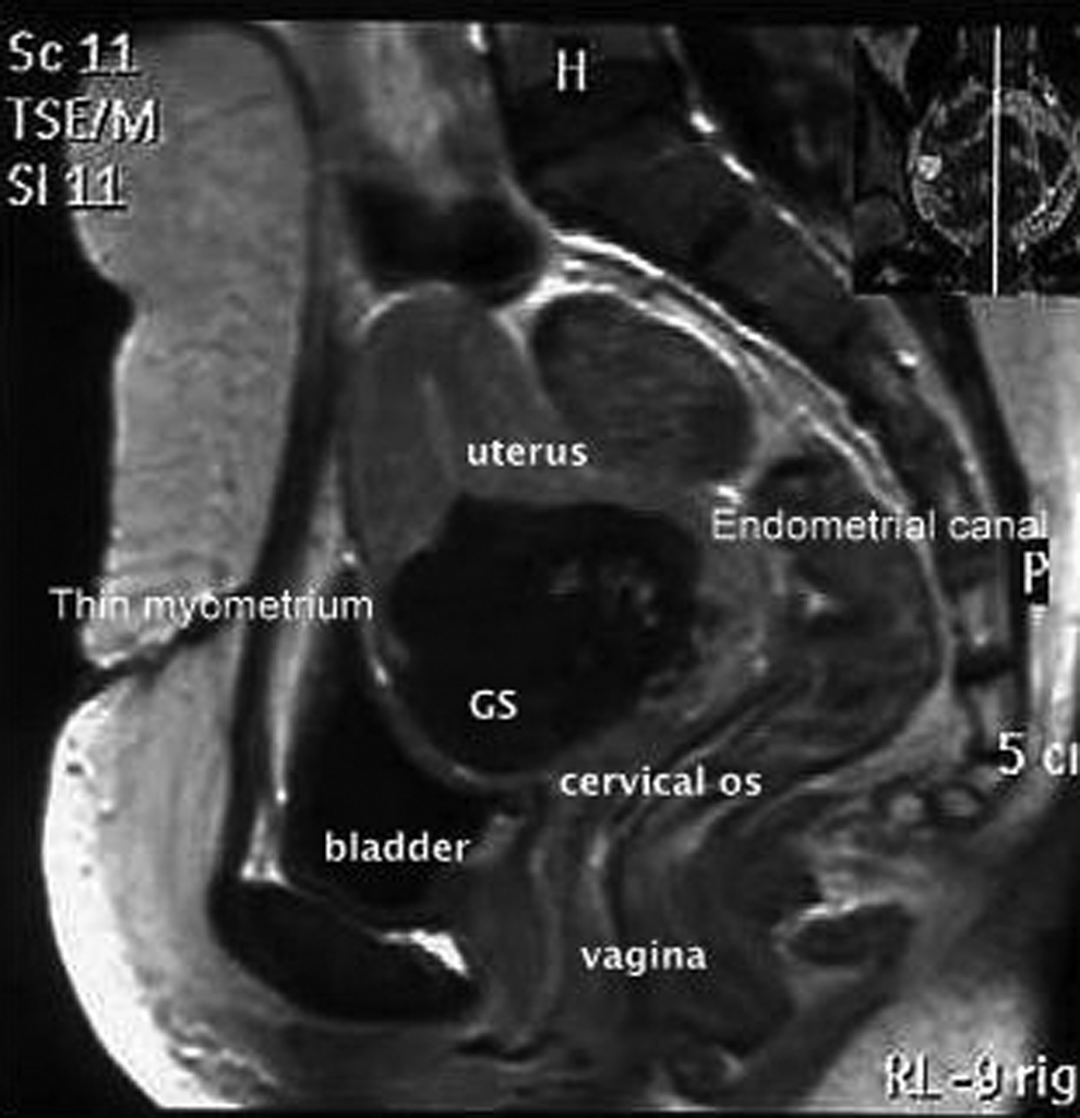
Magnetic resonance imaging T1 W contrast shows a hypointense mass anterior to the endometrial canal with an enhancing central gestational sac and thin myometrium anteriorly.
The patient was counseled and management options were discussed. She agreed to medical management and her informed consent was taken. Systemic intramuscular methotrexate therapy (50 mg/m2) was given in a single dose. Follow-up consisted of weekly outpatient clinical assessments for pain in her abdomen and vaginal bleeding as well as measurements of her serum ß-hCG levels. Her quantitative serum ß-hCG levels began to decrease progressively and fell below 2 IU/L on day 28 post-treatment. However persistence of the mass with decreased vascularity was seen on day 28 on transvaginal ultrasonography (TVUS). Ultrasound examinations were then arranged on a monthly basis until it was confirmed that all pregnancy tissue had been spontaneously expelled or absorbed.
Results
The pregnancy-related mass disappeared completely on day 72 post-treatment. The patient was counseled regarding contraception and was encouraged that, for her next pregnancy, to come for an early scan to determine the exact location of the pregnancy.
Discussion
CSP is the rarest form of ectopic pregnancy. However, with the rising rate of elective cesarean deliveries all over the world, it is probable that this incidence may increase. Thus, it is important to have a high index of suspicion of CSP in patients, especially those with risk factors for the condition in order to diagnose it. The exact cause and pathophysiology are still not well-understood. It has been hypothesized that poor vascularity in the anterior lower uterine segment impairs healing after cesarean procedures in some women, rendering this area vulnerable to formation of small dehiscent tracts or defects into which trophoblasts can implant. 1 Such a tract can also develop from the trauma of other uterine surgery (e.g., curettage, myomectomy, metroplasty, hysteroscopy, and even manual removal of a placenta). 2
It is uncertain if the risk of CSP is related to the number of previous cesarean sections a patient undergoes. Some researchers believe that multiple cesarean section are a strong risk factor for CSP because of the increased scar surface area. These deficient scars are more likely to occur after multiple cesarean sections because of the presence of fibrosis, which leads to poor vascularity of the lower uterine segment and impaired postoperative healing 3 but other researchers argue against such a correlation. A recent systematic review found that 52% cases followed one previous cesarean section, 36% after two cesarean sections, and 12% after three or more previous caesarean sections. 4 Interestingly, cesarean section because of breech presentation has been noted to confer an increased risk of CSP. 5 This may be the result of inadequate formation of the lower uterine segment that leads to poor healing, leading to a defect in the scar. 5
There is insufficient evidence in the literature regarding whether surgical technique (e.g., a single noninverting running suture technique for uterine closure instead of a double-layer closure) might have any role in causing CSP. The impact of the time interval between the previous cesarean sections and the subsequent CSP implantation is also not clear.
Diagnosis of CSP requires a high index of clinical suspicion, as up to 37% of patients may be asymptomatic. Alternatively, 39% women may present with light painless vaginal bleeding 4 and abdominal pain may not be an associated feature, as was seen in the current case. Ultrasound is the first-line imaging modality for evaluation to determine the presence of a CSP; TVUS has a reported sensitivity of 84.6%.
Ultrasound imaging criteria to diagnose a CSP are as follows:
1
(1) An empty uterine cavity and cervical canal (2) Development of the gestational sac in the anterior uterine wall at the isthmus (the presumed site of the previous LSCS scar) (3) Evidence of functional trophoblastic circulation noted on Doppler examination, defined by the presence of an area of increased peritrophoblastic vascularity seen on color Doppler examination
4
(4) The absence of healthy myometrium between the bladder and the sac.
When the gestational sac is seen in the lower part of the uterine cavity, differentiation among spontaneous abortion in progress, cervico–isthmic pregnancy, and CSP is challenging. In cases of spontaneous abortion in progress, the gestational sac should be seen in the cervical canal and appear avascular on Doppler imaging, reflecting the fact that the sac has been detached from its implantation site, whereas, in CSP, the gestational sac would appear well-perfused on color Doppler imaging and would be located in the anterior uterine wall at the isthmus. In cervico–isthmic pregnancy, there is healthy uterine tissue present between the sac and the bladder, while, in CSP, there is an absence of healthy myometrium between the bladder and the sac. 6
The role of MRI is limited to cases in which the TVUS findings are equivocal, or when the obstetrician requires additional information before planning surgery. 7 However, MRI images were more conclusive in the present case.
There are no universal guidelines for treating CSP, owing to its rarity. Generally, termination of pregnancy (TOP) in the first trimester is strongly recommended, as there is a high risk of subsequent uterine rupture, massive bleeding, and life-threatening complications. 8 The patient's clinical symptoms and desire for future fertility; her age; size of the gestation; and the clinician's experience determines which treatment option is appropriate. Systemic administration of methotrexate is a standard treatment for tubal ectopic pregnancy. There should be no reason to doubt methotrexate's efficacy for treating CSP. Furthermore, single-dose systemic administration has not been reported to cause any adverse effects, such as nausea, stomatitis, alopecia, pneumonitis, etc., when the medication is used to treat CSP. Indeed, the likelihood of success with methotrexate is greater with an hCG level of <5000 mIU/L or when the embryo has no cardiac activity. 9 As the current patient was stable and fetal cardiac activity was absent, medical treatment with systemic methotrexate was chosen after the patient gave her informed consent. She was also counseled regarding the possible need of surgical treatment despite starting medical therapy and the prolonged follow-up that was necessary to ensure that the pregnancy-related mass had resolved. She responded well without any complications.
Conclusions
Although cesarean section is a common operation, CSP is comparatively a rare occurrence. However, its incidence is likely to increase along with the increasing worldwide caesarean rate. Every health care professional involved in managing early pregnancy complications should maintain a heightened awareness of the possibility of a CSP. Furthermore, if the location of such an implantation is misdiagnosed, a common gynecologic operation, such as dilatation and curettage, can result in catastrophic hemorrhage. Massive bleeding after procedures such as D&E should raise the suspicion of CSP in high-risk cases. Sonographers should follow strict criteria to assess if any pregnancy is implanted at the site of a previous cesarean scar. MRI can play an adjunct role in evaluating a patient for the presence of a CSP. Early diagnosis and treatment can prevent obstetric emergencies, such as hemorrhage and uterine rupture, and reduce maternal morbidity as well as helping to preserve future fertility in a patient.
Footnotes
Disclosure Statement
No competing financial conflicts exist.