
Abstract
Abstract
Introduction
C
Case
A 24-year-old G4P2 L2A1 was referred to our Emergency Obstetric Department as a case of post-medical termination of pregnancy (MTP) bleeding with shock. Her obstetric history was that she had a spontaneous incomplete abortion at 3 months of pregnancy 6 years ago, which was treated by dilatation and curettage. Her first baby was born 5 years ago by lower segment caesarean section (LSCS) done in view of intrauterine growth restriction with oligohydramnios, and her second baby was born by repeat LSCS 2.5 years ago. Her present obstetric record revealed that she was a case of two previous LSCS at 14 weeks of gestation, Rh negative, and underwent MTP at a private hospital. Dilation and evacuation was attempted after 6 hours of vaginal tablet, most probably misoprostol, and the procedure was stopped as she had hemorrhage and had gone into shock. She was resuscitated and started on dopamine drip and blood transfusion of 1 unit and was shifted to our center after 4 hours. On arrival, she was conscious, oriented, febrile with a pulse of 110/minute and a BP of 90/70 mm Hg, and blood and dopamine on flow. Her hemoglobin was 6.5 g%, bleeding time was 2 minutes and 6 seconds, clotting time was 8 minutes 15 seconds, and the FDP (fibrin degradation product) was positive. Her systemic examination was normal. On per abdominal examination, the uterus was just palpable, there was minimal suprapubic tenderness, there was no guarding and rigidity, and no free fluid. Per speculum examination revealed the cervix and vagina to be healthy and there was minimal bleeding through os. On per vaginal examination, the uterus was soft, anteverted, and 12 weeks in size. Ultrasonogram (USG) in the emergency department reported retained products of conception, and a diagnosis of incomplete abortion was made. She was given two packed cell transfusion and four FFP over 24 hours and was closely monitored. She was hemodynamically stable and had persistent tachycardia and moderate bleeding per vaginum. Her hemogram after 24 hours showed hemoglobin of 8.5 g%, BT was 4 seconds, CT was 4 minutes, PT was 19 seconds, INR was 1.47, and FDP was negative. Repeat USG the next day by the duty consultant was reported as suspicious of fibroid uterus or incomplete abortion with products in the lower pole of the uterus (Fig. 1). Instrumental evacuation was done (after starting 10 units of oxytocin) under sedation and a paracervical block after 35 hours of admission. More than 300 g of old blood clots mixed with fresh blood and few products were removed gently with sponge-holding forceps, and on blunt curettage, the uterine cavity felt irregular, there was no grating sensation, the anterior wall of the uterus was found to be lax and bag like, and the curette was going freely superiorly beyond this bag-like structure even after giving intravenous methergine. Hence, bed-side USG performed by a consultant (who was performing the evacuation) revealed an empty uterine cavity and a bulging and enlarged isthmus, which had a few echogenic areas toward the myometrial–bladder interface. A diagnosis of cesarean section scar pregnancy was made at this stage. Few products were removed under USG guidance, the procedure was stopped, and injection tranexamic acid was started, which was continued for 48 hours. The blood loss was ∼400 mL, and she received one more packed cell transfusion and two FFP. Repeat USG on the same day confirmed cesarean scar pregnancy (Fig. 2). The uterus measured 4.48 cm in length and there was a variable echogenic mass of 4.3×6.3×4.9 cm below the uterus and above the cervix abutting the bladder. The cervix measured 2.86 cm in length. The uterine wall of isthmus adjacent to the bladder measured 6 mm and the posterior wall measured 12 mm.

Transabdominal ultrasonogram (USG) showing an enlarged lower part of the uterus with echogenic contents and the body of the uterus on the top of the mass. Cervix is not seen in the picture. Misinterpreted as cervical phase of incomplete abortion.
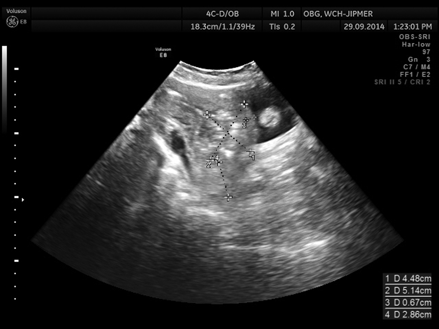
Transabdominal USG—postevacuation. The uterus measured 4.48 cm in length and there was a variable echogenic mass of 4.3×6.3×4.9 cm below the uterus and above the cervix abutting the bladder. Cervix measured 2.86 cm in length. The uterine wall of isthmus adjacent to the bladder measured 6 mm and the posterior wall measured 12 mm. Cesarean scar pregnancy.
She was given one dose of 50 mg methotrexate injection, intravenously, after the repeat hemogram, which was normal. Her β-human chorionic gonadotropin (HCG) dropped from 946 to 301 mIU/mL (on the day of evacuation). Follow-up USG after 10 days of evacuation and 8 days of single-dose methotrexate showed minimal persistent echogenic areas near the isthmus. Her β-HCG was 234.5 mIU/mL on this day and she was given another dose of methotrexate and discharged with a follow-up after 3 weeks.
Discussion
Cesarean scar pregnancy can be called as one of the dangerous types of ectopic pregnancy, as it is often asymptomatic in the early stages of development. It constitutes 6.1% of all ectopic pregnancies in women with a history of one cesarean section. 4 If left unrecognized and untreated in the early stages, it can proceed to the later stages of pregnancy and present as placenta previa with morbid adherence, which has high morbidity and mortality. 5 Hence, it is recommended that it is essential to observe the implantation site during the first trimester scan so as to diagnose it early. 6 The USG criteria to diagnose cesarean scar pregnancy are as follows. (1) The uterine cavity should be empty and the cervical canal should also be empty without contact with the sac. (2) The gestational sac with or without the fetus should occupy the anterior part of the isthmus, which is enlarged. (3) The anterior myometrium of the isthmus should be deficient or thin and should be in very close proximity with the posterior wall of the bladder. (4) Complex adnexal mass with free fluid should be absent.6,7 These criteria are further modified by Timor-Tritsch et al. 8 In the present case in the initial USG (Fig. 1), no attempt was made to trace the cervical canal and the mass that was echogenic appeared to occupy the lower part of the uterus equally bulging anteriorly and posteriorly, thus mimicking the cervical phase of abortion. This may due to the advanced stage of a pregnancy of 14 weeks. The sensitivity of two-dimensional (2D) USG is about 85% 9 and Doppler will improve the diagnostic accuracy in early gestation by revealing the peritrophoblastic ring of blood vessels. A three-dimensional color Doppler finding described by Chou et al. 9 may be of help in doubtful cases. The other diagnostic tools include MRI, hysteroscopy, and laparoscopy, but these are not necessary, as 2D USG with a combination of the abdominal and vaginal approach can help to see the panoramic view of the uterus cervix clearly and the relationship of the gestational sac to the bladder, and the thinned out or absent myometrium between the bladder and the gestational sac can be easily delineated. Doppler was performed with 2D echo in the present case, but it was inconclusive probably because of the advanced gestational age and there was interference with pregnancy in the form of surgical evacuation.
The main risk factor for the development of scar pregnancy is the absence or deficient deciduabasalis at the site of scar on the uterus, and the risk may increase with increasing number of cesarean sections. However, a recent review reported 52% of cases occurring after one cesarean and 36% after two previous cesarean sections. 2 This may be due to the fact that the incidence of the previous two caesarean section (CS) was low. The present case had one dilatation and curettage and two previous CS as risk factors. The β-HCG levels will be raised markedly and will be similar to intrauterine pregnancy. 11 In the present case, because of interference surgically, the levels were low.
There are various approaches to treat CS scar pregnancy such as injection of methotrexate locally into the sac or embryo and systemically or by a combination of systemic and local therapy. A combined intramuscular and intragestational methotrexate therapy, where 25 mg of methotrexate is injected into the embryo/fetus, 25 mg into the placental area, and 25 mg intramuscularly, was evaluated, retrospectively, in 19 cesarean scar ectopic pregnancies. Sonographic follow-up included measurement of volume of the gestational sac and determining the vascularity index. Serum β-HCG was measured weekly for 3 weeks and then bimonthly till undetectable. However, resolution took as long as 140 days, and the women were advised abstinence during this period. 8
Surgical therapies include dilatation and curettage, local resection, and uterine artery embolization. 4 It takes as long as 11 weeks for β-HCG to normalize and 12 to 17 weeks for the USG mass to disappear after methotrexate therapy. Little et al. reported a case of cesarean scar ectopic pregnancy, which did not respond to systemic methotrexate therapy and eventually ended up with hysterectomy due to torrential hemorrhage following ultrasound-guided dilatation and curettage. 12 Dilatation and curettage is not recommended as it may induce torrential bleeding necessating hysterectomy. 9 In the present case, she experienced severe hemorrhage and also developed coagulation abnormality in the form of disseminated intravascular coagulation (DIC). Combined therapy with methotrexate and dilatation and curettage may shorten the time interval for resolution, but Zhuang and Huang 13 found uterine artery embolization followed by suction curettage as a better option in terms of less hemorrhage and hospitalization, compared to methotrexate therapy followed by curettage. Gupta et al. treated a very early cesarean scar pregnancy with transvaginal ultrasound-guided intragestational methotrxate and uterine artery embolization. 14 Hysteroscopic resection of six cesarean scar ectopic pregnancies was reported by Deans and Abbott. This requires skill and is an invasive procedure, which results in hemorrhage, and hence, a time-taking task. However, the authors comment that the hysteroscopic procedure is better than local and systemic methotrxate therapy and there is lack of evidence regarding this. 15 The mode of therapy has to be individualized depending on the gestational age, hemorrhage, and complications at presentation. In general, patients with acute presentation like hemorrhage and rupture can be managed effectively by surgical methods and those asymptomatic women diagnosed during routine ultrasonographic examination may be managed by medical therapy with local and/or systemic methotrexate therapy.
Conclusion
This case illustrates that it is mandatory to subject the patient to USG before performing termination of pregnancy. Every obstetrician should have the USG knowledge of diagnosing CS scar pregnancy and must look for the implantation site during early pregnancy scan in all cases with history of cesarean section.
Footnotes
Disclosure Statement
No competing financial interests exist.