
Abstract
Abstract
Introduction
U
This article reports a rare case of nonpuerperal uterine inversion caused by a submucus myoma, and provides a brief review of relatively recent literature.
Case
A 47-year-old Moroccan woman presented to the gynecology/obstetrics emergency department of the Maternal Teaching Hospital, Rabat, in Rabat, Morroco. She had heavy menstrual bleeding and a protruding vaginal mass. She had a history of four successful vaginal deliveries, with the last one being 9 years prior. However, she did not have any prior medical diseases or surgical interventions.
During examination, she was noted to be pale with polypnea. Her heart rate was 100 beats per minute and her blood pressure was 100/60 mm Hg. Her abdomen was normal. A vaginal examination revealed the presence of a large protruding mass, 12 cm in length, with bleeding (Fig. 1). The cervical os was not identifiable. On bimanual examination, her uterus could not be felt. In addition, a pelvic ultrasound examination failed to identify her uterus. Transvaginal ultrasound could not be performed because of the nonreducible protruding mass from her vagina. Laboratory testing revealed that this patient had low hemoglobin rate of 5.7g/dL.
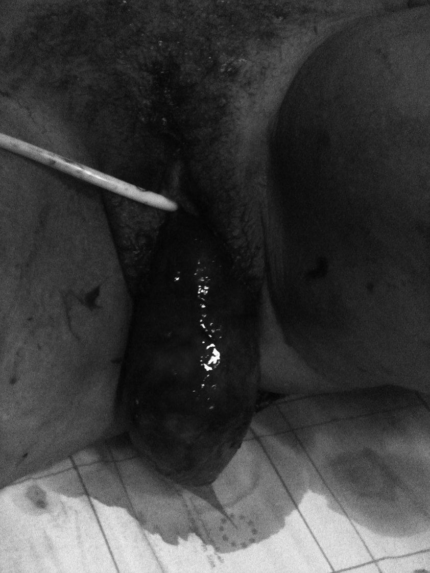
Clinical photograph of the inverted uterus protruding from the vulva.
The patient was brought to the operating theater after a transfusion of three blood packs. After administration of general anesthesia, all attempts to repositioning her uterus failed. Laparotomy with a Pfannenstiel incision was performed. The uterus appeared to be depressed in a narrow pelvic split, with the round and broad ligaments pulled into this pelvic-floor depression. A total abdominal hysterectomy was performed with bilateral salpingo-oophorectomy but without repositioning of the uterus. The free uterus was then pushed down into the vagina and the vaginal cuff was closed.
A macroscopical examination, the inverted uterus was noted to be caused by a white spherical mass that was 6 cm in diameter and attached to the fundus.
Results
This patient's postoperative period was uneventful, and she was discharged home on the 4th day after being admitted to the hospital. A final histopathologic examination of the excised specimen revealed that she had a benign submucous leiomyoma.
Discussion
Uterine inversion is a rare complication of the puerperium, and nonpuerperal uterine inversion is of extremely rare occurrence. 1 There is no specific incidence of this condition, although it comprises one-sixth of all uterine inversions. 2 From 1887 to 2005, 150 cases of nonpuerperal uterine inversion have been identified. 3
A structured search, using the term:
Ref. 3.
Oguri H, Maeda N, Yamamoto Y, Wakatsuki A, Fukaya T. Non-puerperal uterine inversion associated with endometrial carcinoma—a case report. Gynecol Oncol 2005;97:973.
Rosales Aujang E, González Romo R. Non-puerperal uterine inversion: Report of a case [in Spanish]. Ginecol Obstet Mex 2005;73:328.
Cormio G, Loizzi V, Nardelli C, Fattizzi N, Selvaggi L. Non-puerperal uterine inversion due to uterine sarcoma. Gynecol Obstet Invest 2006;61:171.
Pandit U. Prolapsed uterine sarcoma causing non-puerperal uterine inversion in a post menopausal woman. JNMA J Nepal Med Assoc 2006;45:373.
Buyukkurt S, Vardar MA, Zeren H, Ozgunen FT. Non-puerperal inversion of the uterus caused by leiomyosarcoma: A case report and clinical management. J Obstet Gynaecol Res 2007;33:402.
Ref. 4
Fofie CO, Baffoe P. Non-puerperal uterine inversion: A case report. Ghana Med J 2010;44:79.
Ref. 5.
Krissi H, Peled Y, Efrat Z, Goldshmit C. Ultrasound diagnosis and comprehensive surgical treatment of complete non-puerperal uterine inversion. Arch Gynecol Obstet 2011;283(suppl1):111.
Omololu OM, Rabiu KA, Quadri MA, Oyedeko MO, Fatogun YM. Non puerperal uterine inversion due to submucous fibroid: A case report. Niger Postgrad Med J 2011;18:158.
Atalay MA, Demir BÇ, Solak N, Atalay FO, Küçükkömürcü S. An unusual presentation of a submucous leiomyoma accounting to [sic] a non-puerperal uterine inversion: A case report. J Turk Ger Gynecol Assoc 2013;14:116.
Tibrewal R, Goswami S, Chakravorty PS. Non puerperal uterine inversion [in French]. J Obstet Gynaecol India 2012;62:452.
Pelissier-Komorek A, Lucereau-Barbier M, Diab J, Gavillon N, Graesslin O. Acute non-puerperal uterine inversion [sic] the third degree [in French]. Gynecol Obstet Fertil. 2013;41:130.
Umeononihu OS, Adinma JI, Obiechina NJ, Eleje GU, Udegbunam OI, Mbachu II. Uterine leiomyoma associated non-puerperal uterine inversion misdiagnosed as advanced cervical cancer: A case report. Int J Surg Case Rep 2013;4:1000.
Mehra R, Siwatch S, Arora S, Kundu R. Non-puerperal uterine inversion caused by malignant mixed Müllerian sarcoma. BMJ Case Rep 2013;2013:pii.
Turhan N, Simavli S, Kaygusuz I, Kasap B. Totally inverted cervix due to a huge prolapsed cervical myoma simulating chronic non-puerperal uterine inversion. Int J Surg Case Rep 2014;5:513.
Shabbir S, Ghayasuddin M, Younus SM, Baloch K. Chronic non puerperal uterine inversion secondary to sub-mucosal fibroid. J Pak Med Assoc 2014;64:586.
Rathod S, Samal SK, Pallavee P, Ghose S. Non puerperal uterine inversion in a young female—a case report. J Clin Diagn Res 2014;8:OD01-2.
Zhang X, Sun L, Chen X, Hua K. Uterus preserving reposition of non-puerperal uterine inversion under laparoscope: A case report and literature review. Gynecol Obstet Invest 2015;79:206.
–, not applicable.
Nonpuerperal uterine inversion is mostly associated with benign leiomyomas. The remaining cases of tumor-related inversion are associated with uterine sarcomas. Inversion associated with carcinomas is extremely rare. 3
The main symptoms include: vaginal bleeding; abdominal pain; mass protruding from the vagina; and urinary retention. 3 Vaginal bleeding associated with nonpuerperal uterine inversion could be severe enough to cause hemorrhagic shock. 2
The etiology of uterine inversion has not been clearly defined. Possible explanations could be a thin uterine wall, rapid growth of a tumor, tumor size, fundic location of a tumor, tumor attachment to the uterine wall with a thick pedicle, dilatation of the cervix by distension of the uterine cavity, and sudden expulsion of the tumor. 4
Sonographic diagnosis of this condition is challenging. Sonography reveals absence of the uterus. A key imaging feature facilitating diagnosis is that of the “bulls' eye” appearance of the invaginating adnexal structure on both computed tomography and magnetic resonance imaging. 2
Most surgical procedures described in the literature showed that different techniques have been used to reposition the uterus first followed by hysterectomy. 5 In the current case, abdominal hysterectomy was accomplished safely without repositioning the uterus. The abdominal route is preferred over the vaginal one, as the vaginal incision is reduced to a minimum. 5 However, one of the difficulties of this surgical procedure is the close proximity of the ureters to the unfundibulo-ovarian vessels and to the uterine arteries. 5
Conclusions
Nonpuerperal uterine inversion is a rare gynecologic condition that needs to be recognized and treated surgically, because any delay can lead to life-threatening hemorrhage.
Footnotes
Disclosure Statement
No financial conflicts exist.