
Abstract
Abstract
Background:
Dilation and curettage (D&C) is a safe, common surgical procedure. Complications occur rarely, but outcomes depend upon prompt recognition and management of such complications. Acute abdominal pain after D&C, even without a suspicion of hemorrhage, warrants additional evaluation.
Case:
A 36-year-old gravida 5, para 2022, woman underwent a suction D&C in early pregnancy and, postoperatively, had significant pain symptoms and signs of an acute abdomen. Diagnostic laparoscopy revealed an incarcerated, transected fallopian tube drawn into the endometrial cavity through a small uterine perforation. The incarcerated fallopian tube was released from the myometrial defect, and salpingectomy was performed.
Results:
The patient was discharged to go home on the day of surgery, and her recovery was unremarkable.
Conclusions:
Uterine perforation during D&C might involve adnexal structures or the surrounding viscera. This case report is the first report of immediate laparoscopic management of fallopian-tube incarceration after uterine perforation. Laparoscopy is the preferred approach for a patient with suspected uterine perforation who is hemodynamically stable.
Introduction
D
Case
A 36-year-old gravida 5, para 2022, woman presented to a high-volume abortion clinic at 6 weeks and 5 days of gestation requesting surgical termination of this latest pregnancy. Her history was significant for 1 full-term vaginal delivery, 1 full-term low-transverse cesarean delivery, and 2 D&C procedures for miscarriages. The remainder of her medical history was unremarkable.
At the outside clinic, the gestational age of this patient's current pregnancy was confirmed by ultrasound. She was administered propofol sedation, and a bimanual examination confirmed a uterine size appropriate for the gestational age with no palpable abnormalities. The procedure was performed by an experienced attending surgeon. Cervical dilation with rigid dilators was performed in a standard fashion until adequate dilation was achieved to pass a 7-mm rigid suction cannula. Suction aspiration was performed. During the procedure, the cannula advanced further than expected for the size of the uterus, but no intraperitoneal visceral contents were noted in the cannula. The surgeon suspected a uterine perforation and removed all of the surgical instruments. The patient was observed in the recovery room.
This patient remained hemodynamically stable, with minimal vaginal bleeding. As she recovered from the anesthesia, she complained of increasing pain in her lower abdomen. An examination revealed significant tenderness in her left lower quadrant. An ultrasound showed no intra-abdominal fluid suggestive of a hemoperitoneum. However, given the woman's pain, the examination findings, and concern regarding uterine perforation during the procedure, she was transferred emergently to a nearby teaching hospital. Details of the clinical course leading to this transfer were communicated directly by telephone. The outside physician and the receiving attending surgeon discussed the case during the woman's transfer by ambulance. The remainder of the patient's course was managed by the receiving surgical team.
Upon arrival, the patient complained of nausea and lower abdominal pain, which she rated as 10 on a 1–10 pain scale. Her vital signs remained normal. Another physical examination revealed that she had a moderately distended abdomen, marked tenderness to palpation in the left lower quadrant, and guarding and rebound. Vaginal bleeding remained minimal. A Focused Assessment with Sonography in Trauma examination was negative for a hemoperitoneum, and a stat complete blood-cell count was returned with a hemoglobin level of 10.9 g/dL and a hematocrit of 34.6%. These values were identical to this patient's preoperative levels, suggesting that she had minimal surgical blood loss.
Immediate surgery was indicated, based on the severity of this patient's pain and the examination findings that were consistent with an acute abdomen. Laboratory and imaging findings did not suggest significant intra-abdominal hemorrhage, but the severity of pain symptoms warranted operative evaluation. A laparoscopic approach was chosen, rather than laparotomy, because the patient was hemodynamically stable. Type and cross for 2 units of packed red blood cells was ordered.
Laparoscopy, using a standard entry, was performed under general anesthesia. There was no hemoperitoneum. An ∼1-cm perforation was visible in the left aspect of the uterine fundus, just medial to the cornua (Fig. 1). Nearly the entire length of the left fallopian tube was incarcerated into the uterine cavity through this defect. The tube remained attached to the adjacent mesosalpinx and other adnexal structures, which were tethered in close proximity. The remainder of this abdominal survey was unremarkable, including evaluation of the adjacent small bowel.
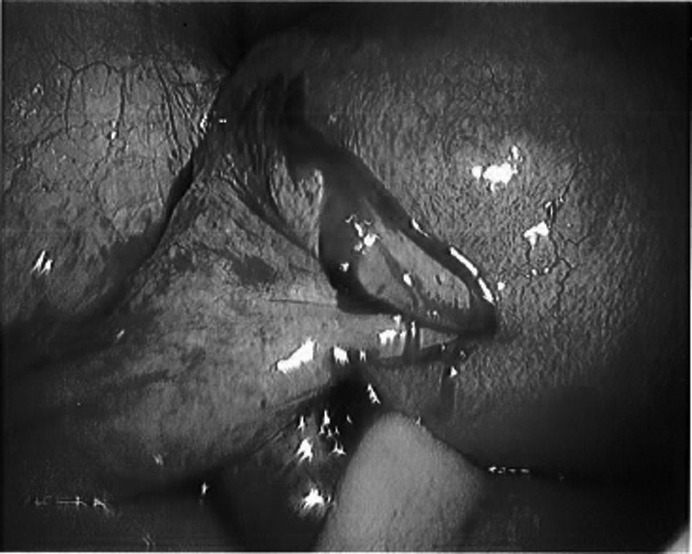
Laparoscopic view of the left fallopian tube incarcerated into the uterine defect.
Using atraumatic graspers, gentle traction was applied to the fallopian tube and the tube was extracted from the myometrial defect. The tube was released easily in its entirety. A full-thickness transection was noted in the ampulla, with active bleeding. The tube appeared edematous and hemorrhagic from the ampulla through the fimbriated distal end. Given the bleeding from the transection and the dilated, edematous appearance of the remainder of the tube, salpingectomy was indicated. A left salpingectomy was performed without difficulty. The uterine perforation site was hemostatic, and the right adnexa appeared to be normal.
It was unclear if the initial D&C procedure was completed prior to the patient's transfer to the current authors' care. To ensure complete evacuation of the uterus and to remove any remaining pregnancy tissue, gentle suction curettage was performed under direct laparoscopic visualization. The perforation was not reentered, and it remained hemostatic, with a decreased pneumoperitoneum.
Results
After this last procedure, this patient was discharged to go home on same day and had an unremarkable recovery.
Discussion
Uterine perforation is a rare but known complication of D&C, recognized at a rate of 0.09–2.8/1000 cases, and is more common at higher gestational ages. 1 Many patients remain asymptomatic, resulting in a clinically unrecognized perforation without long-term sequelae. Other patients require only observation, when the perforation is recognized intraoperatively and the patient is stable and without pain or evidence of hemorrhage. In contrast, in symptomatic women, or in cases with concern for hemorrhage or visceral injury, prompt evaluation via diagnostic laparoscopy or exploratory laparotomy is indicated.
In the current case, an initial evaluation in the hospital prompted concern for uterine perforation and possible injury to adjacent structures, but not for significant bleeding. This patient's vital signs were in the normal range, imaging did not suggest blood in the abdomen, and her hemoglobin and hematocrit levels were normal and stable. The presentation of her acute abdomen alone warranted surgical evaluation.
The choice of initial surgical approach—laparoscopy or laparotomy—depends on the patient's hemodynamic stability, the likelihood of massive intraabdominal hemorrhage, and the suspicion for visceral injury. Additional considerations include surgeon experience and availability of resources for rapid conversion from laparoscopy to laparotomy if necessary. In the current case, diagnostic laparoscopy was the preferred initial method and facilitated completion of the procedure using a minimally invasive approach.
The uterine fundus is a common site of perforation, and conventional teaching describes visualization of the bowel, omentum, or mesenteric fat in the suction cannula as a primary means of intraoperative identification of perforation. Based on the anatomic location of the fallopian tube, it is logical to presume that uterine perforation could involve the tube, but such occurrences are rarely reported.
A few cases of fallopian-tube or ovarian incarceration have been reported after D&C for pregnancy termination or retained placentas after term deliveries. In nearly all of these reports, the diagnosis of tubal or ovarian incarceration was delayed, from 4 days to several months to as long as 5 years after the D&C procedure.2–7 Most frequently, abnormal imaging findings prompted surgical evaluation.2–6 The elapsed time between the initial D&C and final surgical procedure ranged from 18 months to 5 years, in patients presenting with subacute pain, vaginal discharge, infertility, or other symptoms.3–6 In 1 case, an asymptomatic woman underwent diagnostic laparoscopy revealing tubal incarceration 5 days after a D&C for a medical abortion, after histology testing revealed that the specimen was fallopian-tube tissue and not products of conception. 7 One report described uterine perforation during D&C that caused severe pain and an acute abdomen; this patient was managed with an immediate exploratory laparotomy. Salpingectomy was performed for fallopian-tube incarceration, and the patient recovered in the hospital until postoperative day 6. 8
To the current authors' knowledge, immediate laparoscopic management of fallopian-tube incarceration during uterine perforation has not previously been reported. In the current patient, entrapment or ischemia of the fallopian tube caused significant pain and peritoneal signs, resulting in a clinical presentation of acute abdomen. In this case, the patient's symptoms persisted for hours after the original surgery, while, in other reported cases, patients presented days-to-months later or remained asymptomatic with findings only on imaging or pathology testing.
Conclusions
This unique case illustrates that significant pain after uterine aspiration, even at an early gestational age and without signs of ongoing hemorrhage, requires prompt evaluation. In the absence of signs that raise concerns for massive hemorrhage or major visceral or vessel injury, a minimally invasive approach should be favored over laparotomy.
Footnotes
Author Disclosure Statement
No competing financial conflicts of interest exist.