
Abstract
Abstract
Background:
Osseous metaplasia of the nonteratomatous ovary is extremely rare. This condition has been reported in association with endometriosis and certain ovarian neoplasms.
Case:
A 35-year-old nulliparous patient presented with new-onset severe abdominal pain associated with abdominal fullness, nausea, and vomiting. She had a large, tender pelvic mass anterior to the cervix. Computed tomography showed a midline pelvic mass, measuring 13.1 × 12.3 × 9.3 cm, located anteriorly and superiorly to the uterine fundus, with partially calcified septations as well as an eccentric soft-tissue nodule The uterus was enlarged and lobulated, containing multiple fibroids. There was a complex cystic mass superior to the uterus producing low-level internal echoes and having a few septations with areas of calcification. The patient underwent an exploratory laparotomy during which the pelvic mass was identified as an endometrioma arising from the left ovary. The cyst was densely adherent to the superior aspect of a leiomyomatous uterus with a large fundal fibroid measuring ∼7 cm. Intraoperative pathologic evaluation revealed that this cyst was benign. Lysis of adhesions, followed by myomectomy, was performed, removing 19 fibroids weighing 235 g in aggregate.
Results:
The patient had an uncomplicated postoperative course and was referred for infertility evaluation. Pathologically, this lesion was consistent with an endometriotic cyst (endometrioma) with focal osseous metaplasia.
Conclusions:
It has been theorized that chronic inflammation might be a driving force in the formation of bone within these lesions.
Introduction
Osseous metaplasia of the nonteratomatous ovary is rare. A few case reports have previously described ossification of ovarian neoplasms, osseous metaplasia without any associated abnormalities, and osseous metaplasia associated with endometriosis.1–16 Osseous metaplasia should not be confused with pathologic calcification of tissue or with formation of teratomas. This article describes a case of osseous metaplasia within an endometrioma and reviews the literature.
Case
A 35-year-old nulliparous patient presented with a 4-day history of new-onset severe abdominal pain associated with abdominal fullness, nausea, and vomiting. Her history was significant for regular menstrual periods with dysmenorrhea. She had been attempting to become pregnant for the prior 2 years without success. A pelvic examination revealed a large, tender pelvic mass anterior to the cervix. Computed tomography showed a midline pelvic mass, measuring 13.1 × 12.3 × 9.3 cm, located anteriorly and superiorly to the uterine fundus, with partially calcified septations as well as an eccentric soft-tissue nodule (Figs. 1 and 2). The uterus was enlarged and lobulated, containing multiple fibroids. Ultrasonography showed the uterus measuring 14.4 × 7.5 × 8.4 cm with multiple fibroids, and the right ovary measured 3.4 × 1.4 × 2.7 cm. There was a complex cystic mass superior to the uterus producing low-level internal echoes and having a few septations with areas of calcification measuring 9.2 × 13.2 × 11.5 cm (Figs. 3 and 4).
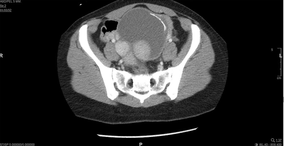
A computed tomographic image on the transverse plane showed a midline pelvic mass, measuring 13.1 × 12.3 × 9.3 cm, with partially calcified septations as well as an eccentric soft-tissue nodule.
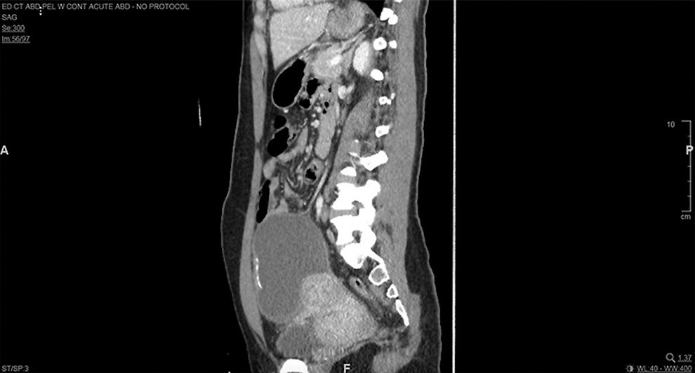
A computed tomography image on the sagittal plane also showed a midline pelvic mass, measuring 13.1 × 12.3 × 9.3 cm, with partially calcified septations as well as an eccentric soft-tissue nodule, which was consistent with the image seen on the transverse plane.

A sagittal midline view of transabdominal ultrasound (US) image showed a complex cystic mass that was superior to the uterus and producing low-level internal echoes. The US also showed a few septations with areas of calcification measuring 9.2 × 13.2 × 11.5 cm, sagittal view.
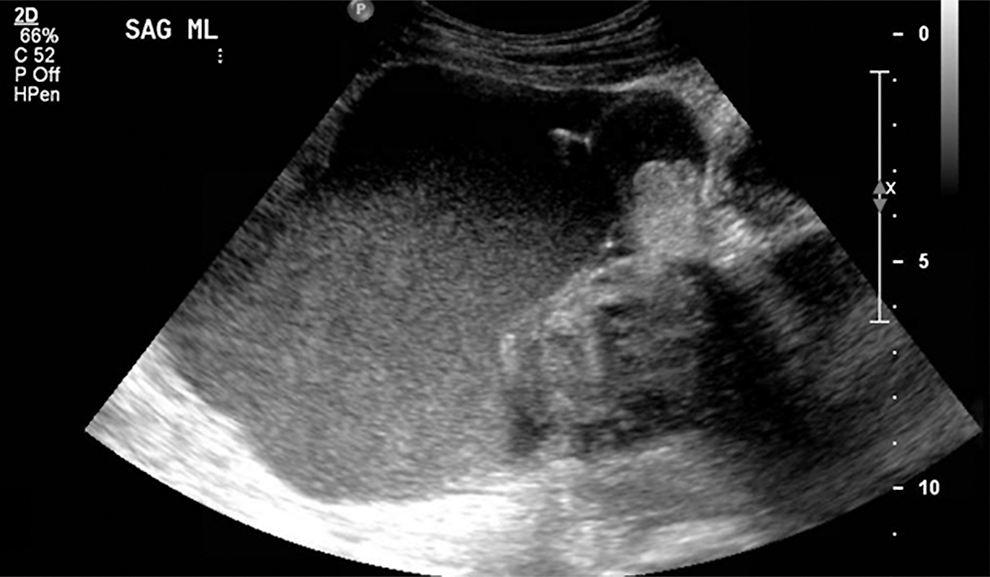
A sagittal midline view of a transabdominal ultrasound (US) image also showed a complex cystic mass that was superior to the uterus and producing low-level internal echoes. The US also showed a few septations with areas of calcification measuring 9.2 × 13.2 × 11.5 cm. This was consistent with the sagittal US view.
The patient underwent an exploratory laparotomy during which the pelvic mass was identified as an endometrioma arising from the left ovary. The cyst was densely adherent to the superior aspect of a leiomyomatous uterus with a large fundal fibroid measuring ∼7 cm. The right fallopian tube and ovary appeared to be normal. The left fallopian tube was partially adherent to the left adnexal mass, which was carefully separated to maintain tubal integrity. The cyst ruptured during dissection and dark brown material was noted within the cyst, consistent with an endometrioma. A fragment of hard tissue measuring 1 × 0.5 × 0.2 cm was noted within the endometrioma. The cyst was excised from the ovary, and intraoperative pathologic evaluation revealed that this cyst was benign. Lysis of adhesions, followed by myomectomy, was performed, removing 19 fibroids weighing 235 g in aggregate.
Results
The patient had an uncomplicated postoperative course and was referred for infertility evaluation.
Additional Pathology
The specimen received in the pathology laboratory was a disrupted red–tan cyst measuring 11 × 6.5 × 0.7 cm. On the outer surface, a focal area of thickening, with grossly recognizable small fragments of bony tissue measuring 1 × 0.5 × 0.2 cm, was seen. The inner surface of the unilocular cyst was hemorrhagic. Histologically, this lesion was consistent with an endometriotic cyst (endometrioma) with focal osseous metaplasia (Fig. 5).

Well-formed bone was seen within the cyst wall. Away from the bone, an endometriotic cyst was noted that was lined by endometrial glandular epithelium with underlying stroma (inset, upper right). Color images are available online
Discussion
Osseous metaplasia is a rare finding in nonteratomatous ovaries, and, in particular, ovarian endometriomas. A review of the literature yielded an article on osseous metaplasia within an endometrioma of a supernumerary ovary 1 and another article showing complete ossification of the ovary; however only microscopic foci of endometriosis were identified. 2 Heterotopic bone in the ovary has also been associated with extensive endometriosis. 3 These researchers theorized that osseous metaplasia was a response to constant inflammatory insults to the tissues secondary to endometriosis. This theory of inflammation was supported further by Campo et al., 4 who described osseous metaplasia within the uterus and ovaries bilaterally in a patient with an intrauterine device.
There are several case reports describing ossification associated with ovarian neoplasms including luteinized thecomas,5,6 Sertoli–Leydig tumors, 7 mucinous cystadenomas,8,9 and a fibroma (Table 1). 10 Other case reports have shown calcification surrounding the ovaries rather than ossifications.11–13 There have also been other isolated case reports of ossification without any associated abnormalities. Rosa e Silva et al. 14 and Shipton et al. 15 both described this phenomena.
Reported Cases of Ossification in Nonteratomatous Ovaries
LSO, left salpingo-oophorectomy; G, gravida; P, para; TAH, total abdominal hysterectomy; BSO, bilateral salpingo-oophorectomy; DM, diabetes mellitus; IUD, intrauterine device; AUB, abnormal uterine bleeding; TVUS, transvaginal ultrasound; CT, computed tomography; EMB, endometrial biopsy; LVI, lymphovascular invasion; US, ultrasound; TGF, tumor growth factor.
Conclusions
Osseous metaplasia is described in the literature infrequently. This condition might be associated with chronic inflammation or irritation.
Footnotes
Author Disclosure Statement
No financial conflicts of interest exist.