
Abstract
Abstract
Yu Botao, Jie Ma, Wenjing Xiao, Qingyu Xiang, Kaihua Fan, Jun Hou, Juan Wu, and Weihua Jing. Protective effect of ginkgolide B on high altitude cerebral edema of rats. High Alt Med Biol 14:61–64, 2013.—Ginkgolide B (GB) is one of the ginkgolides isolated from leaves of the Ginkgo biloba tree. The aim of this study was to investigate whether GB has a protective effect on high altitude cerebral edema (HACE) of rats. HACE was induced by hypobaric hypoxia exposure for 24 hours in an animal decompression chamber with the chamber pressure of 267 mmHg to simulate an altitude of 8000 m. Before the exposure, three doses (3, 6, and 12mg·kg−1) of GB were given intraperitoneally (ip) daily for 3 days. Effects of GB on brain water content (BWC), activity of superoxide dismutase (SOD), concentration of glutathione (GSH) and malondialdehyde (MDA), expression of active caspase-3 and poly(ADP-ribose) polymerase (PARP) were measured. In GB pretreatment groups (6 and 12 mg·kg−1, but not 3 mg·kg−1), BWC, the concentration of MDA, the expression of active caspase-3 and PARP were reduced significantly, while the activity of SOD and concentration of GSH were significantly increased. In conclusion, these results indicate that GB has a protective effect on cerebral edema caused by high altitude in rats. The protective effect of GB might be attributed to its antioxidant properties and suppression of the caspase-dependent apotosis pathway.
Introduction
Ginkgo biloba have been used as a medicine in China for over 5000 years. Ginkgo biloba extracts isolated from leaves of Ginkgo biloba tree contain flavonoid and terpenoid substances. Ginkgolide B (GB, BN52021) is one of the ginkgolides and the major constituent of the terpenoid fraction. A number of studies have shown that GB possesses neuroprotective effects on various brain injuries such as permanent focal ischemia, transient focal ischemia, and ischemia-reperfusion injury by exerting antioxidant effects, diminishing brain edema, or inhibiting cell apoptosis (Huang et al., 2008; Huang et al., 2012; Qi et al., 2010).
As the effect of GB on HACE had not been explored, we carried out our study with the hypothesis that GB pretreatment might produce a protective effect on rats with high altitude cerebral edema via potential antioxidant and caspase-related anti- apoptotic mechanisms.
Materials and Methods
Animals
Male SPF Sprague-Dawley rats (250–280 g) were obtained from the Laboratory Animal Center of Sichuan University (Chengdu, China). Rats were housed under humidity- and temperature-controlled conditions and a 12 h light/dark cycle with free access to food and water. All experiments were carried out in accordance with guidelines of Institutional Animal Care and Use Committee of Chengdu Military General Hospital (Chengdu, China).
Materials
Ginkgolide B (Batch Number: 0701908, Purity >98%) was purchased from Chengdu Herbpurify CO., LTD, China. SOD, GSH, MDA kit (Catalogue Numbers: A001-3, A006 and A003-1); total protein quantification kit (Coomassie Brilliant Blue, Catalogue Number: A045-2) were purchased from Nanjing Jiancheng Bioengineering Institute, China. Rabbit caspase-3 polyclonal antibody was purchased from Wuhan Boster Biological Technology, Ltd., China. Rabbit anti-PARP antibody was purchased from Beijing Boisynthesis Biotechnology Co., LTD, China.
High altitude cerebral edema model
A HACE model was produced according to the methods modified from previously reported (Deng et al., 2005). Briefly, the rats were exposed to hypobaric hypoxia for 24 h in an animal decompression chamber (FLYDWC50-IA Guizhou Fenglei Air Ordnace LTD, CO, China) with the chamber pressure of 267 mmHg to simulate an altitude of 8000 m. The altitude rose up at the speed of 10 m·s−1 according to the vacuum detection. The temperature and humidity in the chamber had 25±2°C and 60±5%, respectively. The rats had free access to food and water.
All rats were randomly divided into 5 groups: control group, HACE group, and 3 different pretreatment groups which were administered GB 3, 6, and 12 mg·kg−1 intraperitoneally (i.p.) daily for 3 days before the hypobaric hypoxia exposure. One hour after the last administration, the rats were placed in the animal decompression chamber.
Brain water content
Rats were anesthetized with 10% chloral hydrate (300 mg·kg−1, i.p.) and sacrificed after 24 h hypobaric hypoxia exposure. After the brains were taken out and placed on ice immediately, the olfactory bulb and cerebellum were removed. Then the brains were separated into the left and right hemispheres. The left hemispheres were weighed to determine the wet weight (WW) and then were dried for 24 h in a vacuum drying oven at 80°C to measure the dry weight (DW). BWC was expressed as the percentage change between WW and DW. BWC=(WW−DW)/WW×100%.
Biochemical analysis
The right hemisphere of the brain was homogenized with cold normal saline (NS), centrifuged (4°C, 12,500 g, 10 min), and the supernatant was collected for assays. The antioxidant status of the brain was assessed by the activity of SOD and concentration of GSH. The lipid peroxidation was determined by the concentrations of MDA, an end product of lipid peroxidation. The detailed procedures for determinations followed the instructions of the kits.
Immunohistochemical assay
Rats were anesthetized with 10% chloral hydrate (300 mg·kg−1, i.p.) and perfused instantly with cold NS and then 4% paraformaldehyde. The brains were taken out and fixed in 4% paraformaldehyde. Brain tissue slices were dehydrated, embedded in paraffin, and sectioned (5 μm). The sections were incubated with rabbit anti-caspase-3 (1:100) or rabbit anti-poly(ADP-ribose) polymerase (PARP) (1:100) primary antibodies overnight at 4°C. Negative controls were carried out using phosphate buffer solution (PBS) instead of primary antibodies. Then the sections were rinsed with 0.01 mol/L PBS and treated with biotinylated goat anti-rabbit IgG for another 20 min at 37°C. After DAB (diaminobenzidine) coloration, hematoxylin afterstain and gradient alcohol dehydration, cells that showed up brown were designated positive. Sections were examined and photographed by OLYMPUS CKX41-A32PH (Japan). The integrated optical density (IOD) of sections was measured using Image-Pro Plus 6.0.
Statistical analysis
All values were presented as mean±S.D. (standard deviation). Statistical analysis was performed using one-way analysis of variance (ANOVA). An effect was considered statistically significant if the p value was less than 0.05 (p<0.05).
Results
Effect of GB on brain water content in HACE rats
As shown in Figure 1A, BWC, a measurement of cerebral edema, was significantly increased in HACE group as compared with control group after 24 hours hypobaric hypoxia exposure (p<0.01). Compared with the HACE group, BWC was reduced significantly in GB treatment group (6 and 12 mg·kg−1) (p<0.05, p<0.01). However, there was no significant difference in BWC between the 3 mg·kg−1 GB pretreatment group and HACE group.
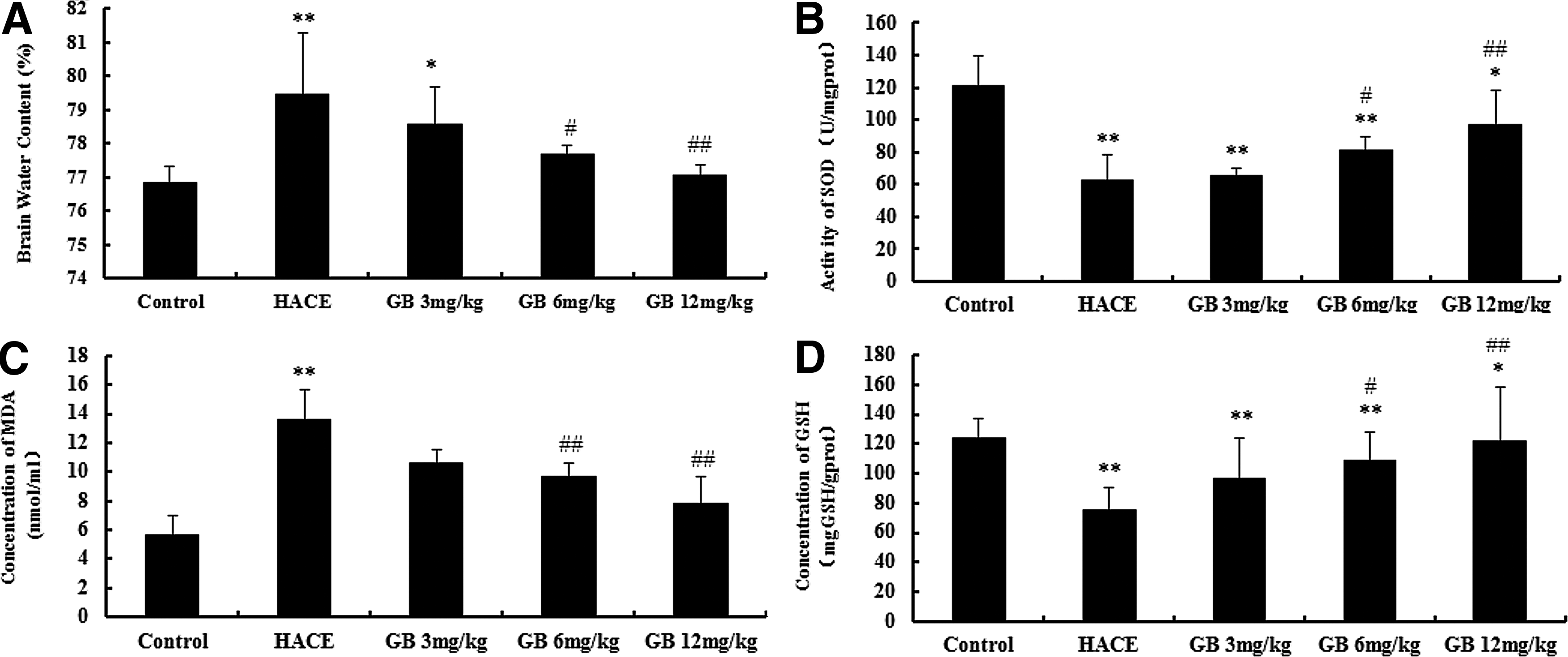
Effect of Ginkgolide B on cerebral edema and activity of SOD, concentrations of GSH and MDA in HACE rats (n=6 for each group).
Effects of GB on activity of SOD, concentrations of GSH and MDA in HACE rats
As shown in Figure 1B–1D, the activity of SOD and the concentration of GSH decreased significantly, and the concentration of MDA increased significantly in the HACE group compared with the control group (p<0.01). Compared with the HACE group, GB pretreatment group (6 and 12 mg·kg−1) increased the activity of SOD and concentration of GSH significantly, and reduced the concentration of MDA (p<0.05, p<0.01). However, there was no significant difference in activity of SOD, and the concentrations of MDA and GSH between the 3 mg·kg−1 GB pretreatment group and the HACE group.
Effect of GB on caspase-3 and PARP expression in HACE rats
As shown in Figure 2, an immunohistochemistry assay showed that in the HACE group, a massive expression of active caspase-3 and PARP in the cortex compared with the control group (p<0.01). Both 6 and 12 mg·kg−1 GB pretreatment could reduce caspase-3 and PARP expression significantly compared with the HACE group (p<0.01). However, 3 mg·kg−1 GB pretreatment group did not show a significant difference from the HACE group. The negative controls did not produce specific staining.

Effect of Ginkgolide B on expression of caspase-3 and PARP in cortex area of HACE rats (n=4 for each group).
Discussion
A growing body of evidence has demonstrated that GB has many beneficial effects such as anti-inflammatory, antioxidant, and neuroprotective effects, so it presents excellent potential for therapeutic action in many diseases. Our study revealed that, in high altitude brain injury, GB pretreatment could exert a protective effect that might be associated with inhibition of cerebral edema, amelioration of biochemical outcomes, as well as suppression of the caspase-dependent apotosis pathway.
Previous studies have shown that the high altitude exposure could cause an increase in reactive oxygen species (ROS) and cellular oxidative stress, which is accompanied by enhanced-formation of free radical and lipid peroxidation in brain tissues (Bakonyi and Radak., 2004; Behn et al., 2007; Maiti et al., 2006). It has also been demonstrated that ROS may play a critical role in HACE (Purushothaman et al., 2008). The production of ROS is increased during hypobaric hypoxia exposure due to low oxygen availability and imbalance between production and scavenging of ROS (Li and Huang, 2008; Martilla et al., 1988). The consequence of excessive production of ROS may affect structure and function of proteins, lipids, and nucleic acids, with subsequent cell injury and dysfunction. Moreover, membrane lipids of brain cells are very rich in polyunsaturated fatty acids, which are especially sensitive to ROS (Finkel et al., 2000). SOD has the ability to specifically detoxify O2- to H2O2, and then scavenged by GSH. Activity of SOD and concentration of GSH can reveal the abilities of ROS being scavenged (Dringen, 2000). It is difficult to measure ROS, because they are short lived. MDA, an end product of lipid peroxidation, can be used as a marker of lipid peroxidation in the brain tissue (Songur et al., 2004). In this experiment, the activity of SOD, concentration of GSH increased significantly, while the concentration of MDA decreased significantly in the GB pretreatment group. These findings suggest that GB may attenuate the brain injury induced by high altitude in rats by enhancing the activity of endogenous antioxidants and depressing lipid peroxidation and oxidative stress. GB might exert its protective effect by its antioxidant property (Maclennan et al., 2002).
In addition, some studies reported that severe hypobaric hypoxia produced abundant, obvious apoptotic damage of neurons in vulnerable areas of the rat brain, moreover hypobaric hypoxia may trigger caspase-dependent neuronal damage (Barhwal et al., 2007; Rybnikova et al., 2005). Caspase-3 acts as a central effector in the execution phase. Its activation leads to cell apoptosis, DNA fragmentation and nuclear morphologic change (Dringen, 2000). Futhermore, we also measured the expression of poly(ADP-ribose) polymerase (PARP), which is a family of proteins primarily involved in DNA repair and programmed cell death. Inactive PARP resides in the nucleus where it can be activated by DNA damage. In the present study, we were able to show a reduction in caspase-3 and PARP expression with GB pretreatment. These results suggested that the protective effect of GB may be partly attributed to the suppression of the caspase-dependent apotosis pathway.
Conclusions
This study suggests that pretreatment with GB can alleviate high altitude cerebral edema, oxidative stress, and expression of active caspase-3 and PARP. The protective effect of GB might be partly attributed to its antioxidant property and suppression of caspase-dependent neuronal damage. All these findings show that GB has potential as a promising preventive medication for HACE. Further studies are necessary to determine the detailed mechanisms underlying the protective effect of GB on HACE.
Footnotes
Author Disclosure Statement
The authors declare no conflict of interest or financial ties to disclose.