




Abstract
Abstract
Gokce, Gokcen, Suleyman Metin, Uzeyir Erdem, Gungor Sobaci, Ali Hakan Durukan, Halil Huseyin Cagatay, and Metin Ekinci. Late hyperbaric oxygen treatment of cilioretinal artery occlusion with nonischemic central retinal vein occlusion secondary to high altitude. High Alt Med Biol. 15:84–88, 2014.—
Introduction
T
In this report we present a case of CRAO with nonischemic CRVO in a 48-year-old woman after a journey to high altitude region and the use of late period hyperbaric oxygen (HBO) therapy successfully.
Case Report
A previously healthy 48-year-old lowlander (Bayrakli, Izmir, Turkey. 25 meters/82 ft above mean sea level) woman was admitted to our department for sudden blurred vision in her right eye, started at 2 weeks prior to our examination. The patient had a history of 1-month exposure to high altitude (journey to Gole, Ardahan, Turkey. 2540 meters/8333 ft above mean sea level, which is only just classed as high altitude) that finished a day before the onset of her complaints (the complaints began at low altitude). The patient had no history of any health problem during her stay at high altitude. She had also no cardiovascular risk factors including smoking, hypertension, family history, or hypercholesterolemia. On admission, the best corrected visual acuity (BCVA) was 10/20 in the right eye (RE) and 10/10 (Snellen Chart) in the left eye (LE). The absence of relative afferent pupillary defect suggested nonischemic nature of the CRVO. Anterior segment examinations and intraocular pressures were normal. Fundus examination of the RE revealed whitening of the retina along the distribution of cilioretinal artery, sparing fovea, flame-shaped hemorrhages, and roth spots with minimally dilated and tortuous retinal veins (Fig. 1). Optic disc examination did not show any evidence of optic nerve drusen or other anomalies. Fluorescein angiography (FA) demonstrated hypofluorescence in the perfusion area of cilioretinal artery and delayed venous filling without any signs of capillary nonperfusion (Fig. 2). Spectral domain optical coherence tomography showed evidence of retinal edema with thickened retinal nerve fiber layer in the hypoperfusion area. Humphrey visual field analysis revealed centrocecal scotoma (Fig. 3). The patient's previous medical history was unremarkable and she was not taking any medication. On systemic examination, her blood pressure was 120/75 mmHg, pulse rate was 85/min and axillary body temperature was 36.7°C. Neurological examination revealed no abnormality. Laboratory work-up revealed normal hematocrit, hemoglobin, white blood cell counts, platelets, and erythrocyte sedimentation rate, C-reactive protein, fasting blood lipids and glucose. Non-organ-specific autoimmune disorder or autoantibody was not found. Carotid ultrasonography and echocardiography demonstrated no abnormality. No systemic therapy of vasodilator and anticoagulant medication was administered. The patient was treated with 2 h daily session of HBO at 2.5 atmosphere absolute (ATA) for 11 days. BCVA rised to a level of 20/20 for the right eye and the scotomas disappeared immediately after using of the HBO treatment (Fig. 4). The scheduled treatment was completed successfully without any complication or coincidental medical event.
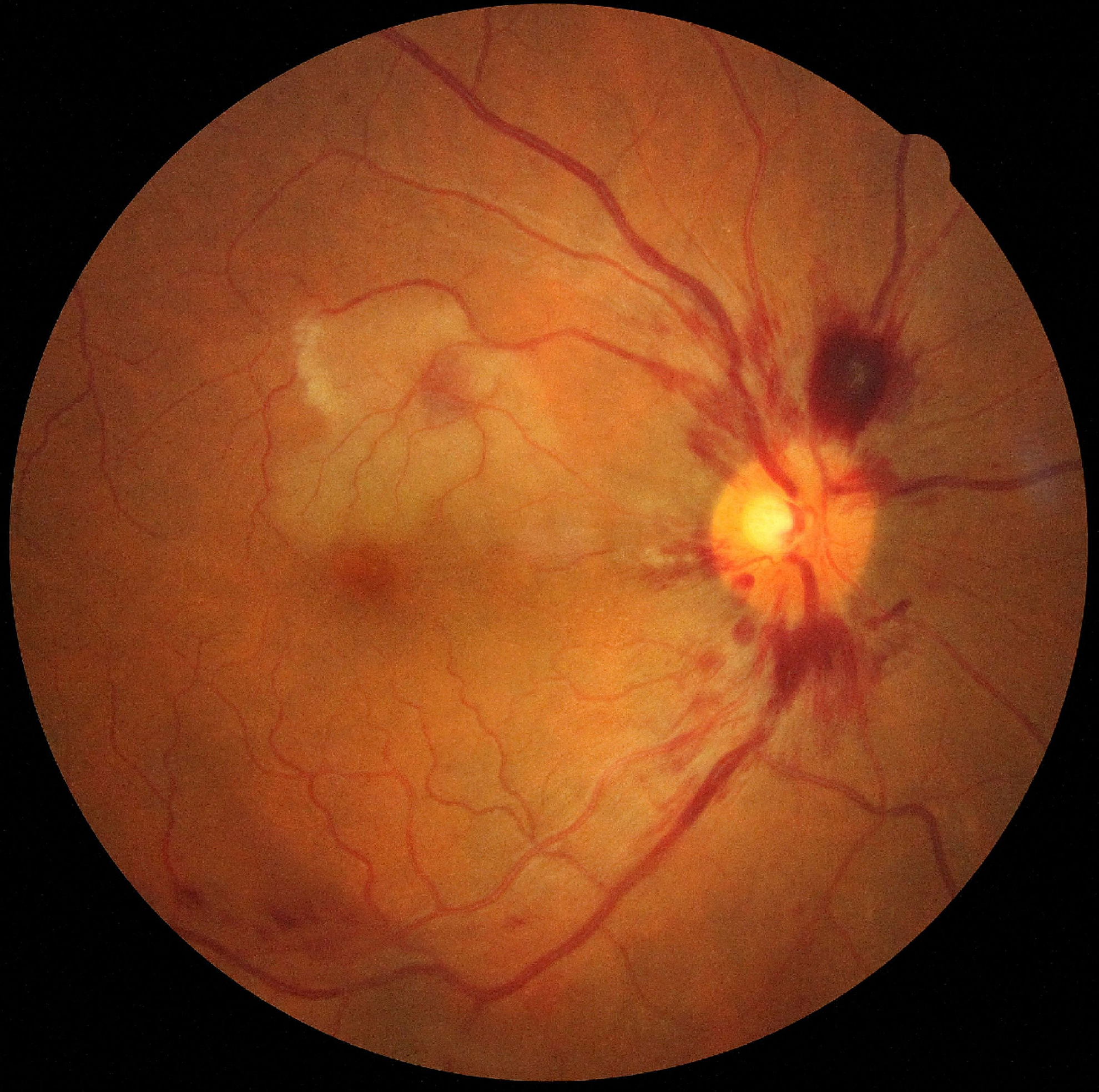
Fundus examination of the right eye revealed whitening of the retina along the distribution of cilioretinal artery, sparing fovea, and flame-shaped hemorrhages, and roth spots with minimally dilated and tortuous retinal veins.

Fluorescein angiography demonstrated hypofluorescence in the perfusion area of cilioretinal artery and delayed venous filling without any signs of capillary nonperfusion.

Humphrey visual field analysis revealed centrocecal scotoma.

Scotomas disappeared immediately after using of the HBO treatment.
Discussion
Coexistance of CRVO and CRAO is a rare disorder. Several hypotheses have been proposed for the simultaneous development of CRVO and CRAO. However, the exact pathogenesis of this combination remains unclear (Bottós et al., 2008; McLeod et al., 2009). The occlusion of the central retinal vein results in backward transmission of increased intraluminal pressure through the retinal capillary bed. This pressure causes transient blockage of the cilioretinal artery and this is the most popular suggested mechanism (Hayreh et al., 2008).
Both CRVO and CRAO are ultimately hypoxic phenomena. The retinal blood flow is mainly related to arterial oxygen (O2) pressure and it was reported that high altitude-related hypoxia causes increased retinal blood flow, dilatation of vessels, and increased venous pressure (Grimm et al., 2012; Maccormick et al., 2012; Müllner-Eidenböck et al., 2000). Although the evidence for hypobaric hypoxia causing thrombosis is weak, atmospherical O2 pressure reduces in high altitude and can precipitate thrombosis due to hypobaric hypoxia, respiratory alkalosis, and its effect on coagulation system. During the acclimatization process at the prolonged stay in high altitude region, systemic compensatory mechanisms begin. Although there is a temporal association with this patient's ascent to moderate altitude and her eye problems, they may be unrelated and this should be suggested—there is no control and evidence within the literature is anecdotal. On the other hand, it is unclear why the main complaint of our patient occurred at the end of her journey to high altitude region. Barthelmes et al. (2011) reported that retinal vasculopathies that were directly related to the degree of hypoxia and time of exposure to high altitude were frequently seen upon descent. It was supposed that as a result of overall degree of hypoxic exposure, reperfusion ischemia upon descent and delayed vasculopathy secondary to hypoxic damage to capillary endothelium might be the possible reasons (Barthelmes et al., 2011). During hypoxia at high altitude, the imbalance of autoregulatory mechanisms that facilitates the releasing of free O2 radicals causing hemodynamic disorder is suggested to be one of the multiple contributing factors for this clinical entity.
As the blockage in CRVO-related CRAO is usually incomplete and transient, the retina retains its functional ability for a longer period of time compared to acute thromboembolic artery occlusions (Weiss et al., 2009). Hence the first step treatment for this clinical entity is conservative management. The eye may well recover without treatment. In most cases, spontaneous resolution may occur in one month and prognosis usually depends on the severity of CRVO (Querques et al., 2008).
In recent studies, HBO was suggested to be safe, easily administered, low-cost and effective treatment method in nonacute, nonarteritic CRAO (Weiss et al., 2009). Furthermore, favorable effects of HBO on CRVO-related retina edema was also reported (Wright et al., 2007). HBO may not only increase oxygenation and perfusion pressure, but also reduces intraocular and episcleral venous pressure which leads to move a thrombus to a more distal site (Weiss et al., 2009; Wright et al., 2007). In addition, the luminal sizes of the retinal veins change with distending pressure as an effect of HBO (Weiss et al., 2009). These changes lead to acceleration of the blood flow in the eye.
In conclusion, CRVO-related CRAO should be regarded as a rare complication of exposure to high altitude, and HBO seems to be the treatment of choice of high altitude related co-occurence of CRVO and CRAO in the late period.
Footnotes
Acknowledgment
We confirm that the patient has given written informed consent to the publication of this original article.
Author Disclosure Statement
None of the authors has any conflicts of interest to disclose.