
Abstract
Abstract
In hypobaric hypoxia (HH) at high altitude, the immune responses are changed probably due to oxidative stress-induced production of free radicals and nonradicals. Vitamin E is an antioxidant and protects the cells from oxidative damage. The present study was carried out to study the antioxidant role of vitamin E on the immune changes induced by oxidative stress in HH at high altitude. Select immune responses (phagocytic activity of white blood cell [WBC], cytotoxic activity of splenic mononuclear cells [MNCs], and delayed type of hypersensitivity [DTH]) and hematological changes (total count and differential count [DC] of WBC) were measured in male rats exposed to intermittent HH (at 5486.4 m in a simulated chamber for 8 hours/d for 6 consecutive days) and in normobaric condition with and without p.o. administration of vitamin E in three different doses (20, 40, and 60 mg/kg body weight). The increase of phagocytic activity of blood WBC, and reduction of cytotoxic activity of splenic MNC and DTH response were observed in rats exposed to HH. After the administration of vitamin E at different doses, the immune changes were blocked in a dose-dependent manner. Exposure to HH also led to the elevation of serum corticosterone (CORT), which was arrested after administration of vitamin E. The results indicate that the immune changes in HH at high altitude are probably mediated by the production of free radicals and nonradicals, and vitamin E can block these immune changes by its reactive oxygen species quenching effects.
Introduction
T
The reduction in oxygen availability in the mitochondrial electron transport chain in hypobaric hypoxia (HH) leads to enhanced production of free radicals and nonradicals, such as superoxide anion (O2•−), hydroxyl (•OH), and hydrogen peroxide (H2O2) etc (Dawson et al., 1993; Duranteau et al., 1998; Schild et al., 2003; Smita et al., 2015; Cobleya et al., 2018; Siques et al., 2018).
Several investigators have reported that immune responses were altered in humans (Chohan et al., 1975; Biselli et al., 1991; Klokker et al., 1993; Quanhui et al., 2002; Klessen et al., 2005) and in animals (sSaiRam et al., 1998; Facco et al., 2005; Ghosh, 2009; Goswami et al., 2012, 2014) exposed to HH at different simulated high altitudes (5000–8000 m). The percentage of CD4 and CD3 T lymphocytes was reduced and the percentage of the natural killer (NK) cell was increased in female volunteers of Italian expedition in Nepal at 5050 m altitude (Biselli et al., 1991). Mirrakhimov et al. (1986) reported that the male subjects exposed to 3600 m height in Eastern Pamir Mountain range showed acute mountain disease along with the deficient T cell-mediated immunity.
The phagocytic activity of peritoneal macrophages and circulating white blood cell (WBC) was increased in HH in rats at 7576 m (SaiRam et al., 1998) and 5486.4 m (Ghosh, 2009). These investigators have also reported that the delayed type of hypersensitivity (DTH) was reduced in HH-exposed rats at same altitude indicating impairment of T cell activity (SaiRam et al., 1998; Ghosh, 2009).
Vitamin E is the most important lipid-soluble antioxidant present in tissues and it is important for normal functioning of the immune cells (Han et al., 2006; Pekmezci, 2011). Park et al. (2003) suggested that the vitamin E played an important role in the reactive oxygen species (ROS) quenching process and thus might protect membranes from ROS-induced damage. Supplementation of vitamin E to male Wistar rats before the exposure to intermittent HH at 5700 m or 6300 m, partially blocked the elevated hemoglobin (Hb) concentration and increased super oxide dismutase (SOD) and catalase (CAT) activity in those conditions (Devi et al., 2007).
The immune changes in HH may be mediated by the direct effect of increased free radicals and nonradicals and/or indirectly by the increased synthesis of prostaglandins (PGs) in different tissues by inducible cyclooxygenase (COX) 2 (Mallat and Chamak, 1994; Aloisi, 2001; Li et al., 2002; Ricciotti and FitzGerald, 2011; Kuo et al., 2013). PGs are potent regulators of immune responses (Terrazas et al., 1999; Walker and Rotondo, 2004; Wehbi and Tasken, 2016), and HH-induced immune changes are prevented by inhibitors of COX (Goswami et al., 2012). Vitamin E as a potent inhibitor of ROS may influence the HH-induced immune changes.
In the present study, the role of vitamin E on free radicals and nonradicals induced changes of some select immune responses (phagocytic activity of blood WBC, cytotoxic activity of splenic mononuclear cells [MNCs] and DTH) and hematological changes (total count [TC] and differential count [DC] of WBC) were measured in male rats exposed to intermittent HH at high altitude (5486.4 m) by administering different doses of vitamin E.
Materials and Methods
Animals
In this study adult (6–8 weeks of age) Charles Foster male albino rats (180–200 g) were used. The animals were fed with standard food pellet and water ad libitum. Animals were housed individually in polypropylene animal cages in standard laboratory condition with the normal temperature, 25°C ± 1°C, and with alternate 12-hour light–12-h dark cycle. The animal studies were performed in accordance with the guidelines for the care to minimize the pain and discomfort of the animals and use of laboratory animals of the Institutional Animal Care and Use Committee (IACUC) and all the animal protocols were approved by the IACUC of the Department of Physiology at the University of Calcutta (IAEC/PROP/TKG1/2010 approved on November 16, 2011).
Experimental design
Animals were randomly divided into eight groups: four groups (in each group six rats were used) of rats in normobaric condition and four groups (in each group six rats were used) of rats in HH. The four groups in normobaric condition were control rats administered with different doses of vitamin E: CVE0 (vehicle only, without vitamin E or 0 mg/kg body weight of Vitamin E), CVE1 (20 mg of vitamin E/kg body weight), CVE2 (40 mg of vitamin E/kg body weight), and CVE3 (60 mg of vitamin E/kg body weight). Similarly four different groups of rats in HH were HHVE0 (without vitamin E, vehicle only), HHVE1 (20 mg of vitamin E/kg body weight), HHVE2 (40 mg of vitamin E/kg body weight), and HHVE3 (60 mg of vitamin E/kg body weight). The immunological (phagocytic activity of blood WBC, leukocyte adhesive inhibition index [LAI], cytotoxic activity of splenic MNC and DTH reaction), serum corticosterone (CORT) concentration, and hematological parameters (TC of WBC and DC of WBC) of the abovementioned groups were measured at the end of the exposure schedule.
Exposure to HH
A metallic decompression chamber was used to expose the rats to simulated HH at 5486.4 m altitude. The rats were exposed to this simulated conditions of HH for 8 hours daily (9:00 am to 5:00 pm) for 6 consecutive days following the method of Ghosh (2009), Goswami et al. (2012), Goswami et al. (2014), and Goswami et al. (2016). A vacuum pump was used to create the hypobaric condition in the chamber while atmospheric air can circulate through the chamber. A mercury manometer was used to monitor the pressure within the chamber. The pressure within the chamber was maintained at 380 ± 3 mmHg by regulating the air flow through the chamber. The chamber was sufficiently lighted during exposure while its temperature was maintained at 25°C ± 1°C. The animals were provided with adequate food and water during exposure. An identical chamber was used to expose the rats to normobaric condition by maintaining the normal atmospheric pressure within the chamber.
Treatment with vitamin E
Vitamin E (LobaChem, India) was dissolved in 70% ethanol–water mixture (v/v) before experiment and it was administered orally through a gastric cannula attached to a 1-mL syringe. The daily dose of vitamin E was divided equally into two parts; one part (half of the total dose, i.e., 10, 20, and 30 mg/kg of vitamin E) was given orally 5–6 minutes before the exposure to HH and the other part (rest half of the total dose, i.e., 10, 20, and 30 mg/kg of vitamin E) was given immediate after the exposure to HH on each day.
Blood collection
Blood (0.5 mL) was collected (between 5.00 and 5.30 pm) at the end of exposure schedule to HH/normobaric condition from the heart of deeply anesthetized rat (Na-thiopentone, 50 mg/kg body weight, i.p.) by a syringe containing 100 μL of Na-citrate (3.8%; Sigma) and it was used to measure the phagocytic activity of blood WBC by fluorescence-activated cell sorting (FACS) analysis. Blood (1.5 mL) was also collected without anticoagulant subsequently and 1 mL of this collected blood was kept for serum collection for the measurement of serum CORT concentration and rest of the blood (0.5 mL) was mixed with anticoagulant ethylenediaminetetraacetic acid (EDTA; Merck) for the measurement of TC of WBC and DC of WBC.
TC of WBC and DC of WBC
Neubauer hemocytometer was used to measure the TC of WBC. After staining the blood film with Leishman stain (Merck) DC of WBC was determined under high power of microscope (WHO, 2000).
Peripheral immune responses
Phagocytic activity of blood WBC by FACS analysis
The method for measuring phagocytic activity of blood WBC has been described earlier (Dutta et al., 2011; Goswami et al., 2012). The blood cells were allowed to engulf fluorescein isothiocyanate (FITC)-tagged bacterial cell membrane, which was prepared by the method of Oben and Foreman (1988) for different durations. The mean fluorescence of FITC-positive cell population in the samples of different time points was obtained from FACS analysis. A regression line was then drawn from mean fluorescence values of samples at different time points using the MINITAB statistical software. The phagocytic index (PI) of the blood WBC was obtained from the slope of the regression line. A negative slope of the regression line was an indicator of greater phagocytic activity, as time-dependent changes in mean fluorescence values indicated the dynamics of the bidirectional component of phagocytosis by the WBC.
LAI
The Leukocyte Adhesive Inhibition (LAI) index of splenic MNCs were measured following the method described earlier (Dutta et al., 2011). The spleens were collected aseptically from deeply anesthetized rats (Na-thiopentone, 50 mg/kg body weight, i.p.) and transferred to a solution containing phosphate-buffered saline (PBS) and 3.8% PBS in a ratio of 10:1 (v/v).Single splenic cell suspension was made from the collected spleens and MNC were separated by Percoll density gradient 1.077. The MNC in the suspension was counted using the Neubauer hemocytometer and then the Neubauer hemocytometer was incubated at 37°C for 30 minutes in a moist environment. After 30 minutes of incubation, the counted field was washed gently with PBS by Pasteur pipette and adhered cells were counted.
The percentage of LAI = (No. of adhered cell after incubation × 100)/(No. of total cell count before incubation). A smear of the isolated MNC suspension was made and was stained with Leishman stain (Merck). The percentage of MNC in the smear was calculated to verify the purity of separation.
Cytotoxicity assay of splenic MNC
The cytotoxicity of splenic MNCs (the effector cells collected from spleen by the procedure described in LAI) against target cells (EAC cells) was tested in an lactate dehydrogenase (LDH) release assay using the LDH-FS Nonradioactive Cytotoxicity Assay Kit (DiaSys Diagnostic Systems GmbH) and has been described earlier by Goswami et al. (2012). The release of LDH from effector/target coculture (C) was measured. The spontaneous release of LDH from the effector cells (E) and target cells (T) was determined. The total target cells' LDH release was also measured (M). All tests were performed in triplicate and the amount of LDH released was calculated according to the following formula:
DTH reaction to bovine serum albumin
DTH reaction to bovine serum albumin (BSA) was measured following the method described by Dutta et al. (2011) and Goswami et al. (2012). The right foot pad of rats were sensitized to BSA by giving a subcutaneous injection of 25 μg of BSA emulsified in 50 μL of Freund's Complete Adjuvant, and 50 μL of sterile saline was injected into the left foot pad (intradermally) of the rats on the day before exposure to HH was started. The thickness of both footpads was measured before injecting BSA or saline using micrometer screw gauge (Model No. SMC 20325; Sterling Manufacturing Co.). After 5 days, the rats were rechallenged with 50 μL of sterile saline containing 38.5 μg of BSA (booster dose) into both footpads. The thickness of both footpads was measured 24 hours after injection of booster dose.
The footpad extension due to inflammation was calculated by taking the difference of footpad thickness in normal condition before injection of first challenge dose of BSA or saline and 24 hours after injection of booster dose.
Serum CORT concentration
Serum CORT concentration was determined by radioimmunoassay using a commercially available kit (125I Rat Corticosterone [COAT-A-COUNT; Siemens Healthcare Diagnostic, Inc.]) and gamma counter. The antisera used for the assay was highly specific for the rat CORT and it had 1.58% crossreactivity with 11-Deoxycorticosterone. The assay sensitivity is ∼5.7 μg/dL and intra–inter assay coefficient variation was <6%. All the experimental samples were run in duplicate.
Statistical analyses
Data are expressed as mean ± standard error of the mean. Two-way analysis of variance (ANOVA) was employed to compare the data of eight groups having four groups with different vitamin E treatment in normobaric condition and in HH followed by LSD post hoc test using the statistical package for social science Software (SPSS software: 20.0.0). p ≤ 0.05 was considered as a significant difference.
Results
TC of WBC and DC of WBC
The TC of WBC and DC of WBC (percentage of neutrophil, lymphocyte, monocyte, eosinophil, and basophil) were not significantly changed in rats exposed to HH (HHVE0) compared with that of normobaric rats (CVE0). The TC of WBC and DC of WBC remained unaltered in vitamin E-administered groups in HH (HHVE1, HHVE2, and HHVE3) compared with normobaric rats with corresponding doses of vitamin E (CVE1, CVE2, and CVE3) and without vitamin E (CVE0) (Table 1).
Vitamin E was administered in control rats in normobaric condition without vitamin E (vehicle only i.e., 0 mg/kg body weight of vitamin E [CVE0]) and with three doses of vitamin E at 20 mg/kg body weight (CVE1), 40 mg/kg body weight (CVE2), and 60 mg/kg body weight (CVE3) and in HH without vitamin E (vehicle only i.e., 0 mg/kg body weight of vitamin E [HHVE0]) and with three doses of vitamin E at 20 mg/kg body weight (HHVE1), 40 mg/kg body weight (HHVE2), and 60 mg/kg body weight (HHVE3). Values are expressed as mean ± SEM (n = 6 in each groups). There is no significant difference noted in any groups.
DC, differential count; HH, hypobaric hypoxia; SEM, standard error of the mean; TC, total count; WBC, white blood cell.
Phagocytic activity of circulating blood granulocytes by FACS analysis
The phagocytic activity of circulating WBC was increased (as indicated by lower PI) in HH-exposed rats without vitamin E (HHVE0) compared with that of normobaric rats without vitamin E (CVE0) [F(7, 40) = 4.388, p < 0.01]. The phagocytic activity was not changed in normobaric rats treated with different doses of vitamin E (CVE1, CVE2, and CVE3) compared with that of CVE0 (normobaric rats without vitamin E). But the HH-induced enhanced phagocytic activity was gradually inhibited with increasing doses of vitamin E in rats exposed to HH and the HH-induced enhanced phagocytic activity returned to normal level at the dose of 40 mg of vitamin E/kg body weight and remained at normal level at the dose of 60 mg of vitamin E/kg body weight (Fig. 1).
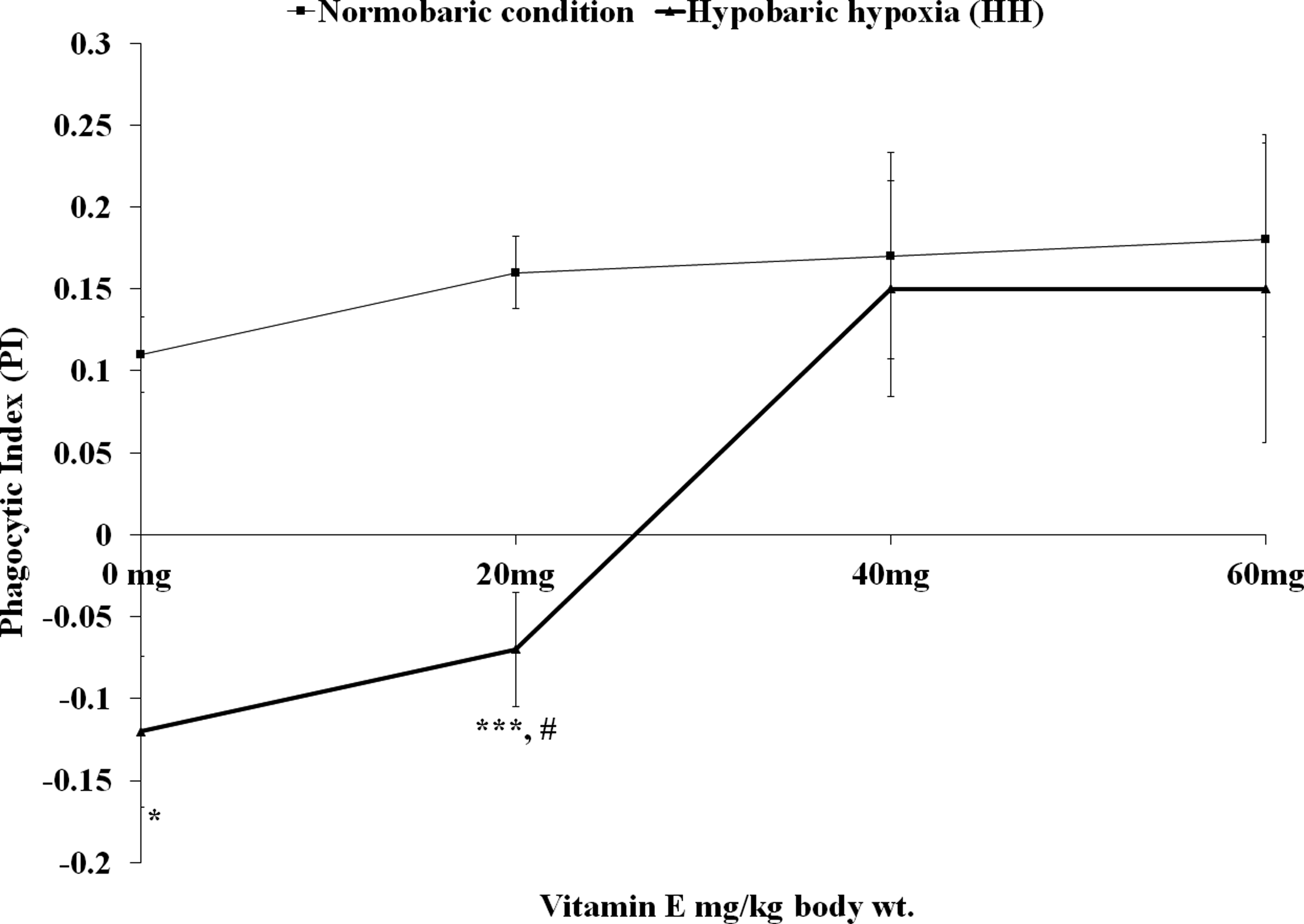
The PI of circulating blood WBC in rats exposed to normobaric condition and HH with three doses (20, 40, and 60 mg/kg body weight) of vitamin E and without vitamin E (vehicle only, i.e., 0 mg/kg body weight of vitamin E). The PI of hypobaric hypoxic (HH) rats (vehicle only, i.e., 0 mg/kg body weight and 20 mg/kg body weight of vitamin E) was significantly low compared with that of corresponding doses in normobaric rats *p ≤ 0.01 and #p ≤ 0.01. The PI was also significantly decreased in HH rats with 20 mg/kg body weight of vitamin E compared with normobaric rats without vitamin E (vehicle only, i.e., 0 mg/kg body weight of vitamin E). ***p ≤ 0.05. Values are expressed in mean ± SEM (n = 6 in each group). HH, hypobaric hypoxia; PI, phagocytic index; SEM, standard error of the mean; WBC, white blood cell.
The PI was significantly low in HHVE1 compared with CVE1 [F(7, 40) = 4.388, p < 0.05] and CVE0 [F(7, 40) = 4.388, p < 0.01], but it was not significantly different from HHVE0. The PI of HHVE2 and HHVE3 were not significantly changed compared with normobaric rats with corresponding doses of vitamin E (CVE2 and CVE3) and without vitamin E (CVE0).
Serum CORT concentration
The serum CORT level was increased in HH-exposed rats without vitamin E (HHVE0) compared with that of normobaric rats without vitamin E (CVE0; [F(7, 40) = 2.512, p < 0.01]). The serum CORT level remained unaltered in normobaric rats with graded doses of vitamin E (i.e., in CVE1, CVE2, and CVE3) compared with that of CVE0 (normobaric rats without vitamin E). The serum CORT level remained higher in HHVE1 compared with that of CVE1 [F(7, 40) = 2.512, p < 0.05] and CVE0 [F(7, 40) = 2.512, p < 0.05], but it was not significantly different from HHVE0 (Fig. 2). The serum CORT level was not changed in HHVE2 and HHVE3 compared with that of the corresponding doses of vitamin E (CVE2 and CVE3) and without vitamin E (CVE0).
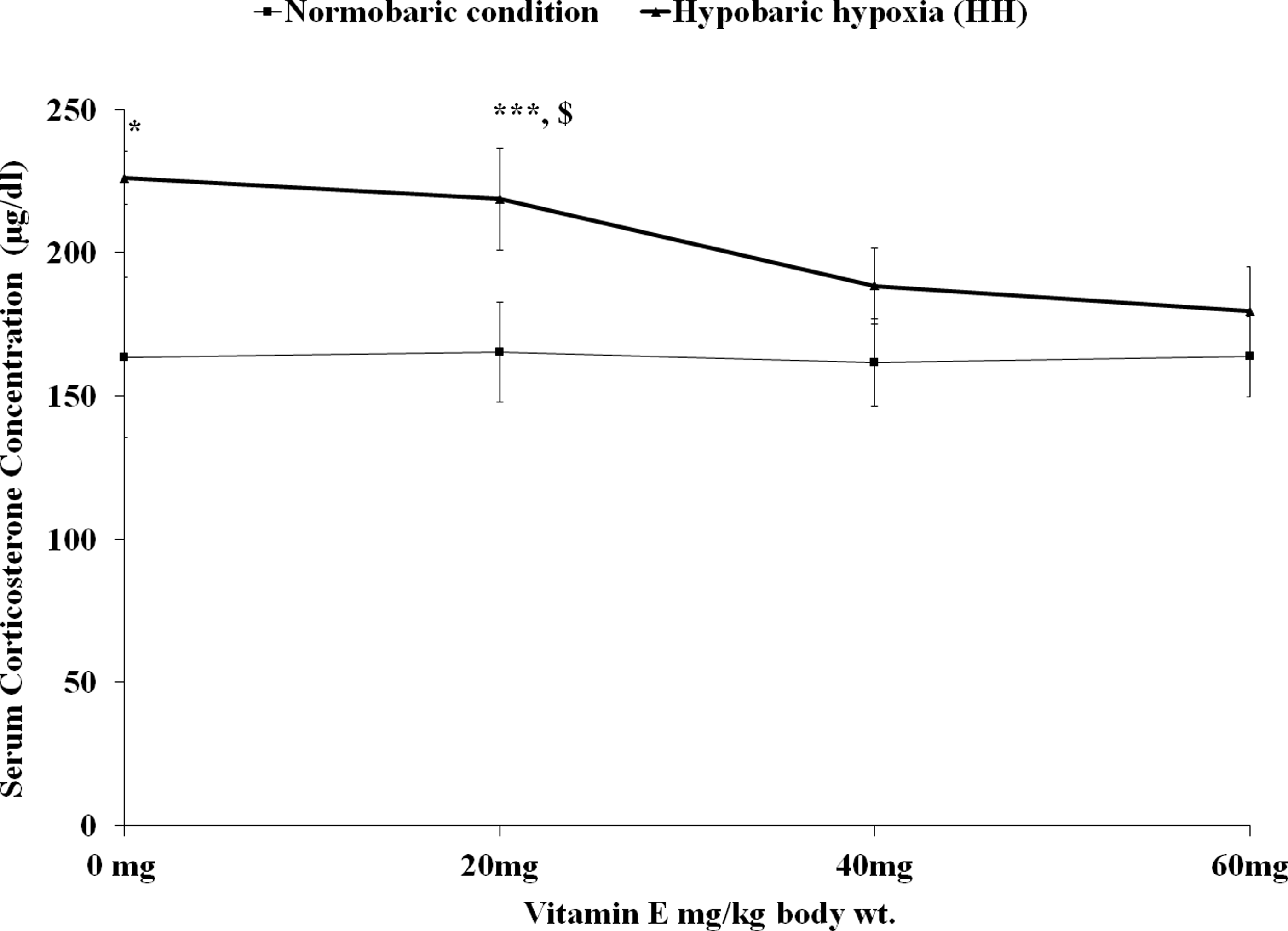
The serum CORT concentration (μg/dL) in rats exposed to normobaric condition and HH with three doses (20, 40, and 60 mg/kg body weight) of vitamin E and without vitamin E (vehicle only, i.e., 0 mg/kg body weight of vitamin E). The serum CORT concentration (μg/dL) of HH rats (vehicle only, i.e., 0 mg/kg body weight and 20 mg/kg body weight of vitamin E) significantly increased compared with that of corresponding doses in normobaric rats, *p ≤ 0.01 and $p ≤ 0.05. The serum CORT concentration was also significantly raised in HH rats with 20 mg/kg body weight of vitamin E compared with normobaric rats without vitamin E (vehicle only, i.e., 0 mg/kg body weight of vitamin E) ***p ≤ 0.05. Values are expressed in mean ± SEM (n = 6 in each group). CORT, corticosterone.
Percentage of LAI
The percentage of LAI remained unaltered in rats exposed to HH (HHVE0) compared with that of rats in normobaric condition (CVE0). The percentage of LAI was also not significantly changed in vitamin E-administered rats exposed to HH (HHVE1, HHVE2, and HHVE3) compared with that of rats in normobaric condition with corresponding doses of vitamin E (CVE1, CVE2, and CVE3) and without vitamin E (CVE0) (Fig. 3).
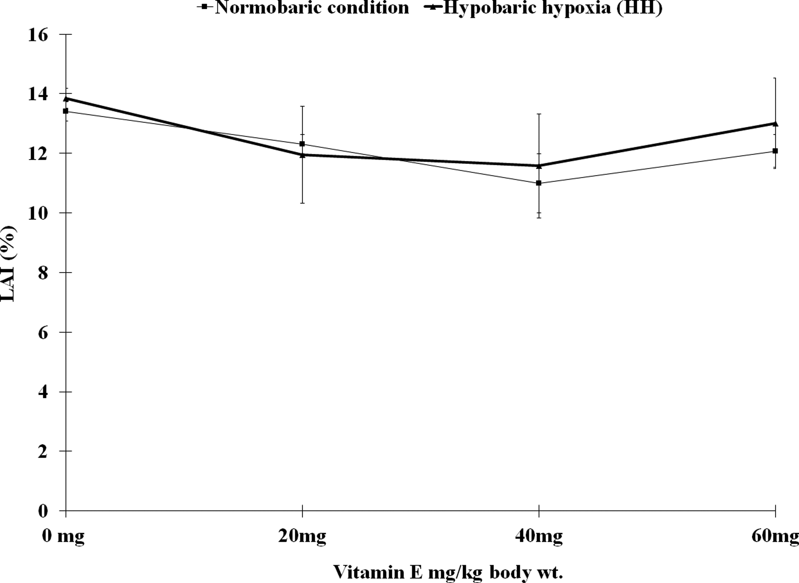
The percentage of LAI index in rats exposed to normobaric condition and HH with three doses (20, 40, and 60 mg/kg body weight) of vitamin E and without vitamin E (vehicle only, i.e., 0 mg/kg body weight of vitamin E). There is no significant difference noted in any groups. The values of LAI are expressed as mean (mean values of three subgroups) ± SEM. (Each subgroup provides pooled values of three animals.) LAI, leukocyte adhesive inhibition.
Cytotoxicity assay of splenic MNC
The cytotoxic activity of splenic MNC was decreased significantly in HH exposed rats without vitamin E (HHVE0) compared to normobaric rats without vitamin E (CVE0; F(7, 16) = 4.500, p < 0.01]). The cytotoxic activity of splenic MNC cells was not changed at any doses in normobaric rats (CVE1, CVE2 and CVE3) compared to normobaric rats without vitamin E (CVE0). But the HH-induced reduction of cytotoxic activity of splenic MNC cells was blocked gradually after administration of increasing doses of vitamin E and the HH-induced reduction of this activity returned to the normal level at the doses of 40 mg of vitamin E/kg body weight. The normal level of MNC cytotoxicity was maintained in rats treated with vitamin E at the dose of 60 mg/kg body weight (Fig. 4).

The cytotoxic activity of splenic MNC in rats exposed to normobaric condition and HH with three doses (20, 40, and 60 mg/kg body weight) of vitamin E and without vitamin E (vehicle only, i.e., 0 mg/kg body weight of vitamin E). The cytotoxic activity of splenic MNC was reduced significantly in HH rats (vehicle only, i.e., 0 mg/kg body weight and 20 mg/kg body weight of vitamin E) compared with that of corresponding doses in normobaric rats *p ≤ 0.01 and #p ≤ 0.01. The cytotoxic activity of splenic MNC was also significantly lowered in HH rats with 20 mg/kg body weight of vitamin E compared with normobaric rats without vitamin E (vehicle only, i.e., 0 mg/kg body weight of vitamin E), *p ≤ 0.01. Values are expressed in mean ± SEM (n = 6 in each group). MNC, mononuclear cell.
The cytotoxic activity of splenic MNC cells remained decreased in HHVE1 (HH-exposed rats with 20 mg vitamin E/Kg body weight) compared with CVE1 [F(7, 16) = 4.500, p < 0.01] and CVE0 [F(7, 16) = 4.500, p < 0.01], but it was not significantly different from HHVE0. The cytotoxic activity of splenic MNC cells was not changed significantly in HHVE2 and HHVE3 compared with CVE2 and CVE3, respectively and without vitamin E (CVE0).
Delayed hypersensitivity reaction to BSA
The hypersensitivity reaction as measured by swelling of right footpad after injection of booster dose of BSA in HHVE0 (HH-exposed rats without vitamin E) was decreased compared with that of normobaric rats without vitamin E (CVE0; [F(7, 40) = 4.160, p < 0.001]). There was no significant change in hypersensitivity reaction of right footpad after injection of booster dose of BSA at any doses in normobaric rats (CVE1, CVE2, and CVE3) compared with that of normobaric rats without vitamin E (CVE0). The hypersensitivity reaction of right footpad in HHVE1 (HH-exposed rats with 20 mg vitamin E/Kg body weight) remained depressed compared with that of CVE1 [F(7, 40) = 4.160, p < 0.05] and CVE0 [F(7, 40) = 4.160, p < 0.01], but it was not significantly different from HHVE0. This HH-induced suppression of swelling of right footpad returned to the normal level in HHVE2 and HHVE3 when compared with CVE2, CVE3, or CVE0 (Fig. 5).
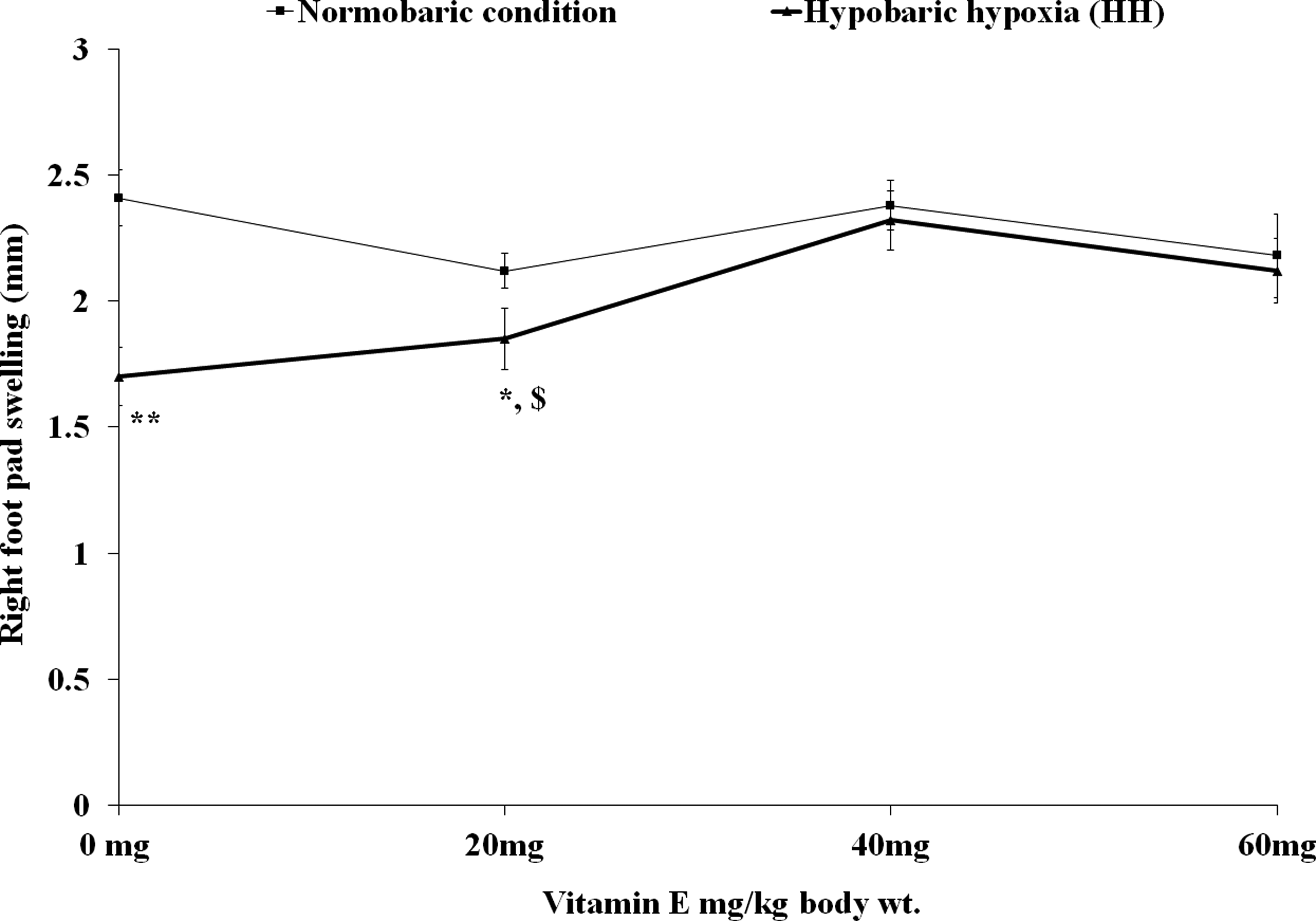
The right footpad swelling in rats exposed to normobaric condition and HH with three doses (20, 40, and 60 mg/kg body weight) of vitamin E and without vitamin E (vehicle only, i.e., 0 mg/kg body weight of vitamin E). The right footpad swelling of HHc rats (0 and 20 mg/kg body weight of vitamin E) are significantly decreased compared with that of corresponding doses in normobaric rats, **p ≤ 0.001 and $p ≤ 0.05, respectively. The right footpad swelling was also significantly decreased in HHc rats with 20 mg/kg body weight of vitamin E compared with normobaric rats without vitamin E (vehicle only, i.e., 0 mg/kg body weight of vitamin E), *p ≤ 0.01. Values are expressed in mean ± SEM (n = 6 in each group).
The DTH reaction in left footpad with saline as challenge dose and followed by BSA as booster dose did not show any significant change among eight groups of rats (data are not shown).
Discussion
The increased phagocytic activity of blood WBC, and reduced cytotoxic activity of splenic MNC and DTH reaction response observed in rats exposed to HH at 5486.4 m simulated altitude of the present study, have been reported previously by others (Ghosh, 2009; Goswami et al., 2012, 2014) in rats in similar experimental condition. The increased phagocytic activity was also observed in reticuloendothelial cells of rats after chronic exposure to a simulated high altitude of 5000 m (Cherdrungsi, 1989) and in peritoneal macrophages of rats exposed to HH at 7576 m (SaiRam et al., 1998).
While enhanced NK cell activity of blood mononuclear cells (BMNCs) was observed in humans exposed to HH at 380 Torr (5800 m) for 20 minutes (Klokker et al., 1993), others reported unaltered BMNC in humans exposed to high altitude at 7620 m (Meehan et al., 1988) and 5050 m (Facco et al., 2005). The reduced cytotoxic activity of splenic MNC in rats of the present study appears to be different from that of the NK cell activity in HH-exposed humans at altitudes of 5800 m (Klokker et al., 1993), 7620 m (Meehan et al., 1988), and 5050 m (Facco et al., 2005). The suppression of DTH type IV observed in the present study has also been reported by SaiRam et al. (1998) in HH at 7576 m.
These immunological changes are probably due to the oxidative stress in high altitude (Edmonds and Blake, 1994). In high altitude, free radicals and nonradicals are generated due to oxygen deficiency (Sarada et al., 2002). The hypoxia is associated with the reduction of oxygen availability and may result in reduced activity of mitochondrial electron transport chain, which can lead to enhanced production of free radicals and nonradicals (Dawson et al., 1993; Duranteau et al., 1998; Schild et al., 2003). The free radicals and nonradicals may act on the membrane of the immune cells and can affect their activity. Even free radicals and nonradicals were generated from the neutrophils of the human subjects exposed to simulated HH (Hitomi et al., 2003).
The T cell activity is suppressed by increased free radicals and nonradicals in HH and may be the underlying cause of depressed DTH reaction in rats (SaiRam et al., 1998). The NK cell activity may also be inhibited by the free radicals and nonradicals, such as superoxide anion and hydrogen peroxide (Mellqvist et al., 2000).
In the present study, vitamin E has been used to decrease the free radicals' level as it is a well-known quencher of these components (Kaminski and Boal, 1992; Park et al., 2003; Moreira et al., 2014; Nimse and Pal, 2015). Vitamin E is the most important lipid-soluble antioxidant present in body tissues that protects membranes from free radical-induced damage and it is important for the normal functions of immune cells (Han et al., 2006; Pekmezci, 2011).
The results showed that the increased phagocytic activity of blood WBC in HH attained normal level after administration of vitamin E at the doses of 40 and 60 mg/kg body weight but not at a dose of 20 mg/kg body weight. The phagocytosis and chemotaxis in peritoneal macrophage were declined in other studies after supplementation of vitamin E in stressful condition (Izgüt-Uysal et al., 2004). Some investigators reported that the vitamin E protects leukocytes from the toxic effects of linden in vitro (Podstawka et al., 1991). However, vitamin E was not able to influence the phagocytic activity in normobaric condition of the present study. In normobaric condition, the free radicals are less due to the absence of any oxidative stress, and as a result, the phagocytic activity was not affected. The presence of vitamin E at this condition is ineffective due to this low level of free radicals.
When the levels of free radicals and nonradicals, such as superoxide anion and hydrogen peroxide, were increased in HH at high altitude, the phagocytic activity was facilitated. Vitamin E as an antioxidant can reduce the production of free radicals in HH and upregulate vitagenes, such as Hsp70 and heme oxygenase 1 (Calabrese et al., 2010). The lower level of the free radicals in the presence of vitamin E at this condition helped the phagocytic activity to attain the status of normobaric condition. Probably the hermetic role of free radicals on phagocytic activity in normobaric and HH at altitude is evident here. Thus, it appears from the present study that the HH-induced increased phagocytic activity of circulating blood WBC has been recovered after administration of vitamin E due to its ability to lower production of free radicals and its quenching effect on these components in that condition.
The decreased cytotoxic activity of splenic MNC in HH of the present study showed recovery after administration of vitamin E at the doses of 40 and 60 mg/kg body weight but not at the dose of 20 mg/kg body weight. The efficacy of vitamin E on cytotoxic activity of splenic MNC in HH has not been studied earlier but another antioxidant vitamin C at lower dose (200 and 400 mg/kg body weight) was able to prevent the HH-induced depression of cytotoxic activity in rats exposed to HH (Goswami et al., 2014). It appears that vitamin E helps to regain the cytotoxic activity of splenic MNC by reduction of free radicals and nonradicals as Mellqvist et al. (2000) reported that the inhibition of the NK cell cytotoxicity was mainly mediated by the production of free radicals and nonradicals, such as superoxide anion and hydrogen peroxide.
The graded effect of vitamin E on the cytotoxic activity of splenic MNC in HH of the present study probably supports the link between free radicals/nonradicals level and cytotoxic activity. In normobaric condition vitamin E did not induce any change of cytotoxic activity of splenic MNC at any dose as the level of free radicals was low. After administration of vitamin E in HH at high altitude, the cytotoxic activity of splenic MNC showed recovery probably due to its lower production and quenching effect on free radicals as mentioned earlier. A biphasic action of free radicals on the cytotoxic activity of splenic MNC in normobaric and HH at high altitude is probably indicated from the results. However, vitamin E administration in mice at the dose of 100 mg/d showed enhancement of NK cell activity of its splenic MNC in vitro with murine YAC-1 as target cells (Ashfaq et al., 2000). In the present study, vitamin E was administered at relatively lower dose in rats and these differences in experimental condition may account for the difference in two studies.
The T cell-mediated DTH type IV reaction, which is reduced in HH showed recovery after administration of vitamin E at the doses of 40 and 60 mg/kg body weight but not at the 20 mg/kg body weight. The DTH reaction type IV depends on the TH1 cells and cytotoxic T cell (CTL), which are inhibited by increased free radicals and nonradicals (SaiRam et al., 1998). As some free radicals and nonradicals generated in HH is responsible for the inhibition of DTH reaction, vitamin E can recover this immune response probably by lowering the free radicals and nonradicals.
The regulators of DTH-like cytokines, IL-2 and IFN-γ (Goldsby et al., 2003), which are produced from TH cell, may be inhibited by the vitamin E due to its free radical- and nonradical-lowering effect. The gradual recovery of the DTH type IV reaction with increasing dose of vitamin E probably indicates that the observed effects are not nonspecific effects of vitamin E. The antioxidant vitamin C, which has the ability to decrease free radicals also showed similar effects on DTH reaction of rats in HH (Goswami et al., 2014). In the present study, the vitamin E did not induce any change of DTH at any dose in normobaric condition, probably because of low free radicals at that condition or the applied dose was low to produce any effect at normobaric conditions.
The higher concentration of serum CORT usually increases the number of neutrophils and decreases lymphocyte count. The results of the present study indicate that DC of WBC was not altered in rats exposed to HH. Probably the increased level of CORT observed in the study is not sufficient to cause the changes of DC. Similarly, the LAI was not inhibited at this level of CORT, although increased level of glucocorticoids inhibits the adhesivity of splenic MNC (Goulding and Roderick, 1993). The decreased cytotoxic activity and DTH in HH may be explained on the basis of elevated level of CORT (Goulding and Roderick, 1993). The phagocytosis of blood WBC is inhibited by increased CORT level but a small increase of CORT can increase it (Goulding and Roderick, 1993). Therefore, the observed immune responses are resultant effects of many factors, such as free radicals and nonradicals, serum CORT, and probably other factors such as proinflammatory cytokines.
The oxidative stress in HH may affect the hypothalamic–pituitary–adrenal (HPA) axis. Previous investigators have reported an increased cortisol level in humans exposed to high altitude (Ramirez et al., 1995; Mazzeo, 2005; Richalet et al., 2010). There are reports that serum CORT level was increased in rats exposed to simulated altitude (Chen et al., 2007; Fan et al., 2009; Baitharu et al., 2011; Goswami et al., 2012, 2014). In the present study, the serum CORT level was also increased in HH, which may be due to the stimulation of CRH-secreting neurons present in the paraventricular nucleus of hypothalamus by the increased PGs and proinflammatory cytokines, such as IL1β, and TNFα in HH (Hartmann et al., 2000). The increased free radicals and nonradicals in HH is probably the stimulator of PGs and proinflammatory cytokines.
After the administration of vitamin E in rats exposed to HH, the serum CORT level returned back to normal. It was found from the results of the present study that vitamin E at the doses of 40 and 60 mg/kg body weight was effective in preventing the increased serum CORT level in rats exposed to HH but vitamin E at dose of 20 mg/kg body weight was not able to inhibit HH-induced increased level of serum CORT. The dose-related effects of vitamin E on serum CORT level in rats exposed to HH probably indicate the gradual elimination of free radicals and nonradicals by increasing concentration of vitamin E.
Another ROS quencher vitamin C (at the dose of 200 and 400 mg/kg body weight) was also effective in decreasing the serum CORT level in rats exposed to simulated altitude but serum CORT was increased with higher dose of vitamin C (600 mg/kg body weight) in HH of that study (Goswami et al., 2014). Vitamin C probably acts as pro-oxidant at a higher dose in the later study, but the vitamin E showed the ROS quencher activity at all of the three doses used in the study. It was also reported that inhibition of PG synthesis by nonspecific COX blocker, naproxen in rats exposed to HH, also resulted in the prevention of the HH-induced increased serum CORT level (Goswami et al., 2012).
The free radicals and nonradicals generated in oxidative stress may initiate a chain of processes that can lead to the alteration of serum CORT and immune responses observed in the present study. These processes may be blocked at different steps to prevent HH-induced changes and the present study indicates that vitamin E as a potent eliminator of free radicals and nonradicals is effective in preventing HH-induced rise of serum CORT and immune responses.
Conclusion
The present study indicates that the HH-induced immune changes are probably mediated by production of excess free radicals and nonradicals in HH at high altitude. The recovery of these immune responses after administration of vitamin E is probably due to the ROS quenching effects of vitamin E.
Footnotes
Acknowledgments
The research work was supported by fund from the Defense Institute of Physiology and Allied Sciences (DIPAS), Delhi, India, Defense Research Development Organization (DRDO), Government of India [Task No. TC/290/TASK-114(TKG)/DIPAS/2006]. The authors also extend their thanks to Dr. Syed N. Kabir, Scientist, Cell Biology & Physiology Division, Indian Institute of Chemical Biology (IICB), Kolkata, India; Dr. Tapan Jyoti Chowdhury, Associate Professor, Rammohan College, Kolkata, India; Dr. Santi Mohan Mondal, Technical Officer, Central Research Facility (CRF), IIT, Kharagpur, India; Mr. Nilotpal Mandal, Research Scholar, Department of Human Physiology with Community Health, Vidyasagar University, Midnapore, Paschim Medinipur, West Bengal, India; and Dr. Goutam Dutta, Department of Physiology, Prabhat Kumar College, Purba Medinipur, West Bengal, India for their technical assistance.
Author Disclosure Statement
No competing financial interests exist.