




Abstract
Abstract
We examined the hypothesis that an expiratory resistance mask containing a small amount of dead space (ER/DS) would reduce the apnea–hypopnea index (AHI) during sleep, attenuate the severity of acute mountain sickness (AMS), and offset decrements in cognitive function compared with a sham mask. In a double-blinded, randomized, sham-controlled, crossover design, 19 volunteers were exposed to two nights of normobaric hypoxia (FIO2 = 0.125), using a ER/DS mask (3.5 mm restrictive expiratory orifice; 125 mL DS volume) and sham mask (zero-flow resistance; 50 mL DS volume). Cognitive function, AMS, and ventilatory acclimatization were assessed before and after the 12-hour normobaric hypoxia exposure. Polysomnography was conducted during sleep. AHI was reduced using the ER/DS sleep mask compared with the sham (30.1 ± 23.9 events·hr−1 vs. 58.9 ± 34.4 events·hr−1, respectively; p = 0.01). Likewise, oxygen desaturation index and headache severity were reduced (both p < 0.05). There were also benefits on limiting the hypoxia-induced reductions in select measures of reaction speed and attention (p < 0.05). Our study indicates that a simple noninvasive and portable ER/DS mask resulted in reductions (49%) in AHI, and reduced headache severity and aspects of cognitive decline. The field applications of this ER/DS mask should be investigated before recommendations can be made to support its benefit for travel to high altitude.
Introduction
Acute mountain sickness (AMS) affects 25% of individuals who venture above 2500 m, with the incidence increasing at higher altitudes (Honigman et al., 1993). Although the exact mechanism(s) are unclear, it is widely postulated that mild swelling in the brain due to cerebral hypoxia plays a significant role in the development of AMS (Schoene, 2008). Several preventative strategies exist for limiting AMS, including gradual ascents and a range of pharmacological remedies (Luks et al., 2014), all of which facilitate improved oxygen uptake and/or delivery. Expiratory resistance (ER), a nonpharmacological intervention, has been investigated with encouraging results, both in improving the level of hypoxemia and in treating and preventing AMS (Launay et al., 2004; Lipman et al., 2015; Nespoulet et al., 2013; Savourey et al., 1998; Schoene et al., 1985). The mechanism(s) by which ER acts to improve blood oxygenation and prevention of AMS, is believed to be attributable to an interplay of a shifting of pulmonary interstitial fluid into the capillaries, an increase in lung volume through preventing expiratory airspace collapse, a decrease in airway resistance, and improvements in ventilation to perfusion ratio (Wayne, 1976). However, with minimal evidence and randomized control trials to support its course of action, it is not yet clear the mechanistic basis of ER on AMS prevention.
Cheyne–Stokes respiration (i.e., periodic breathing) is another common problem universally encountered by those traveling to high-altitude environments. Periodic breathing is characterized by alternating periods of apnea and hyperventilation, and this condition often leads to arousals during sleep, impaired sleep quality, and has also been associated with AMS symptoms in some (Burgess et al., 2004; Eichenberger et al., 1996), but not all studies (Nussbaumer-Ochsner et al., 2012). Rebreathing, or dead-space (DS) masks have been shown to be effective at reducing periodic breathing in conditions of normobaric hypoxia, most likely by reducing the ventilatory overshoot after an apneic event, increasing CO2 reserve and/or increasing cerebral blood flow (Burgess et al., 2018; Lovis et al., 2012; Patz et al., 2013). Although a recent study did not see an improvement in periodic breathing following the use of adaptive servo-ventilation (Orr et al., 2018), whether the addition of ER with DS will further mitigate hypoxemia, AMS, and favorably alter the frequency of apneic episodes and the architecture of sleep remains unknown.
In a randomized and sham-controlled design, the goal of the current study was to evaluate the effectiveness of an ER and DS mask in altering AMS and periodic breathing during sleep. We hypothesized that, when compared with a placebo sham mask, wearing an ER/DS mask during sleep would reduce the apnea–hypopnea index (AHI) and attenuate the severity of AMS upon awakening. The secondary hypothesis was that wearing an ER/DS mask during sleep, if effective, would offset some of the hypoxia-induced decrements in neurocognitive functioning and improve measures of early ventilatory acclimatization.
Methods
Participants
Nineteen healthy young volunteers (n = 10 males and n = 9 females; 25 ± 3 years; 71 ± 12 kg; 174 ± 7 cm; 23 ± 3 kg/m2) were recruited. Exclusion criteria included obesity (body mass index >30 kg/m2), hypertension (systolic >130 mmHg; diastolic >85 mmHg), history of smoking, diabetes, or pregnancy. In addition, participants were excluded if they had any previous history of cardiovascular, neurological, or respiratory diseases. Female participants were tested within 4 ± 2 days, in an attempt to keep test days within a similar phase of their respective menstrual cycles, whereas males were tested within 7 ± 3 days. Aside from oral contraceptives and an intrauterine device, participants were not taking any over-the-counter medications, and all participants abstained from caffeine and/or alcohol consumption for at least 12 hours before experimentation.
Participants had a mean cumulative Epworth Sleepiness Score of 6.5 ± 3.2 (range 2–13), falling under the normal and mild sleepiness range (Johns, 1991). Since there were no subjective reports of sleep apnea, we attribute those with classification of mild sleepiness (4/19) to being university students with exam/internship pressures. Approval of this study was obtained by the Clinical Research Ethics Board at the University of British Columbia (H1-02687), and all procedures conformed to the Declaration of Helsinki. All participants provided written, informed consent before completion of any data collection.
Experimental design
Following baseline questionnaires, assessment of cognitive function and ventilatory acclimatization were performed before entering the hypoxic (fractional inspired oxygen concentration, FIO2 = 0.125) chamber between 6:00 and 7:00 pm. Once in the chamber, participants were free to watch television or work on their laptops for ∼3 hours, after which time they were instrumented for sleep using full polysomnography (Somtè PSG, Compumedics, NC), according to standard format as described in detail elsewhere (Burgess et al., 2013, 2018).
Following the sleep setup, participants were fitted with a sham facemask (zero-flow resistance; 50 mL DS) or a facemask with ER and DS (3.5 mm restrictive orifice; 125 mL DS; Trudell Medical International* Altimate* High-Altitude Sleep Mask, Ontario, Canada; Fig. 1), in a randomized fashion. Following 20–30 minutes of normal rest to allow participants to familiarize with the sleep setup, AMS-LL and headache questionnaires were repeated, the chamber lights were turned off, and the participants were encouraged to sleep. Participants were tested in the hypoxic chamber each night with real-time data acquisition and monitoring. All studies were scored post hoc by an automated scoring software (Version 2.0; Profusion Sleep Software, Compumedics) and verified by the same experienced technologist.
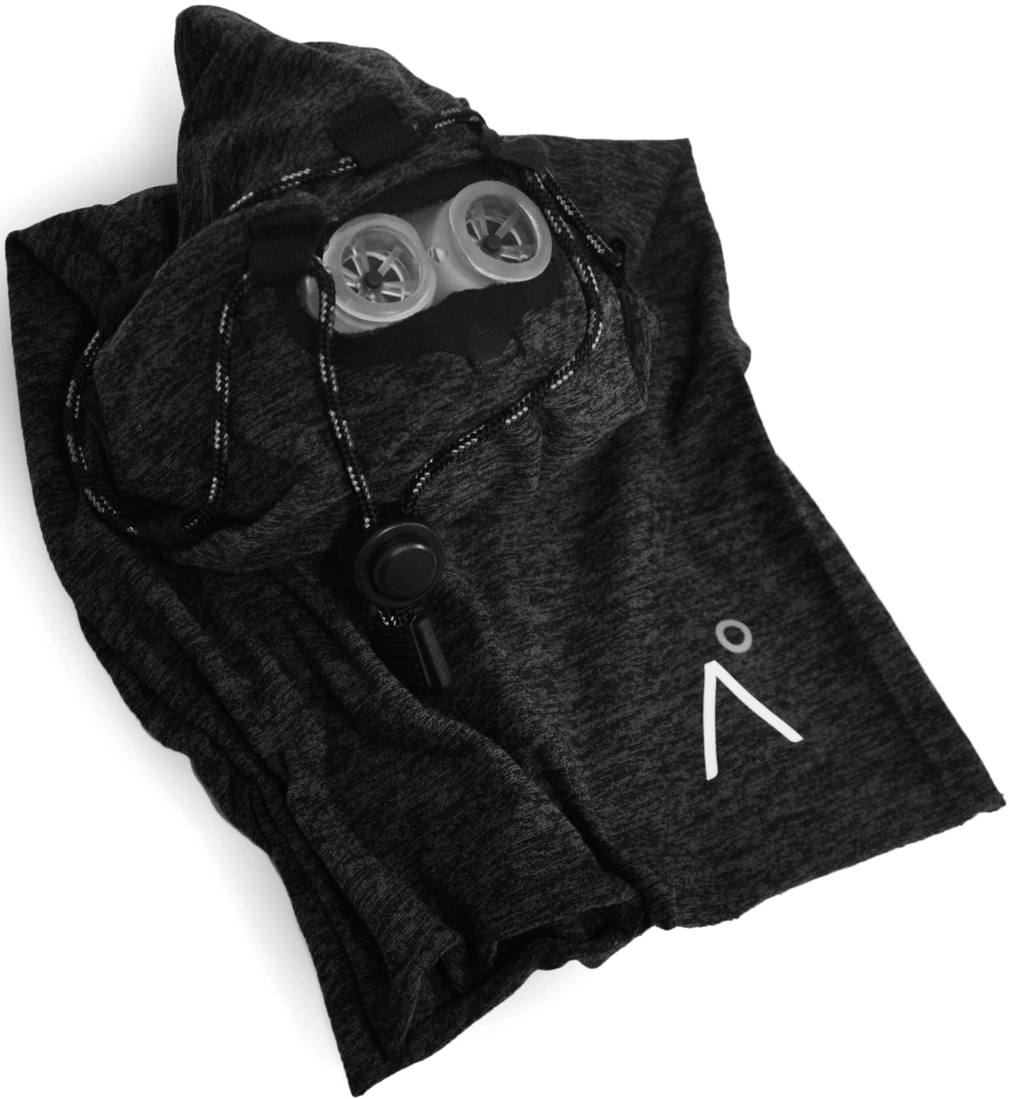
The expiratory resistance (ER; 3.5 mm restrictive expiratory orifice) mask containing a small amount of DS (125 mL DS volume; High-Altitude Sleep Mask, Trudell Medical International, Ontario, Canada) with necktube for securing and creating a seal on the face. The identical-looking sham mask, comprised however of a zero-flow resistance orifice and 50 mL DS. DS, dead space.
Apnea and hypopneas are defined as a reduction in flow for at least 10 seconds, a relative amplitude drop of 20% and 50%, respectively, based on the preceding three to six breaths, and a corresponding ≥3% drop in SpO2. Oxygen desaturation index (ODI) is based on the number of desaturations ≥3% per hour. Six participants were unable to complete the full protocol, and voluntarily removed themselves from the chamber after 3–5 hours of exposure to normobaric hypoxia (due to varying symptoms of altitude illness, anxiety, and/or inability to sleep; n = 2 both nights, n = 4 one night). The latter four participants were equally divided on the first or second night, and consisting of three on the ER/DS mask and one on the sham device.
In the subgroup of the volunteers (n = 12), mouth pressure and VE were measured before sleep and upon awakening while wearing the mask. Mouth pressure was assessed through a portable manometer that was connected directly inside the mask. Although the pressures were slightly higher in the evening when compared with the morning in the ER/DS mask (4.2 ± 1.3 vs. 3.4 ± 1.5 cmH2O; p = 0.05), they were elevated compared with the sham mask (0.2 ± 0.3 vs. 0.1 ± 0.2 cmH2O) in the evening and morning, respectively (p < 0.01). There is no positive inspiratory pressure with the ER/DS mask, only positive expiratory pressure. Altogether, these data indicate that the ER/DS mask functioned as designed to maintain ER when compared with the sham mask. The presleep tests were repeated following 12 hours (i.e., in the morning) of the hypoxic exposure.
Main outcomes and questionnaires
The primary outcome was the difference in AHI between the ER/DS sleep mask compared with the sham. Secondary outcomes included differences in AMS and sleep quality as indexed by: (1) the Lake Louise (LL) questionnaire (Roach et al., 1993); (2) the Environmental Symptoms Questionnaire Cerebral Symptoms (ESQ-C) (Sampson et al., 1983); and (3) a clinically validated headache visual analog scale (VAS; 0–100 mm; Lundqvist et al., 2009). The details of these AMS questionnaires have been provided elsewhere (Bailey et al., 2009). The tertiary outcomes included the assessment of cognition and ventilatory acclimatization.
Cognitive function
Cognitive performance was assessed using Cogstate software (Cogstate Ltd., Melbourne VIC, Australia), which is an automated and standardized battery of cognitive tests performed on a computer (Cole et al., 2013). Three cognitive tests were selected from the battery for this study: (1) detection test (to assess psychomotor performance), (2) identification test (to assess attention), and (3) two-back test (to assess working memory).
Each test required the participant to respond to playing cards that turn over one card at a time; and all tests calculated both reaction time and accuracy performance scores. Higher accuracy scores indicate improved performance; whereas, higher reaction time scores indicate worsened performance. The detection test required the participant to respond as quickly and accurately as possible with “yes” as soon as a new card was turned over. The identification test is a choice reaction time test, with the participant being required to decide whether the playing card presented was red (“yes”) or black (“no”). The two-back test required the participant to respond either “yes” or “no” dependent on whether the playing card was the identical card shown two cards previously. Participants were familiarized to the tests before each experimental session.
Ventilatory acclimatization
Early ventilatory acclimatization to normobaric hypoxic exposure was assessed in two ways: First, ventilatory acclimatization is evident by a progressive increase in minute ventilation (VE) and alveolar ventilation over the first days and/or weeks at altitude. This increase in VE leads to a progressive reduction in arterial carbon dioxide tension (PaCO2) and a partial mitigation of the drop in arterial oxygen tension (PaO2; Rahn and Ortis, 1949). Thus, acclimatized individuals will have a higher PaO2 and lower PaCO2 compared with unacclimatized individuals during or following any given exposure to hypoxia (Hoiland et al., 2018; Rahn and Ortis, 1949). In the current study, resting VE and end-tidal gases (CO2 and O2) were used to index the degree of ventilatory acclimatization following 4 and 12 hours exposure to normobaric hypoxia.
Second, the isocapnic hypoxic ventilatory response (HVR) was also assessed before and following 12 hours in the hypoxic chamber. The HVR can be considered as one of the most important aspects that mediates early ventilatory acclimatization to hypoxia (reviewed in: Hoiland et al., 2018). All respiratory parameters were acquired at 200 Hz using an analog-to-digital converter (PowerLab/16SP ML880; ADInstruments, Inc., CO) interfaced with a computer and analyzed with commercially available software (LabChart; ADInstruments, Inc.).
Throughout all procedures, participants breathed through a mouthpiece (with nose clip) or face mask with an attached bacteriologic filter and a two-way nonrebreathing valve (2600 series; Hans Rudolph, Inc., KS). Expired gas was sampled at the mouth and analyzed for PETO2 and PETCO2 by a calibrated gas analyzer (ML206; ADInstruments, Inc.). Respiratory flow was measured near the mouth using a pneumotachograph (HR 800L; Hans Rudolph, Inc.) and a differential pressure amplifier (ML141; ADInstruments, Inc.). Following 5 minutes of resting breathing, isocapnic hypoxia (PETO2 = 45 mmHg) was maintained for 10 minutes using a dynamic end-tidal forcing system as described elsewhere (Tymko et al., 2015). Estimated arterial oxygen tension (PaO2) was calculated from measured PETO2 values (Tymko et al., 2015) to estimate arterial oxygen saturation (SaO2) (Severinghaus, 1979).
Statistical analyses
Statistical analyses were performed in SPSS (Version 24; IBM Corp.). Data were checked for normality using Shapiro–Wilks tests and the following tests were conducted: Wilcoxon signed-rank test for sleep analysis and linear mixed model for AMS, headache scores, cognitive performance scores, and ventilatory acclimatization. Two levels of repeated measures were applied for both condition (Sham vs. ER/DS) and time (baseline vs. posthypoxia). Following a significant main effect and interaction, a paired samples t-test was employed to make post hoc comparisons within subjects at each level. Between-group comparisons were assessed using paired sample t-tests. The relationship between selected variables was identified using a Pearson product moment correlation. Data are reported as mean ± standard deviation (SD) and a p-value <0.05 was deemed statistically significant.
Results
Sleep (n = 12)
Sleep data from one participant were lost due to technological issues with the PSG recording. Six other participants were excluded due to an absence of sleep (i.e., <30 minutes) and hence an unreliable period for comparison for assessment of the interventions, or the early voluntary removal from the chamber during one or both nights. Therefore, the results of sleep data are based on 12 participants, all of whom obtained full nights (with an overall 462 ± 56 minutes of recording) on both interventional conditions. From these 12 participants, n = 8 were male (80.6 ± 6.9 kg; 178.6 ± 5.8 cm; 25.3 ± 2.5 kg/m2) and n = 4 were female (62.3 ± 4.3 kg; 168.5 ± 6.0 cm; 21.9 ± 0.6 kg/m2).
The AHI was reduced significantly by 49% with the ER/DS mask when compared with the sham (Table 1 and Fig. 2), which was reflected by a reduction in AHI during non-REM sleep. There was also no evidence of obstructive events during sleep. Although the average nocturnal SpO2 was not different between the ER/DS mask versus sham (71.1% vs. 69.8%, respectively; p = 0.45), ODI and the number of desaturations (in all categories) were lower on the ER/DS mask (p = 0.02; Table 1). Although sleep quality (assessed by difficulty sleeping score from AMS-LL score) was similar between ER/DS (1.9 ± 0.9 U) and sham mask (1.9 ± 0.6 U; p = 1.0), those with the ER/DS mask averaged 34 minutes less sleep. This reduction in sleep time, however, was not significant between groups (p = 0.16).

Polysomnographic Recordings of Sleep in Normobaric Hypoxic Chamber
Data are mean ± SD (n = 12; Wilcoxon signed-rank test).
Sleep categorized by polysomnography into nonrapid eye movement (NREM; stages I-III) and rapid eye movement (REM).
p < 0.05.
p < 0.01.
AHI, apnea–hypopnea index; ODI (oxygen desaturation index) based on the number of desaturations ≥3%; AI, arousal index.
AMS-LL and headache scores (n = 19)
As expected, participants had negligible symptoms of AMS-LL or headache before entry into the chamber, which increased upon leaving the chamber (Table 2). The elevation in AMS-LL and headache following sleep in both sham and ER/DS conditions are outlined in Table 2. Although AMS-LL was not different between conditions (p = 0.26), there was a tendency for overall ESQ-C symptom improvement (p = 0.07) and a significant improvement in headache-only VAS scale (p < 0.01; Table 2) when wearing the ER/DS mask compared with sham. Although there was not a relationship between AMS-LL and AHI, there was a tendency between the AMS-LL and ODI (r = 0.40; p = 0.05).
Acute Mountain Sickness and Headache Before and Following 12 Hours of Normobaric Hypoxia
Data are mean ± SD for n = 19 (linear mixed model and paired t-test).
Between condition difference (p < 0.05).
Between time difference (p < 0.01).
AMS, acute mountain sickness; LL, Lake Louise score; ESQ-C, environmental symptoms questionnaire cerebral symptoms score in arbitrary units (AU); VAS, Visual Analog Scale for headache score (0–100); Δ, delta value (post minus pre).
Cognitive function (n = 12)
Due to varying AMS symptoms and subsequent early voluntary removal from the chamber, pre- and posthypoxic exposure cognitive tests were obtained in 12 participants. Of these 12 participants, n = 8 completed their first condition on the ER/DS and n = 4 completed their first condition on the sham mask.
For the absolute changes, reaction time during the psychomotor performance test was higher (i.e., decreased performance) during posthypoxia testing for both the ER/DS and sham masks (main effect: p < 0.01; Fig. 3). Reaction time during this task was lower (i.e., improved performance) for the ER/DS versus sham masks (main effect: p < 0.01; Fig. 3). There was no significant interaction pre- versus posthypoxia exposure for both the ER/DS and sham masks (p = 0.06; Table 3). Reaction time during the attention test was lower (i.e., improved performance) with the ER/DS mask versus sham (main effect: p < 0.01); however, there was no significant interaction pre- versus posthypoxia exposure for both the ER/DS and sham masks (p = 0.47; Table 3).

Percent changes between pre- and posthypoxic exposure of the two of six psychomotor performance tests which showed statistically significant effects, n = 12. Mean and individual data for
Cognitive Performance Scores Pre- and Post-Sham and ER/DS Mask Conditions
Data are mean ± SD for n = 19 (linear mixed model) and n = 12 (paired t-test). *p < 0.05 main effect of condition compared with sham.
p < 0.05 main effect of time across conditions.
p < 0.05 pre- versus postpercent change.
DET-LMN, detection test–reaction time; DET-ACC, detection test–accuracy; IDN-LMN, identification test–reaction time; IDN-ACC, identification test–accuracy; TWOB-LMN, two-back test–reaction time; TWOB-ACC, two-back test–accuracy.
Importantly, there was no significant difference between the pretesting values for the results discussed above (i.e., psychomotor performance test reaction time: p = 0.42; attention test reaction time: p = 0.46)–which is indicative of a fair pretest comparison. There was no significant influence of pre- versus posthypoxia exposure for either the ER/DS or sham masks on psychomotor performance accuracy, attention test accuracy, and working memory reaction time and accuracy (all interaction effects p > 0.05; Table 3).
Relative change scores (i.e., pre- vs. posthypoxia exposure on detection test) for reaction time during the psychomotor performance test were lower (i.e., improved performance) for the ER/DS mask versus sham (0.73% ± 1.13% vs. 1.89% ± 1.74%, respectively; p = 0.02; Fig. 3). Relative change scores for accuracy during the identification test were higher (i.e., improved performance) for the ER/DS mask versus sham (6.10% ± 8.48% vs. 0.47% ± 9.28%, respectively; p = 0.03; Fig. 3). In contrast, there was no significant difference in relative change between pre- versus posthypoxia exposure for either the ER/DS or sham masks for psychomotor performance accuracy, attention test reaction time, and working memory reaction time and accuracy (all p > 0.05). Lastly, although still a meaningful outcome, the abovementioned improvements in cognitive function values likely indicate a preservation in cognition rather than an actual clinical improvement per se.
Ventilatory acclimatization
Resting PETO2 and PETCO2 during normoxia were elevated and reduced, respectively, following normobaric hypoxic exposure (n = 19; main effect p < 0.01; Table 4); but these changes were not different between masks over the repeated time points (interaction effect p > 0.05). Resting eupneic VE was significantly reduced following the hypoxic exposure (main effect p < 0.01), and exaggerated with the ER/DS mask (interaction effect p < 0.01). Due to varying AMS symptoms and subsequent early voluntary removal from the chamber, the HVR tests were obtained in 12 participants both pre- and posthypoxic exposure. There were no significant between-mask differences in the HVR, including SaO2 during HVR (72.2% ± 2.7% and 71.3% ± 1.0% pre- and post-sham mask respectively; 72.4% ± 2.6% and 71.2% ± 2.3% pre- and post-ER/DS mask, respectively; interaction effect p = 0.7).
Resting Ventilation and End-Tidal Gases at Pre-, at Four Hours Exposure, and Post-12 Hours Hypoxia Exposure, and the Hypoxic Ventilatory Response Pre- and Post-12 Hours Hypoxia Exposure for Sham and ER/DS Mask Conditions
Data are mean ± SD (linear mixed model). The preresting condition (n = 19) was under eupneic normoxic breathing conditions without either mask and the 4-hour (n = 19; immediately before lights out) and post (n = 12; after 8 hours of sleep) resting conditions were with the mask during hypoxic exposure. HVR, isocapnic hypoxic ventilatory response test was performed before and immediately upon exiting chamber (n = 19; PETO2 = 50 mmHg). VE, minute ventilation; PETCO2, partial pressure of end-tidal carbon dioxide; PETO2, partial pressure of end-tidal oxygen.
p < 0.05 main effect of condition compared with sham.
p < 0.05 main effect of time compared with pre- across conditions.
p < 0.05 interaction effect compared with pre- within conditions.
Discussion
The main finding of this randomized and sham-controlled study was that a simple noninvasive and portable ER/DS mask resulted in marked reductions (49%) in the AHI during non-REM sleep. Moreover, the findings provide preliminary evidence that a portable ER/DS mask may offset some of the neurological symptoms–especially headache–of early altitude illness and improves selected aspects of cognitive function (reaction time and attention). Moreover, early ventilatory acclimatization to hypoxia was not impacted by use of the ER/DS mask. These main findings, along with relevant methodological considerations, are outlined below in the context of previous research in this area.
Sleep
Following ascent to high altitude by otherwise healthy individuals, periodic breathing during sleep is almost universal, occurring in >90% of people above 4000 m (Ainslie et al., 2013; Burgess et al., 2014). The development of such periodic breathing is the cause of central sleep apnea (CSA). The common trigger of CSA in both heart failure and high-altitude exposure is a transient reduction in PaCO2 (Dempsey, 2005) below the apneic threshold during light non-REM sleep (Dempsey & Skatrud, 1986). The magnitude of the required PaCO2 reduction to initiate CSA depends on the awake values, the ventilatory response to PaCO2 below eupnea, and the position of the isometabolic line (Dempsey, 2005; Dempsey and Skatrud, 1986). The degree of CSA has also been associated with AMS symptoms in some (Burgess et al., 2004; Eichenberger et al., 1996) but not all studies (Nussbaumer-Ochsner et al., 2012). Broadly consistent with these former findings, we observed a tendency for a relationship between the severity of AMS (through the AMS-LL) with ODI (r = 0.40; p = 0.05).
Rebreathing or masks with significant DS (e.g., 500 mL) have been reported to be effective at reducing CSA in conditions of normobaric hypoxia (Lovis et al., 2012; Patz et al., 2013). Although Johnson et al. (2010) employing a variable positive airway pressure device, saw an improvement in CSA at 3800 m, a recent report using adaptive servo-ventilation at the same altitude found no influence on CSA (Orr et al., 2018). Although comparisons between studies is problematic due to the different interventions, it is important to note that none of these aforementioned studies employed a randomized, double-blinded, and sham-controlled design as used in the current study.
The results from the current study, broadly consistent with the majority of the aforementioned studies above (Johnson et al., 2010; Lovis et al., 2012; Patz et al., 2013), reveal that administration of a simple noninvasive and portable ER/DS mask was reflected in marked reductions (49%; p < 0.01) in the AHI. Specifically, AHI was reduced during non-REM sleep when compared with the sham control (56.3 ± 38.9 vs. 31.0 ± 25.3 events·hr−1, respectively). Although the average sleeping SpO2 was not different between the ER/DS vs sham masks, ODI was significantly lower with the ER/DS. Taken together, although more involved field studies are needed first, these findings support the use of this employed ER/DS mask on mediating reductions in AHI and offsetting the number of desaturations during sleep in normobaric hypoxia.
The finding of a 49% reduction in AHI is particularly noteworthy as similar reductions have been reported using well-established pharmacological interventions (e.g., Acetazolamide; reviewed in: Ainslie et al., 2013; Liu et al., 2017). The intriguing question that arises, however, is whether or not periodic breathing at altitude is detrimental or may be a protective/adaptive mechanism [reviewed in (Ainslie et al., 2013)]. For instance, over time at a stable altitude, the incidence of periodic breathing becomes more severe while sleep quality improves, and altitude illness resolves. Moreover, it has been shown that for a given stimulus (either normobaric or hypobaric) mean nocturnal SpO2 can be higher in subjects exhibiting a large amount of apneas compared with those who do not (Hackett et al., 1987; Nespoulet et al., 2012).
The counterargument to the protective/adaptive advantages or disadvantages of periodic breathing at altitude is that Tibetan Sherpa, a population well-adapted for superior performance at high altitude, tend to have little to no period breathing during sleep (Hackett et al., 1980, Lahiri et al., 1983). It should be noted, however, that the current study only examined periodic breathing during a 12-hour hypoxic exposure that was combined with AMS; therefore, extrapolation of these findings in normobaric hypoxia to a more prolonged exposure at high altitude, where periodic breathing is maximized and the mechanisms of acclimatization markedly differ (Ainslie et al., 2013; Burgess et al., 2013; Heinzer et al., 2016), should be done with obvious caution.
Acute mountain sickness
Although symptoms of AMS-LL were not different between conditions (p = 0.26), there was a tendency for symptoms to be improved when assessed using the ESQ-C (p = 0.07), and were significantly reduced when using the clinically validated headache visual analog scale (p < 0.01; Table 2). At least during acute exposure (<12 hours), the main symptoms of AMS are predominantly in the form of headache (Bailey et al., 2009). During exposure to high altitude, the AMS-LL questionnaire is likely more meaningful as it incorporates questions related to gastrointestinal distress, ataxia, edema, etc. Altogether, although future studies are required, these data highlight preliminary evidence of a portable ER/DS to improve some of the headache symptoms of altitude illness.
Since the degree of CSA has also been associated with the development of AMS symptoms (Burgess et al., 2004; Eichenberger et al., 1996), it would seem reasonable that the marked reduction in AHI and ODI in the ER/DS intervention may, at least in part, be responsible for some of the improvements in the recorded headache symptoms.
Cognitive function
Exposure to normobaric and hypobaric hypoxia has been reported to impair reaction time, attention, memory, learning, and executive function (reviewed in: Virués-Ortega et al., 2004). While neuropsychological impairment (e.g., psychomotor speed) has been reported at moderate altitudes (McFarland, 1937; Zhang et al., 2011), this is not a universal finding at different altitudes (e.g., 3000 and 4500 m; Pavlicek et al., 2005). It has been suggested that rate of ascent, exposure time, and severity of altitude may help to explain these inconsistent findings (Caldwell et al., 2018). Irrespective of some of these inconsistencies, oxygen reversal or enrichment at high altitude has been reported to reverse or improve many aspects of neuropsychological impairment (e.g., Gerard et al., 2000; Luks et al., 1998; Moraga et al., 2018).
In the current study, when compared with the sham mask, there were some benefits of the ER/DS mask on selected aspects of cognitive function (i.e., reaction time and attention). Despite a limitation that the cognitive results were not consistent across all cognitive measures (i.e., twice as many participants included in the final analyses started on the ER/DS mask–due to early voluntary removal from the chamber) and the potential that cognitive performance of university/college may be influenced by diurnal variability (e.g., IDN-ACC; Bennett et al., 2008), these preliminary data lend support for further investigation, especially for the clinical and practical relevance.
Although the average sleeping SpO2 was not significantly different between the ER/DS and sham (71.1% vs. 69.8%, respectively), the AHI and ODI during sleep were less on the ER/DS mask. At least at sea level, the intermittent hypoxia associated with obstructive sleep apnea has been associated with cognitive impairment (Davies and Harrington, 2016; Kerner et al., 2017); therefore, it would seem reasonable that the reductions in AHI on the ER/DS mask may reduce the cerebrovascular consequences of intermittent hypoxia associated with CSA in hypoxia and help preserve cognitive function.
Ventilatory acclimatization
The HVR, as an index of peripheral chemoreflex sensitivity, can be considered as one of the most important aspects that mediates early ventilatory acclimatization to hypoxia through a progressive rise in VE that leads to reductions in PaCO2 and a partial mitigation of arterial hypoxemia. Typically, acclimatized individuals will have a higher PaO2 and lower PaCO2 than less acclimatized individuals during or following any given exposure to hypoxia (Hoiland et al., 2018). In the current study, resting PETO2 and PETCO2 during normoxia were respectively elevated and reduced following normobaric hypoxic exposure, these changes (including HVR) were not different between masks. Therefore, it seems that despite the ER/DS mask having a marked influence on the AHI, ODI, and resting VE, it did not impact early ventilatory acclimatization to hypoxia. This is perhaps not surprising, since the average SpO2 during sleep or SaO2 was not different between the ER/DS mask and sham.
Methodological considerations
Although a strength of the study was the employment of a randomized, double-blinded and sham-controlled design, a relevant consideration is that the amount of DS differed slightly between the masks (50 mL vs. 125 mL). In an early study by Patz et al., it was reported that out of the five volunteers (from the 16 tested) who developed moderate CSA (AHI >20 events·hr−1) at a simulated altitude of 3658 m, >500 mL (up to 2.1 L) of DS was required to nearly abolish the central apneas (Patz et al., 2013). This finding is broadly consistent with a study by Lovis and colleagues who reported that 500 mL of dead space only had an influence on reducing AHI (70 ± 26 vs. 29 ± 7 events·hr−1) in those (n = 5) with a high AHI (>30 events·hr−1) at 3500 m versus those with a low or absent AHI (n = 7; Lovis et al., 2012).
When participants were pooled together, however, there was no influence of the dead space (Lovis et al., 2012). Therefore, it would seem unlikely that a difference of 75 mL would fully explain the impact of the ER/DS mask, hence suggesting that the restrictive expiratory orifice may be mediating much of the influence on reducing AHI. The mechanism(s) by which a small amount of ER acts to stabilize breathing requires further study. However, since the ER resulted in marked reductions in ventilation (Table 4) but comparable end-tidal gases compared with the sham mask, one potential influence of ER would be to stabilize breathing (and hence AHI) through changes in loop gain and the interplay between controller gain (i.e., chemosensitivity) and plant gain (i.e., ΔPaCO2/VE; Ainslie et al., 2013; Dempsey et al., 2010). An improvement in plant gain, at least in someone without airway obstruction, likely comes from improved V/Q matching.
Although not experimentally established in the current ER/DS mask, the ER when compared with the sham device likely mediates an increase in end-expiratory lung volume hence promoting alveolar recruitment and potentially improved V/Q relationships. Another possibility is that the reduction in ventilation might be caused by an increased work of breathing through the ER/DS mask. A final methodological consideration was that six out of the 19 participants were excluded due to an absence of sleep (i.e., <30 minutes) or early voluntary removal from the hypoxic chamber, were due to symptoms of AMS. Although there was no obvious treatment effect in these six participants, the inclusion of an additional night where no mask was worn would help provide valuable information for considering the efficacy of this tool for altitude safety.
In conclusion, while noting the importance that these findings should be viewed as preliminary, further research incorporating an ER/DS mask under field conditions are clearly warranted where periodic breathing, AMS, and impairment in cognitive function are likely to occur. Although the chamber results are encouraging, further testing in real-world environments is required to ascertain the benefit of the ER/DS mask for clinical and recreational use.
Understanding these events has important practical implications for both military and recreational personnel upon rapid ascent to high altitude. The use of an ER/DS approach could potentially be used in conjunction with pharmacological interventions or as an alternative when such interventions are not possible or warranted (e.g., acute/rapid exposure, sulfa allergies, etc.). Moreover, employing the ER/DS mask at high altitude when CSA/periodic breathing is typically higher, especially over time, when compared with normobaric hypoxia (Ainslie et al., 2013; Burgess et al., 2013; Heinzer et al., 2016) will provide important insight into the efficacy and consequences of treating periodic breathing. Understanding these mechanisms may have important clinical implications in the context of CSA/Cheyne/Stokes Respiration accompanying congestive heart failure, as well as other etiologies. Until the necessary long-duration field studies have been completed, this mask should not be used as users sole prophylactic strategy against AMS, or to add safety to altitude travel.
Footnotes
Acknowledgment
The authors would like to thank the participants of the study.
Author Disclosure Statement
The study was sponsored by Trudell Medical International*, the company manufacturing and selling the Altimate* High-Altitude Sleep Mask. The authors have no competing financial interests.