
Abstract

Approximately three-quarters of Japan's land area is mountainous, and mountains are familiar to the Japanese population. Mt. Fuji is an important cultural symbol of Japan. “The One Hundred Mountains” is known as a masterpiece (Fukada, 1964), and mountaineering is a popular hobby, with ∼6.5 million climbers.
More than 20 mountain clinics are set up during the mountaineering season, where doctors who enjoy mountaineering provide medical services on a voluntary basis. In general, medical equipment is not available in mountain clinics, and doctors can only provide medication and first aid, such as suturing injuries and external fixation. There are significant limitations to diagnosis owing to the lack of medical equipment. Diagnostic imaging equipment cannot be installed because of the difficulty in transporting large equipment, lack of a stable and sufficient power supply, and difficulty in maintaining the medical equipment.
Some of these problems can be solved with the advancement of medical equipment, and the diagnostic abilities of mountain clinics can be improved. Although there have been some reports on the use of ultrasonography in mountain clinics (Canepa and Harris, 2019), no reports on the availability of compact and lightweight X-ray units in high altitude has been reported. In this study, we report our experience of using a compact and lightweight X-ray unit in a mountain clinic.
Taroudaira hut is located in Toyama Prefecture, 2,330 m above sea level, on the Yakushi-dake (Mt. Yakushi) route, one of the 100 mountains in Japan (Fig. 1). It has 150 sleeping quarters, a dining room, and a mountain clinic. The portable radiography machine weighs ∼3.5 kg, is rechargeable, and has sufficient power saving for wilderness use (Fig. 2). It is used in combination with a flat-panel detector and laptop computer to display the image. A total of six patients were included for X-ray imaging at the lodge clinic. The imaging sites were the chest, abdomen, shoulder joint, wrist, knee, lower leg, clavicle, and lumbar spine. The photographs taken show that the image quality is sufficient to aid in diagnosis (Fig. 3). No abnormal findings were observed in the lumbar spine, and an abdominal ultrasonogram was performed using a portable ultrasound device to diagnose hydronephrosis.

Taroudaira hut.
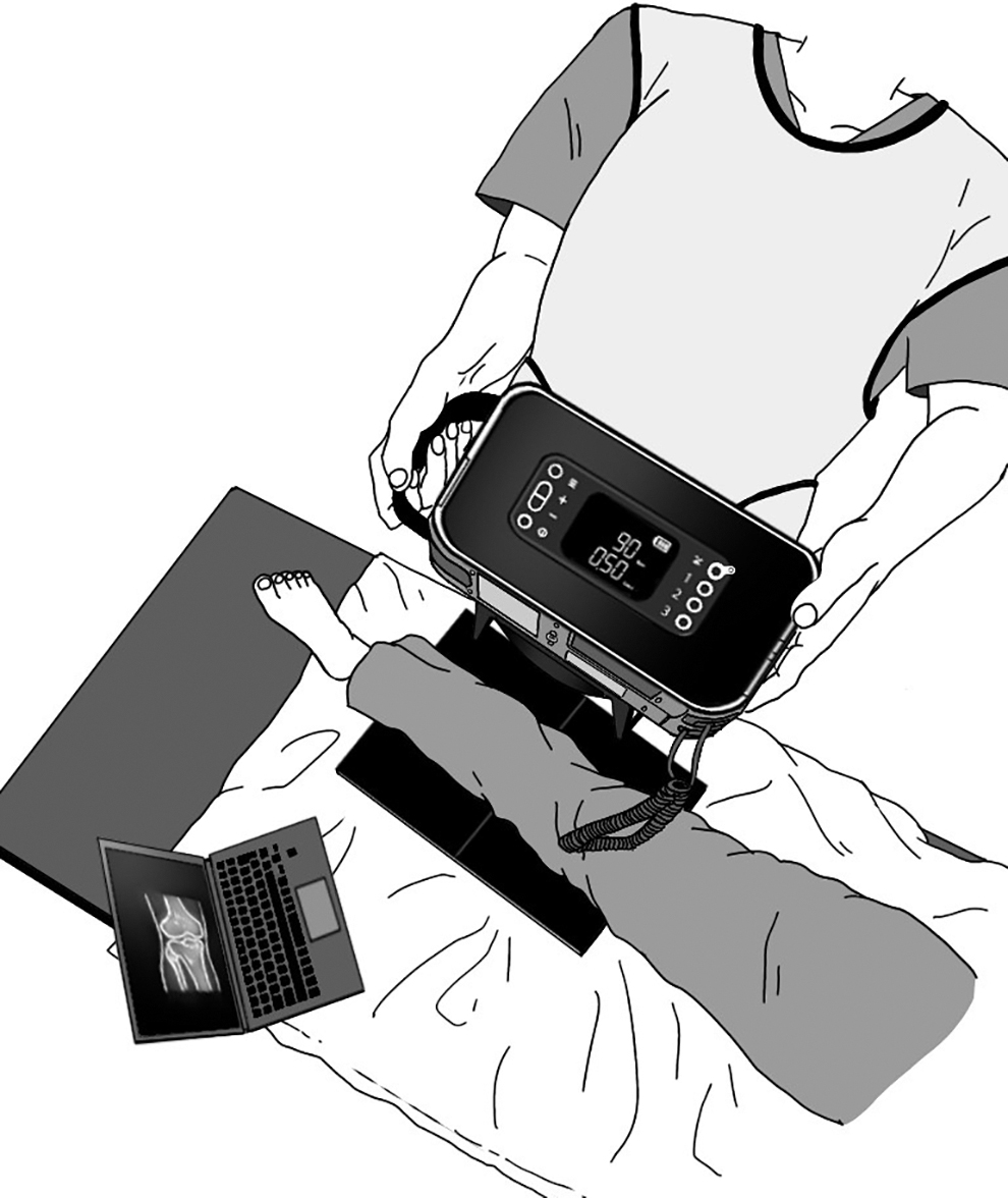
Setting for the compact and lightweight X-ray unit.
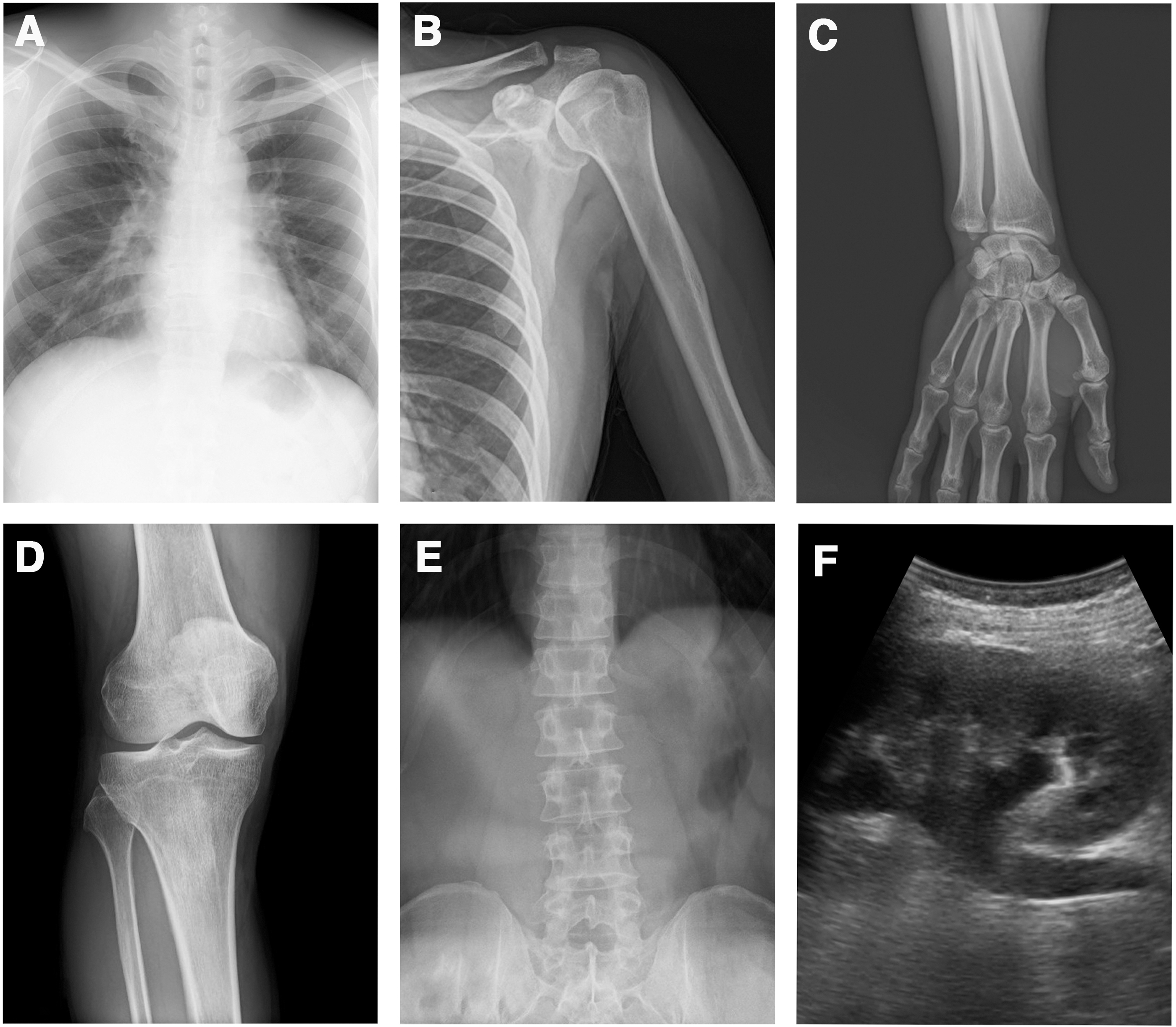
Some of the photographs taken at the mountain clinic using a lightweight portable X-ray system (CALNEO Xair, FUJIFIM Corporation, Tokyo, Japan).
X-rays are particularly beneficial in the diagnosis of fractures, dislocations, foreign bodies, and lung diseases, including high-altitude diseases. For example, trauma accounted for 14% of the patients seen at the Everest Clinic, and respiratory diseases were the most common (Nemethy et al., 2015). In addition, the number of middle-aged and elderly climbers is increasing, and they have the potential to develop diseases unrelated to mountaineering. The use of ultracompact radiography equipment and portable echoes may improve the diagnostic abilities of mountain clinics and influence appropriate decisions regarding rescue and descent.
Footnotes
Authors' Contributions
This study was conceptualized by R.K., T.M., and Y.I. R.K. provided medical care at the mountain clinic and collected data. K.H. provided technical support on a portable X-ray unit. Y.I. provided project management. S.Y. supervised this study. R.K. and Y.I. wrote the article. All authors read and approved the final article.
Author Disclosure Statement
A compact and lightweight X-ray system (CALNEO Xair) and ultrasound system (iViz air) were provided free of charge by FUJIFILM Corporation to conduct this study. The author (Y.I.) also received consulting fees from FUJIFILM Corporation.
Funding Information
No funding was received for this article.