
Abstract

O
Chest stiffness is defined as a chest that cannot be compressed or expanded with ventilations. Therefore, during CPR, no sufficient flow and oxygen can be provided to the arrested patient. Objectifying a stiff chest can be difficult out of hospital. Weather conditions (e.g., light reflection, night, snow, and wind), the patient's garment, chest anatomy, a previous chest surgery, and partial stiffness because of a freezing environment may influence the evaluation of chest stiffness. In a patient who is not resuscitated because the chest seemed stiff and noncompressible, survival may be impaired. Thus, correct evaluation of orthograde blood flow during chest compressions is crucial in situations when the chest may be considered too stiff for sufficient CPR.
The 2021 European Resuscitation Council guidelines for special circumstances of CA propose using ultrasound (US) to assess the presence or absence of vital signs (Lott and Truhlar, 2021). Nowadays in developed countries, US is available in several emergency medical services, and its implementation is rapidly advancing. Basic US skills are easy to learn, and they are often sufficient for a basic diagnostic assessment. Point-of-care ultrasound (POCUS) is a noninvasive tool, which may be used during CPR (Soar et al, 2021). Evaluation of blood flow with US can be performed in both common carotid arteries. These structures are usually easily identifiable on the neck. Using POCUS can help in deciding whether compressions on a seemingly stiff chest are effective.
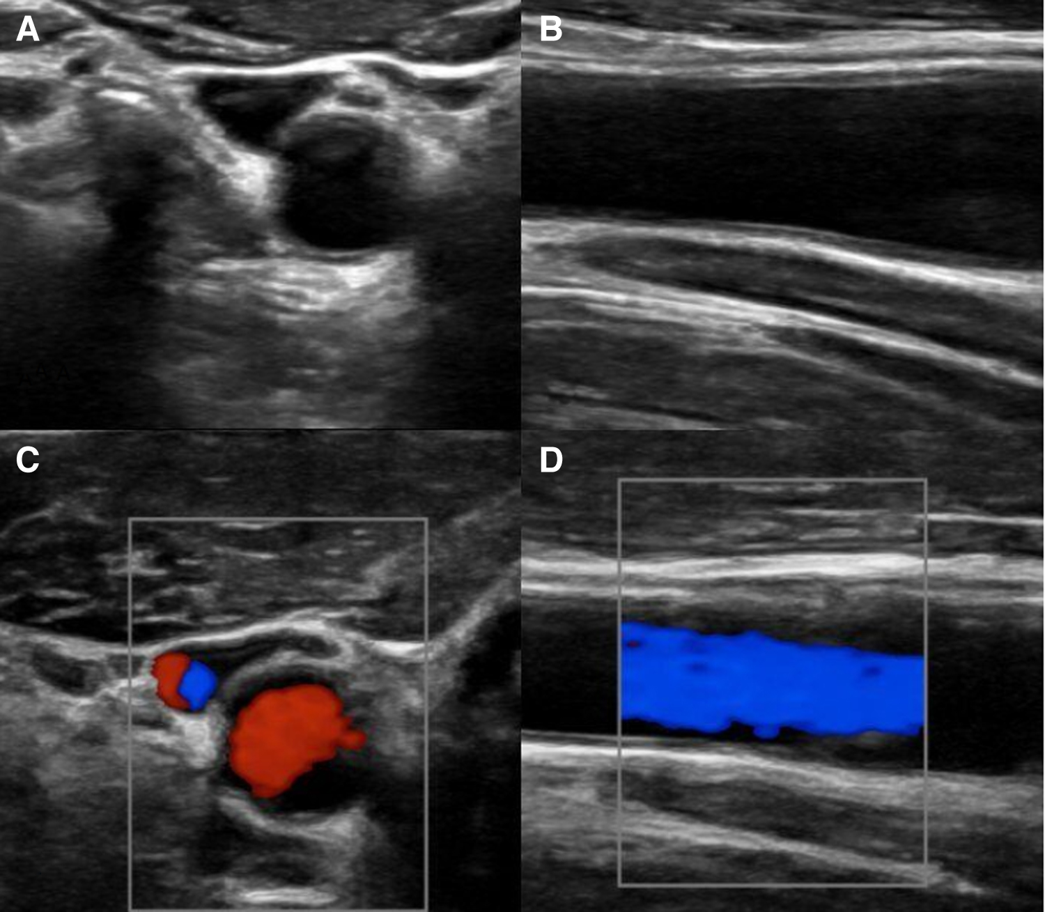
When applying a linear US probe to the neck in a cross-sectional or longitudinal plane (Fig. 1) during chest compressions, a color Doppler or pulsed wave signal can provide helpful information on whether there is orthograde flow to the brain. Color Doppler shows information in terms of flow presence or absence, whereas the pulsed wave signal can help in distinguishing between flow type, direction, and velocity. Changes in the diameter and flow dynamics in the common carotid artery also offer precious details on the quality of CPR (Koch et al, 2022). Ultrasound imaging may be limited because of body movements during CPR, a hypovolemic collapsed carotid artery, major neck trauma, and a cervical collar.
In conclusion, in hypothermic CA patients with a potentially stiff chest, using POCUS, orthograde blood flow in the common carotid arteries in synchrony with chest compressions can be quickly and reliably assessed. The blood flow is indicative of the effectiveness of chest compressions and can help in the decision-making regarding continuation or termination of CPR in case of a patient with a potentially stiff chest.
Footnotes
Authors' Contributions
All authors made substantial contributions, read, and approved the final article.
Author Disclosure Statement
No competing financial interests exist.
Funding Information
No research grants were used to support this work.