
Abstract

A Jumpstart for Ailing Mitochondria
page: 897

In this issue, Keeney and colleagues describe a novel technology to restore bioenergetic function to cells through mitochondrial gene transfer. The technique is named ProtoFection, which stands for protein-mediated transfection, and is in development by Gencia Corporation. Mitochondrial defects can lead to a wide array of diseases that affect the energy metabolism of the cell. High-energy, postmitotic tissues such as muscle, retina, and the brain are particularly sensitive to mitochondrial defects. Mutations or deletions in mitochondrial DNA (mtDNA) can lead to inborn defects; however, mtDNA mutations have also been implicated in aging and neurodegeneration. The importance of these somatic mtDNA mutations in aging or disease remains uncertain but findings in the substantia nigra of a cohort of individuals with Parkinson disease (PD) indicated an increased level of deleted mtDNA with different clonally expanded deletions in cells.
To obtain mitochondrial transduction, the authors engineered a chimeric recombinant protein that combines a mitochondrial transcription factor (TFAM), an N-terminal protein transduction domain, and a mitochondrial localization signal to constitute the ProtoFection reagent. In proof-of-concept experiments, a PD-derived cybrid (i.e., a eukaryotic cell line originating from a fusion between a cytoplast and a whole cell) line was treated with either the reagent alone or admixed with human mtDNA. Both conditions led to increased mitochondrial gene content and gene expression; yet only supplementation with mtDNA (i.e., ProtoFection and DNA) was able to restore the respiration rate of this cell line to normal. (lv)
Gutless Adeno and the Art of Genome Maintenance
page: 883
Several of the commonly used methods of gene transfer lead to persistent expression in vivo from episomal DNA. Three components of this phenomenon have fascinated researchers for decades. First, how and in which conformations is the genome maintained? Second, which of the various conformations serves as a template for efficient transcription that leads to maintained transgene expression? And finally, is replication of the genome necessary in order for it to persist?
In this issue,
Tweaking Tumors with MicroRNAs
page: 831
Many molecular pathways are affected in cancer, including both canonical protein-coding genes as well as noncoding genes. MicroRNAs (miRNAs) are a class of noncoding small RNAs (17 to 27 nucleotides in length) that control gene expression by regulating mRNA translation via partial Watson–Crick interactions with complementary sequences within the 3′ untranslated regions of targeted transcripts. An estimated one-third of protein-coding human mRNAs are regulated by miRNAs. An aberrant miRNA expression profile is a hallmark of several diseases, including cancer. Previous studies have shown that changes in the abundance of a single miRNA can affect the expression levels of hundreds of different proteins and push cells into a transformed state. Two new reports now demonstrate that using gene therapy-based approaches to modulate microRNA activity may be of therapeutic value for the treatment of cancers.
In the study by
Torrisani and colleagues
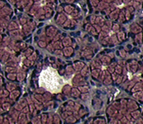
, which appears in this issue (see page 831), the authors confirm previous results showing that let-7 miRNA expression is decreased in pancreatic ductal adenocarcinoma (PDAC) cells of patients compared with normal pancreatic acini adjacent to the tumor. Given that dysregulation of let-7 expression affects mitotic signaling, cell cycling, angiogenesis, and cell adhesion in other models of cancer, the authors hypothesized that restoring let-7 miRNA levels would alter tumor progression in PDAC as well. In vitro, the authors found that transducing PDAC-derived cells with let-7-expressing lentivirus leads to the inhibition of cell proliferation, K-ras expression, and mitogen-activated protein kinase activation. In vivo, however, subcutaneous injection of let-7-expressing lentivirus restored let-7 miRNA expression levels but did not halt tumor growth in a mouse model of PDAC. As to why the treatment did not work in vivo,
The study by
According to