




Abstract
We reported the induction of tumor-selective iodide uptake and therapeutic efficacy of 131I in a hepatocellular carcinoma (HCC) xenograft mouse model, using novel polyplexes based on linear polyethylenimine (LPEI), shielded by polyethylene glycol (PEG), and coupled with the epidermal growth factor receptor-specific peptide GE11 (LPEI-PEG-GE11). The aim of the current study in the same HCC model was to evaluate the potential of biodegradable nanoparticle vectors based on pseudodendritic oligoamines (G2-HD-OEI) for systemic sodium iodide symporter (NIS) gene delivery and to compare efficiency and tumor specificity with LPEI-PEG-GE11. Transfection of HCC cells with NIS cDNA, using G2-HD-OEI, resulted in a 44-fold increase in iodide uptake in vitro as compared with a 22-fold increase using LPEI-PEG-GE11. After intravenous application of G2-HD-OEI/NIS HCC tumors accumulated 6–11% ID/g 123I (percentage of the injected dose per gram tumor tissue) with an effective half-life of 10 hr (tumor-absorbed dose, 281 mGy/MBq) as measured by 123I scintigraphic gamma camera or single-photon emission computed tomography computed tomography (SPECT CT) imaging, as compared with 6.5–9% ID/g with an effective half-life of only 6 hr (tumor-absorbed dose, 47 mGy/MBq) for LPEI-PEG-GE11. After only two cycles of G2-HD-OEI/NIS/131I application, a significant delay in tumor growth was observed with markedly improved survival. A similar degree of therapeutic efficacy had been observed after four cycles of LPEI-PEG-GE11/131I. These results clearly demonstrate that biodegradable nanoparticles based on OEI-grafted oligoamines show increased efficiency for systemic NIS gene transfer in an HCC model with similar tumor selectivity as compared with LPEI-PEG-GE11, and therefore represent a promising strategy for NIS-mediated radioiodine therapy of HCC.
Introduction
To investigate an innovative, alternative therapeutic approach, in an earlier study we examined the feasibility of 131I therapy of HCC after stable transfection with the sodium iodide symporter (NIS), using a mouse α-fetoprotein (AFP) promoter construct to target NIS expression to HCC cells (Willhauck et al., 2008b). NIS is a transmembrane glycoprotein that mediates the uptake of iodide into thyroid follicular cells (Dai et al., 1996; Smanik et al., 1996). The presence of NIS at the basolateral membrane of thyroid follicular cells has been exploited for many years for diagnostic imaging purposes as well as for ablative therapy of differentiated thyroid cancer using radioactive iodide (131I). This noninvasive therapy has proven to be a safe and effective treatment for thyroid cancer, even in advanced metastatic disease (Van Nostrand and Wartofsky, 2007). To extend the use of NIS-mediated radioiodine therapy to other types of cancer, we have proven the feasibility of extrathyroidal radioiodine therapy after induction of iodide uptake by ex vivo NIS transfection or local adenoviral in vivo NIS gene transfer using tissue-specific promoters, such as the prostate-specific antigen (PSA) promoter, the carcinoembryonic antigen (CEA) promoter, and the calcitonin promoter, to specifically target NIS expression to prostate, colon, and medullary thyroid cancer, respectively (Spitzweg et al., 1999, 2000, 2001, 2007; Cengic et al., 2005; Scholz et al., 2005; Willhauck et al., 2007, 2008a). In the liver cancer model we applied the AFP promoter for HCC-specific delivery of the NIS gene and demonstrated tumor-specific iodide uptake activity that allowed a therapeutic effect of 131I in stable NIS-expressing HCC xenografts and after local adenoviral NIS gene transfer (Willhauck et al., 2008b; Klutz et al., 2011b).
After proof of principle of the NIS gene therapy concept in liver cancer in our studies outlined above and by other investigators after retroviral or local/regional adenoviral NIS gene delivery (Faivre et al., 2004; Chen et al., 2006; Herve et al., 2008), the next crucial step toward clinical application must be the evaluation of gene delivery vehicles that allow tumor-selective transgene expression in the presence of a sufficiently high transduction efficiency after systemic application to be able to reach disseminated tumor manifestations. In the evaluation of systemic application of gene delivery vectors, the dual function of NIS as therapy and reporter gene provides the advantage of detailed characterization and direct monitoring of in vivo vector biodistribution as well as localization, level, and duration of transgene expression, which have been recognized as critical elements in the design of clinical gene therapy trials (Spitzweg and Morris, 2002; Dingli et al., 2003; Baril et al., 2010). Several research groups, including our own, have demonstrated the potential of NIS as reporter gene in various applications, showing that in vivo imaging of radioiodine accumulation by 123I or technetium-99m (99mTc) scintigraphy as well as 123I single-photon emission computed tomography computed tomography (SPECT CT) fusion or 124I positron emission tomography (PET) imaging correlates well with the results of ex vivo gamma counter measurements as well as NIS mRNA and protein analysis (Spitzweg et al., 1999, 2000, 2001, 2007; Dingli et al., 2003; Groot-Wassink et al., 2004; Dwyer et al., 2005; Blechacz et al., 2006; Carlson et al., 2006, 2009; Goel et al., 2007; Merron et al., 2007; Willhauck et al., 2007, 2008a–c; Klutz et al., 2009; Baril et al., 2010; Li et al., 2010; Penheiter et al., 2010; Trujillo et al., 2010; Watanabe et al., 2010).
With the aim of systemic delivery of the NIS gene we have reported a nonviral NIS gene delivery approach in the HCC xenograft model, in which linear polyethylenimine (LPEI) was shielded by attachment of polyethylene glycol (PEG) and coupled with the synthetic epidermal growth factor receptor (EGFR)-specific peptide GE11 for actively targeting the NIS gene to EGFR-overexpressing HCC cells (Klutz et al., 2011a). We have demonstrated high transduction efficiency of LPEI-PEG-GE11/NIS resulting in significant EGFR-specific iodide accumulation in vitro and in vivo, which was high enough for a significant delay of tumor growth associated with prolonged survival after three or four cycles of polyplex application followed by 131I therapy (Klutz et al., 2011a).
Specifically for systemic delivery of therapeutic genes we have further developed biodegradable synthetic vectors based on low molecular weight polycations cross-linked either via ester or disulfide bonds (Kloeckner et al., 2006; Russ et al., 2008a,b). In a syngeneic neuroblastoma mouse model we have demonstrated the high potential of synthetic biodegradable polymeric vectors based on pseudodendritic oligoethylenimine (OEI)-grafted polypropylenimine dendrimers (G2-HD-OEI) for tumor-specific delivery of the NIS gene. After intravenous application of NIS-conjugated polyplexes NIS-mediated radioiodine accumulation was restricted mainly to the tumor and sufficiently high for a significant delay of tumor growth associated with improved survival (Klutz et al., 2009). The syngeneic Neuro2A mouse model develops exceptionally well-vascularized tumors and is an ideal animal model to demonstrate the effect of passive tumor targeting using macromolecular drug carriers. Because of the imperfect and leaky tumor vasculature combined with inadequate lymphatic drainage, gene carriers can efficiently accumulate in the tumor tissue (Zintchenko et al., 2009). Before pursuing application of G2-HD-OEI in a clinical setting it is therefore essential to confirm their potential for systemic gene delivery in clinically more relevant human xenograft models. Hence, in the current study we have applied the concept of systemic NIS gene delivery with biodegradable OEI-grafted polypropylenimine dendrimers in a human HCC xenograft mouse model and compared transduction efficiency, tumor specificity, and therapeutic efficacy with the results obtained with LPEI-PEG-GE11 (Klutz et al., 2011a). On the basis of its dual function as reporter and therapy gene, NIS was used for noninvasive imaging of vector biodistribution by 123I scintigraphy and 123I SPECT-CT imaging followed by assessment of the therapy response after application of 131I.
Materials and Methods
Cell culture
The human hepatoma cell line (HuH7) (0403; Japanese Collection of Research Bioresources [JCRB] Cell Bank, Tokyo, Japan) was cultured in Dulbecco's modified Eagle's medium (DMEM)–F12 medium (Invitrogen, Karlsruhe, Germany) supplemented with 10% fetal bovine serum (v/v) (PAA, Cölbe, Germany), 5%
Plasmids and polymers
The expression vector CMV-NIS-pcDNA3 (pCMV-NIS) carrying the full-length NIS cDNA coupled to the cytomegalovirus (CMV) promoter was kindly provided by S.M. Jhiang (Ohio State University, Columbus, OH). As a control, NIS cDNA was removed with EcoRI and religated into the same expression vector in the antisense direction (pCMV-antisense-NIS).
G2-HD-OEI was synthesized as described previously and used as a 5-mg/ml stock solution (Russ et al., 2008b). LPEI-PEG-GE11 was synthesized as described previously (Klutz et al., 2011a).
Polyplex formation
Plasmid DNA was condensed with polymers at the indicated conjugate-to-plasmid (c/p) ratios (w/w) in HEPES-buffered glucose (HBG: 20 mM HEPES, 5% glucose [w/v]; pH 7.4) and incubated at room temperature for 20 min prior to use described previously. Final DNA concentrations of polyplexes for in vitro studies were 4 or 2 μg/ml; for in vivo studies it was 200 μg/ml (Russ et al., 2008b; Klutz et al., 2011a).
Transient transfection
For in vitro transfection experiments, HuH7 cells were grown to 60–80% confluency. Cells were incubated for 4 hr with polyplexes in the absence of serum and antibiotics followed by incubation with growth medium for 24 hr. Transfection efficiency was evaluated by measurement of iodide uptake activity as described below.
125I uptake assay
After transfections, iodide uptake of HuH7 cells was determined under steady state conditions as described previously (Spitzweg et al., 1999). Results were normalized to cell survival measured by cell viability assay (see below) and expressed as counts per minute (cpm) divided by the absorbance at 490 nm (A 490 nm).
Cell viability assay
Cell viability was measured with a commercially available MTS [3-(4,5-dimethylthiazol-2-yl)-5-(3-carboxymethoxyphenyl)-2-(4-sulfophenyl)-2H-tetrazolium, inner salt] assay (Promega, Mannheim, Germany) according to the manufacturer's recommendations as described previously (Willhauck et al., 2007).
Establishment of HuH7 xenografts
HuH7 xenografts were established in female CD-1 nu/nu mice (Charles River, Sulzfeld, Germany) by subcutaneous injection of 5×10⁶ HuH7 cells suspended in 100 μl of phosphate-buffered saline (PBS) into the flank region. Animals were maintained under specific pathogen-free conditions with access to mouse chow and water ad libitum. The experimental protocol was approved by the regional governmental commission for animals (Regierung von Oberbayern).
NIS gene transfer and radioiodine biodistribution studies in vivo
Experiments started when tumors had reached a tumor size of 8–10 mm and after a 10-day pretreatment with
SPECT CT imaging
For SPECT CT imaging the same groups of mice were prepared as outlined above, and 24 hr after polyplex application mice were injected intraperitoneally with 50 MBq (1.35 mCi) of 123I, followed by monitoring of radioiodine biodistribution by serial imaging (1, 3, and 5 hr after 123I application) on a NanoSPECT/CT (Mediso, Budapest, Hungary). CT scans were taken covering the same field of view (FOV) as the SPECT scans. Total scan time was between 30 and 40 min, with 48 projections and a scan time of 20 sec per projection in the case of the SPECT scans.
The SPECT component of the NanoSPECT/CT uses four detector heads, each comprising a thallium-activated sodium iodide [NaI(Tl)] crystal with a size of 262 mm×255 mm×6.35 mm. It gives an axial field of view of 20 mm and, with the medium-resolution aperture/collimator (nine pinholes per head) that was used in this study, has a spatial resolution of about 1.2 mm. The CT component employs a continuously operating miniature microfocus X-ray tube with a maximal anode current of less than 0.2 mA. The detector has an active area of 98.6×49.2 mm and consists of 1024×2048 pixels. It provides an axial field of view of 45 mm and a maximal spatial resolution of 48 μm. The color scale was adjusted so that the reference tissue had the same appearance in both images and the uptake in the tumors could be easily compared visually.
Analysis of radioiodine biodistribution ex vivo
For ex vivo biodistribution studies, mice were injected with G2-HD-OEI/NIS (n=24) or G2-HD-OEI/antisense-NIS (n=9) as described above followed by intraperitoneal injection of 18.5 MBq of 123I 24 hr later. A subgroup of NIS-transduced mice (n=9) was treated with sodium perchlorate before 123I administration as an additional control. Two, 6, and 12 hr after 123I injection, mice were killed and tumors as well as organs of interest were dissected and weighed, and radioiodine uptake was measured with a gamma counter (five NIS-transduced animals per time point [G2-HD-OEI/NIS] and three mice of each control). Results are reported as the percentage of injected dose per organ (% ID/organ).
Analysis of NIS mRNA expression using quantitative real-time PCR
Total RNA was isolated from HuH7 tumors or other tissues, using an RNeasy mini kit (Qiagen, Hilden, Germany) according to the manufacturer's recommendations, and quantitative real-time PCR (qPCR) was performed as described previously (Klutz et al., 2009).
Immunohistochemical analysis of NIS protein expression
Immunohistochemical staining of frozen tissue sections derived from HuH7 tumors after systemic NIS gene delivery was performed with a mouse monoclonal antibody directed against amino acid residues 468–643 of human NIS (kindly provided by J.C. Morris, Mayo Clinic, Rochester, MN) as described previously (Spitzweg et al., 2007).
Radioiodine therapy study in vivo
After a 10-day
Indirect immunofluorescence assay
Indirect immunofluorescence staining was performed on frozen tissues with an antibody against human Ki67 (Abcam, Cambridge, UK) and an antibody against mouse CD31 (BD Biosciences, Heidelberg, Germany) as described previously (Willhauck et al., 2007).
Statistical methods
All in vitro experiments were carried out in triplicate. Results are represented as mean±SD of triplicates. Statistical significance was tested by Student t test.
Results
Iodide uptake studies in vitro
Transfection conditions using G2-HD-OEI/NIS were optimized in HuH7 cells by measurement of perchlorate-sensitive iodide uptake activity 24 hr after polyplex application. We found that the optimal conjugate-to-plasmid (c/p) ratio was 2, which resulted in the highest transfection efficiency at low cytotoxicity (Fig. 1A). This ratio was used in all subsequent experiments. Twenty-four hours after transfection with G2-HD-OEI/NIS, HuH7 cells showed a 44-fold increase in 125I accumulation as compared with cells preincubated with the competitive NIS inhibitor perchlorate (Fig. 1B). Furthermore, no perchlorate-sensitive iodide uptake above background levels was observed in cells transfected with the control vector G2-HD-OEI/antisense-NIS (Fig. 1B). In comparison, the optimal c/p ratio for LPEI-PEG-GE11/NIS was 0.8, which resulted in a 22-fold increase in radioiodide accumulation 24 hr after transfection with LPEI-PEG-GE11/NIS (Fig. 1A) as compared with cells treated with perchlorate. Results of iodide uptake were normalized to cell survival measured by MTS assay, which was not altered after polyplex-mediated NIS gene transfer (Fig. 1A and B).

In vivo radioiodine biodistribution studies
To investigate the iodide uptake activity in HuH7 xenografts after systemic in vivo NIS gene transfer, 123I distribution was monitored by gamma camera imaging in tumor-bearing mice 24 hr after G2-HD-OEI/DNA administration. Whereas no radioiodine accumulation was detected in tumors after application of G2-HD-OEI/antisense-NIS (Fig. 2C), significant radioiodine uptake was observed in 80% (12 of 15) of HuH7 tumors after systemic injection of G2-HD-OEI/NIS (Fig. 2A), in addition to physiological radioiodine accumulation in thyroid, stomach, and bladder (Fig. 2A and C). As determined by serial scanning, 6–11% ID/g 123I (percentage of the injected dose per gram of tumor tissue) was accumulated in NIS-transduced xenograft tumors with a biological half-life of 11 hr. Considering a tumor mass of 1 g and an effective half-life of 10 hr for 131I, a tumor-absorbed dose of 281 mGy/MBq of 131I was calculated (Fig. 2D). To confirm that tumoral radioiodine uptake was indeed NIS mediated, a subset of G2-HD-OEI/NIS-injected mice (n=9) received sodium perchlorate. In all experiments a single injection of sodium perchlorate completely blocked tumoral radioiodine accumulation in addition to abolishing physiological iodide uptake in stomach and thyroid gland (Fig. 2B). Moreover, no significant radioiodine uptake was observed in nontarget organs, including lungs, liver, kidney, or spleen, which confirms the tumor specificity of nanoparticle-mediated NIS gene delivery. In comparison, after LPEI-PEG-GE11-mediated NIS gene transfer HuH7 xenografts accumulated slightly less radioiodine (6.5–9% ID/g 123I) with a shorter effective half-life of 6 hr, which resulted in a significantly lower tumor-absorbed dose of 47 mGy/MBq of 131I (Klutz et al., 2011a).
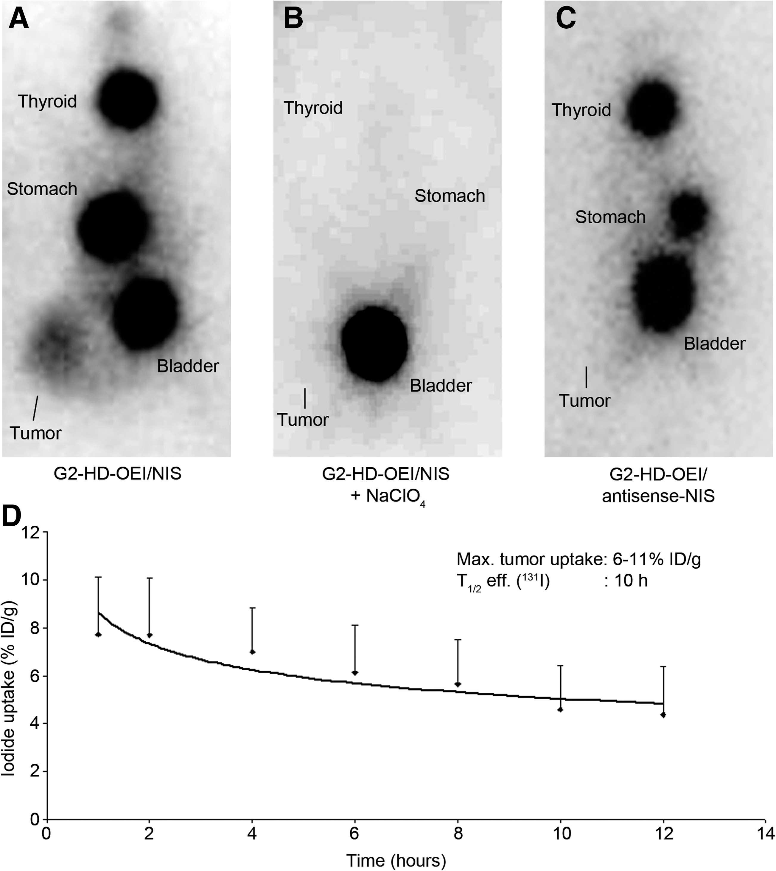
123I scans of nude mice harboring HuH7 tumors 4 hr after intraperitoneal injection of 18.5 MBq of 123I after G2-HD-OEI-mediated NIS gene delivery
In a subset of mice, radioiodine biodistribution was also monitored by 123I SPECT CT imaging after intraperitoneal injection of 50 MBq of 123I (Fig. 3). Tumor-selective iodide accumulation was confirmed after systemic G2-HD-OEI/NIS application (Fig. 3A) whereas no significant radioiodine accumulation was detected in tumors after application of G2-HD-OEI/antisense-NIS (Fig. 3B). Moreover, SPECT CT imaging allowed a more detailed three-dimensional analysis of tumoral iodide accumulation, revealing inhomogeneous iodide accumulation appearing as clusters of iodide uptake throughout the tumor. A maximal tumoral iodide uptake of approximately 7% ID/g was measured in mice after systemic application of G2-HD-OEI/NIS, as compared with the control groups injected with the control polyplexes G2-HD-OEI/antisense-NIS (2.5% ID/g) or after application of sodium perchlorate (3.2% ID/g) (Fig. 3C).
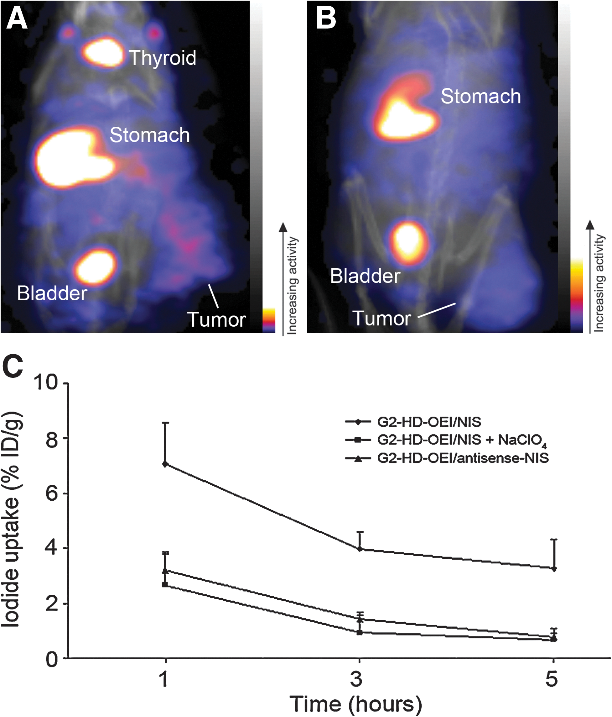
123I SPECT CT scan of tumor-bearing nude mice 5 hr after intraperitoneal injection of 50 MBq of 123I after G2-HD-OEI-mediated NIS gene delivery
In addition to tumoral iodide uptake, significant radioiodine accumulation was observed in tissues physiologically expressing NIS, including stomach, bladder, and thyroid.
Ex vivo radioiodine biodistribution studies
Ex vivo biodistribution analysis confirmed significant iodide uptake in tumors after systemic NIS gene transfer using G2-HD-OEI/NIS (Fig. 4A–C). While NIS-transduced HuH7 tumors accumulated approximately 3.5% ID/organ 123I even 12 hr after radioiodine injection, mock-transduced tumors showed no significant radioiodine uptake. In comparison, after LPEI-PEG-GE11-mediated NIS gene transfer, 12 hr after radioiodine application only 1.5% ID/organ 123I remained in the tumor (Klutz et al., 2011a). It is noteworthy that the average tumor weight in these experiments was approximately 0.9 g. In both groups, thyroid gland and stomach accumulated approximately 41 and 38% 123I ID/organ, resulting from endogenous expression of NIS in thyroid and stomach. It is important to point out that, because of the exquisite regulation of thyroidal NIS expression by thyroid-stimulating hormone (TSH), 123I accumulation in the thyroid gland can effectively be downregulated by thyroid hormone treatment as shown in humans (Wapnir et al., 2004). In addition, iodide accumulation in the stomach is mostly a result of pooling of gastric juices, which is more prominent in mouse experiments than is usually seen in humans because of anesthesia for a prolonged period during imaging procedures (Spitzweg and Morris, 2002; Klutz et al., 2009). Administration of perchlorate in mice injected with G2-HD-OEI-NIS significantly blocked iodide uptake in tumors and in physiologically NIS-expressing tissues including thyroid gland and stomach (Figs. 2B and 4). In addition, no significant radioiodine uptake above the background level was observed in nontarget organs, including lung, liver, kidney, and spleen, confirming the tumor specificity of G2-HD-OEI (Fig. 4; see also Figs. 2A and 3A).

Evaluation of iodide biodistribution ex vivo after injection of 18.5 MBq of 123I. Whereas tumors in NIS-transduced mice showed high perchlorate-sensitive iodide uptake activity (up to 6.6±1.95% ID/organ), nontarget organs revealed no significant radioiodine accumulation. No radioiodine accumulation was measured after injection of control vectors. Results are reported as the percentage of injected dose per organ±SD.
Analysis of NIS mRNA expression by quantitative real-time PCR analysis
NIS mRNA expression levels were analyzed in various tissues by quantitative real-time PCR (qPCR) 24 hr after NIS gene transfer (Fig. 5A). Only a low background level of NIS mRNA expression was detected in untreated tumors or tumors after application of G2-HD-OEI/antisense-NIS. In contrast, a significant level of NIS gene expression was induced in HuH7 tumors after systemic injection of G2-HD-OEI/NIS (Fig. 5A). As expected, administration of the competitive NIS inhibitor sodium perchlorate had no influence on NIS mRNA expression in NIS-transduced tumors. Furthermore, no significant NIS mRNA expression above background levels was detected in nontarget organs, such as liver and lung, after systemic application of G2-HD-OEI/NIS or G2-HD-OEI/antisense-NIS.
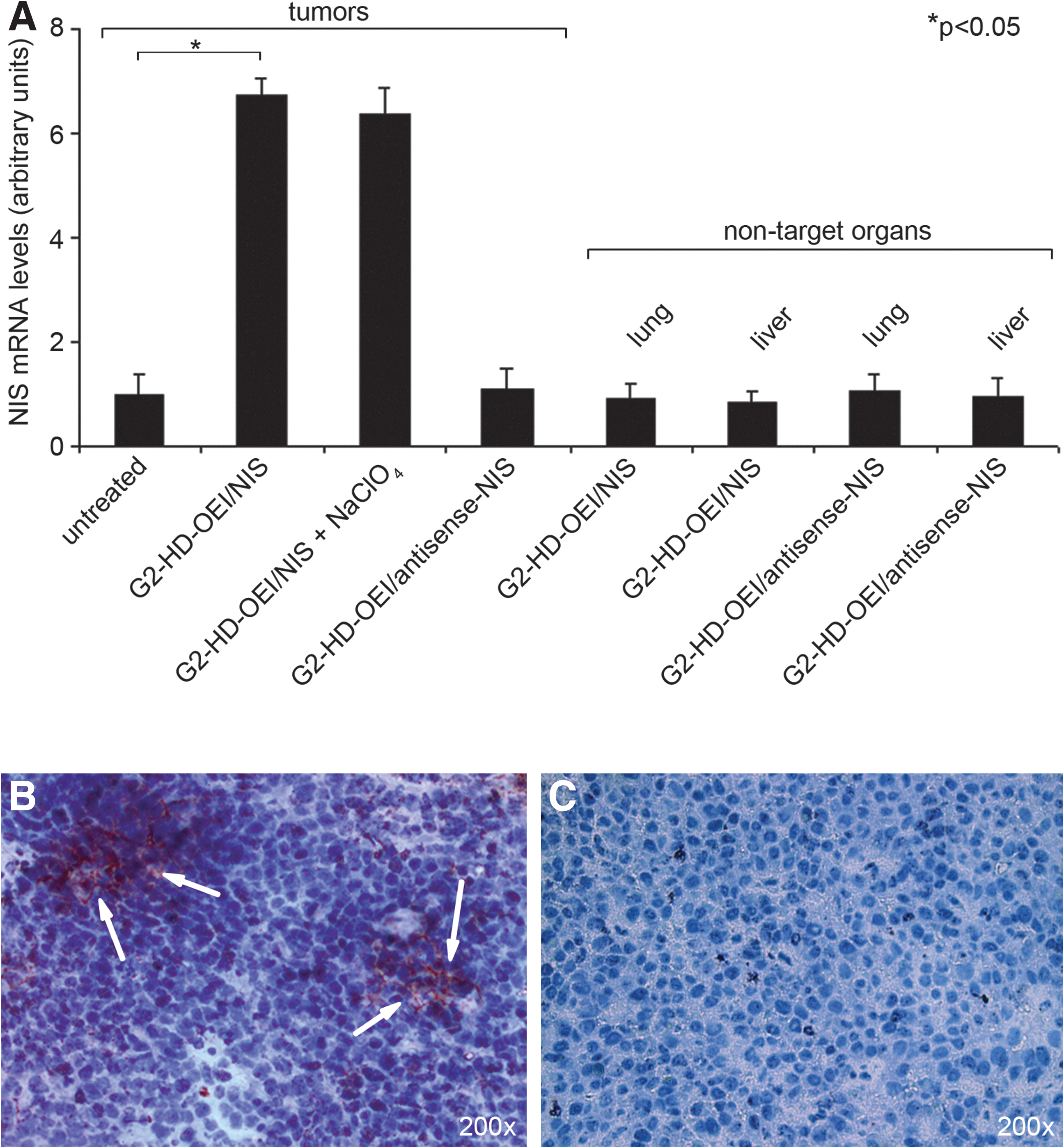
Analysis of NIS protein expression in HuH7 xenografts
Immunohistochemical analysis of HuH7 tumors revealed a heterogeneous staining pattern with clusters of primarily membrane-associated NIS-specific immunoreactivity in tumors after systemic application of G2-HD-OEI/NIS (Fig. 5B, arrows). In contrast, tumors treated with G2-HD-OEI/antisense-NIS (Fig. 5C) or untreated tumors (data not shown) showed no NIS-specific immunoreactivity. Parallel control slides with the primary and secondary antibodies replaced in turn by PBS and isotype-matched nonimmune immunoglobulin were negative (data not shown).
Radioiodine therapy studies after in vivo NIS gene transfer
Twenty-four hours after systemic administration of G2-HD-OEI/NIS or G2-HD-OEI/antisense-NIS polyplexes, a therapeutic dose of 55.5 MBq (1.5 mCi) of 131I was injected intraperitoneally. As control, saline was injected instead of radioiodine. This cycle consisting of systemic NIS gene transfer followed by radioiodine or saline administration was repeated once on days 7 and 8 (Fig. 6A). As an additional control, tumor growth of mice injected only with saline was assessed.
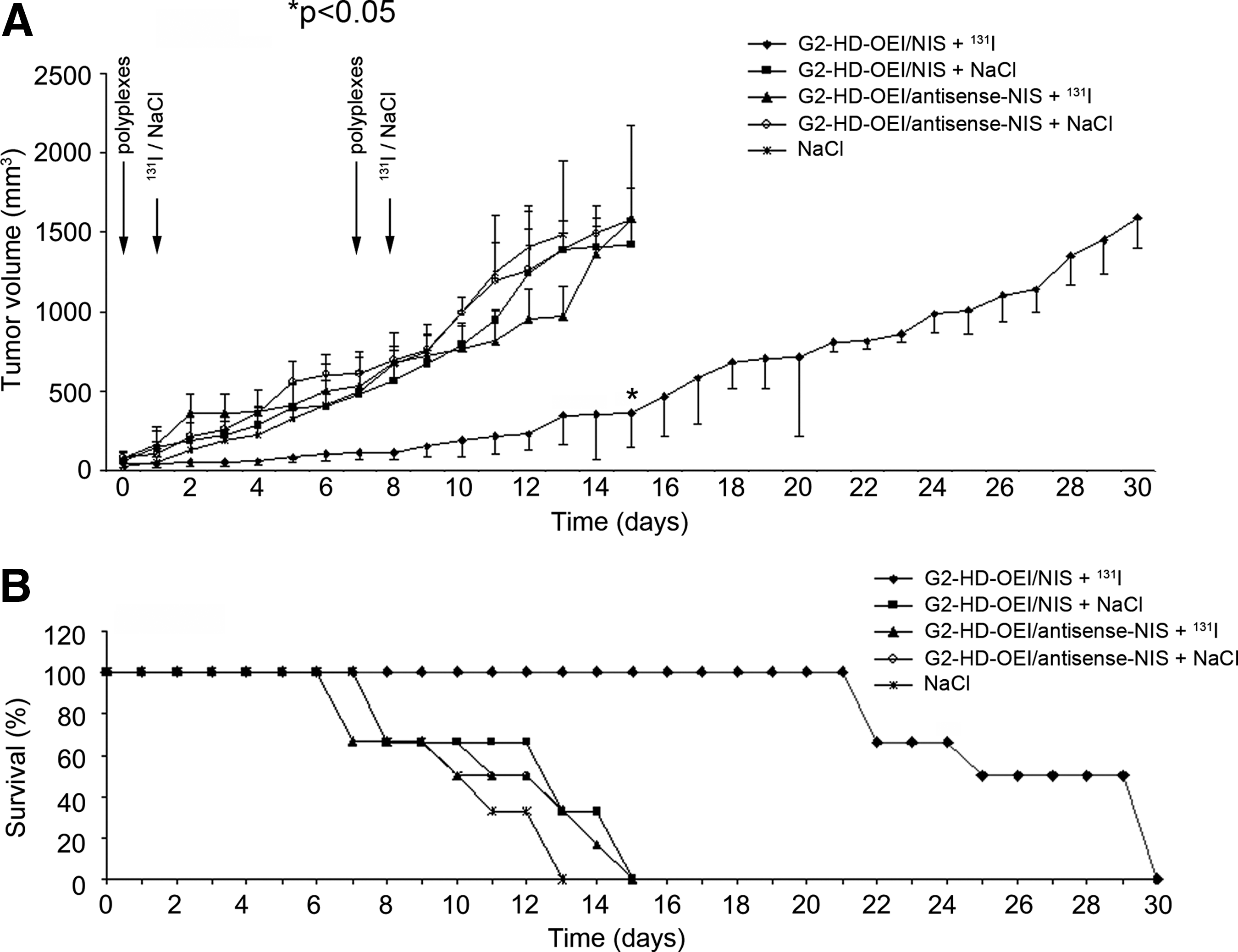
Radioiodine treatment of HuH7 tumors after systemic polyplex-mediated NIS gene transfer in vivo. Twenty-four hours after intravenous polyplex injection (long arrows), 55.5 MBq of 131I or saline was injected (short arrows). This treatment cycle was repeated once on days 7 and 8. 131I therapy after systemic G2-HD-OEI/NIS application resulted in a significant delay in tumor growth (
Mice treated with G2-HD-OEI/NIS or G2-HD-OEI/antisense-NIS followed by application of saline and mice treated with G2-HD-OEI/antisense-NIS followed by application of 131I, as well as saline-treated mice, showed exponential tumor growth. In contrast, NIS-transduced (G2-HD-OEI/NIS) and 131I-treated tumors showed a significant delay in tumor growth (Fig. 6A). Whereas all mice in the control groups had to be killed within 2 weeks of the onset of experiments, because of excessive tumor growth, 100% of the mice treated with 131I after injection of G2-HD-OEI/NIS survived approximately 3 weeks and 50% survived up to 4 weeks (Fig. 6B). Importantly, none of these mice showed major adverse effects of radionuclide or polyplex treatment in terms of lethargy or respiratory failure. However, a minor body weight loss of 3–5% was observed in mice after systemic administration of polyplexes.
In comparison, after LPEI-PEG-GE11-mediated NIS gene delivery, four cycles of LPEI-PEG-GE11/NIS followed by 131I application were needed to achieve a similar degree of tumor growth inhibition as after two cycles of G2-HD-OEI/NIS combined with 131I (Klutz et al., 2011a).
Immunofluorescence analysis
Three to 4 weeks after treatment immunofluorescence analysis using a Ki67-specific antibody (green) and an antibody against CD31 (red, labeling blood vessels) showed significantly lower intratumoral blood vessel density and proliferation index in NIS-transduced tumors (G2-HD-OEI/NIS) (Fig. 7A and C) as compared with mock-transduced (G2-HD-OEI/antisense-NIS) tumors (Fig. 7B and D) after 131I therapy.
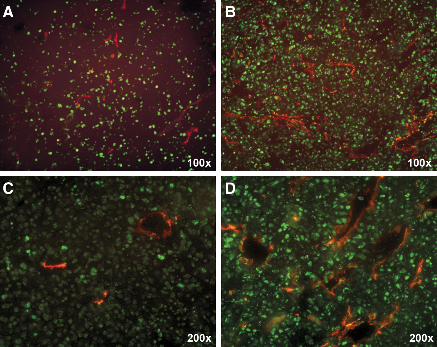
Immunofluorescence analysis using a Ki67-specific antibody (green) and an antibody against CD31 (red, labeling blood vessels) showed significantly decreased proliferation and blood vessel density in NIS-transduced tumors
Discussion
In the present study we investigated the efficacy of synthetic nanoparticle vectors (G2-HD-OEI) to achieve tumor-selective NIS-mediated radioiodine accumulation in an HCC mouse model. After confirmation of high transduction efficiency in vitro, intravenous application of NIS-conjugated G2-HD-OEI in nude mice carrying HCC xenografts was demonstrated to result in tumor-selective radioiodine accumulation, which was high enough for a significant therapeutic effect after application of 131I.
NIS represents one of the oldest and most successful targets for molecular imaging and targeted radionuclide therapy. Cloning and characterization of the NIS gene have therefore allowed the development of the NIS gene therapy concept based on NIS gene transfer into nonthyroidal tumor cells, followed by diagnostic and therapeutic application of radioiodine (Dai et al., 1996; Smanik et al., 1996; Hingorani et al., 2010).
One of the major challenges on the way to efficient application of the NIS gene therapy concept in the clinical setting of metastatic cancer concerns optimal tumor targeting in the presence of low toxicity and sufficiently high transduction efficiency after systemic administration of gene delivery vectors. Only a limited number of studies have investigated systemic NIS gene delivery approaches with the aim of NIS-targeted radionuclide therapy of metastatic disease (Dingli et al., 2004; Goel et al., 2007; Klutz et al., 2009; Liu et al., 2010).
In the present study we used a promising nonviral gene delivery system for tumor-targeted NIS gene transfer in a human HCC xenograft model. Branched polycations based on OEI-grafted polypropylenimine dendrimers (G2-HD-OEI) have been characterized as biodegradable synthetic gene delivery vectors with high in vivo transduction efficiency and remarkable intrinsic tumor affinity in the presence of low toxicity (Russ et al., 2008b; Klutz et al., 2009).
G2-HD-OEI complexed with the human NIS cDNA under the control of the unspecific CMV promoter revealed high transfection efficiency in vitro, resulting in a 44-fold increase in iodide uptake activity in HuH7 cells. After systemic application of NIS-conjugated G2-HD-OEI via the tail vein in vivo, 80% of HuH7 tumors showed tumor-specific iodide accumulation as determined by 123I scintigraphic gamma camera imaging, with accumulation of approximately 6–11% ID/g and an effective half-life of 10 hr for 131I. In contrast, mice pretreated with the competitive NIS inhibitor sodium perchlorate or mice injected with control vectors showed no tumoral iodide uptake, confirming that the observed radioiodine accumulation in the tumors was mediated by functional NIS expression. One explanation for the remarkable tumor selectivity of G2-HD-OEI used in this study is the so-called “enhanced permeability and retention” (EPR) effect. Because of large endothelial fenestrations in tumor vasculature combined with poor lymphatic drainage, circulating macromolecules can preferentially accumulate in solid tumors (Matsumura and Maeda, 1986; Iyer et al., 2006). We and others also observed an intrinsic affinity of well-vascularized tumors for polycations, as their removal from tumor tissue is prevented because of their affinity for tumor cells and tumor matrix (Smrekar et al., 2003; Dufes et al., 2005; Schwerdt et al., 2008). We demonstrated that polyplex organ distribution and transgene expression do not necessarily correlate (Navarro et al., 2010). Although considerable amounts of polyplexes can be entrapped in nontarget organs such as lung and liver, expression is limited to tumor tissue. This can be explained by additional selectivity being achieved by the mitotic activity of tumor cells, which is advantageous for polyplex-mediated transgene expression (Brunner et al., 2000). In a recent study, in the same HCC mouse model we used GE11, an EGFR-specific ligand, for additional active tumor targeting by coupling GE11 to PEG-shielded LPEI (LPEI-PEG-GE11), as the “gold standard” of PEI-based gene carriers (Klutz et al., 2011a). After systemic application of LPEI-PEG-GE11/NIS polyplexes, HuH7 xenografts accumulated slightly lower amounts of iodide (6.5–9% ID/g 123I), but with a lower effective half-life of only 6 hr, which resulted in a smaller tumor-absorbed dose of 47 mGy/MBq of 131I (Klutz et al., 2011a). This difference in tumor-absorbed dose is a result of the lower effective half-life for 131I, and might also be caused by decreased transduction efficiency of LPEI-PEG-GE11. LPEI per se shows excellent transduction efficiency due to an intrinsic endosomolytic mechanism, the so-called “proton sponge effect” (Akinc et al., 2005). The polymer protonation results in chloride influx and osmotic swelling of the endosome, followed by membrane destabilization, which in concert is responsible for the high transfection efficiency (Sonawane et al., 2003). Not only LPEI but also oligoethylenimine (OEI)-based polymers are characterized by this intrinsic endosomolytic mechanism (Sonawane et al., 2003). The major drawback of LPEI, however, is its significant toxicity after systemic application, a result of acute and long-term toxic effects (Chollet et al., 2002) caused at least in part by its high transgene expression in the lungs, due to pronounced aggregation with erythrocytes (Russ et al., 2008a; Klutz et al., 2011a). A technique to reduce the unspecific toxicities and prolonged blood circulation time is shielding of polyplexes by PEGylation. Unfortunately, at the same time as shielding improves the properties of polyplexes for systemic application it appears to reduce its cell transfection activity (Oupicky et al., 2002; Ogris et al., 2003). The reduced cell surface interaction by PEGylation has been overcome by the attachment of targeting ligands. However, the transfection efficiency of the targeted PEG vectors often still does not reach the level achieved with untargeted gene vectors, most probably due to an attenuated endosomolytic effect (Guo et al., 1999; Rudolph et al., 2002), which at least in part explains the lower transduction efficiency of LPEI-PEG-GE11 as compared with G2-HD-OEI, resulting in decreased tumoral iodide accumulation with significantly reduced iodide retention time. On the other hand, LPEI-PEG-GE11 offers the advantage of additional active tumor targeting by coupling the tumor-specific ligand GE11, which could theoretically also be attached to G2-HD-OEI. Taken together, both systems are in principle suitable for targeting the NIS gene to experimental tumors implanted in mice. For the G2-HD-OEI polymer a hypervascularized tumor is a precondition for successful tumor accumulation, whereas the LPEI-PEG-GE11 conjugate is in principle able to promote efficient receptor-mediated uptake into tumor cells by active ligand-mediated tumor targeting, which could be of even greater importance in tumors with a lower level of vascularization.
In addition to 123I gamma camera imaging we have used small-animal whole body SPECT CT imaging in a subset of animals, using 123I as a radiotracer. The cross-sectional fusion imaging techniques such as SPECT CT provide a useful means to improve three-dimensional spatial resolution and separate the overlapping regions of radioiodine uptake in vivo, thereby allowing a more robust biodistribution analysis. In our study 123I-SPECT CT imaging allowed a more detailed three-dimensional analysis of NIS-mediated radioiodine accumulation, which appeared inhomogeneous in clusters of iodide uptake throughout the tumor. The examination of all projections of the SPECT CT images failed to detect any other NIS gene transfer-related signals, suggesting that systemic NIS gene transfer using G2-HD-OEI is highly tumor specific. In addition, our data are consistent with several studies demonstrating the sensitivity of micro-SPECT CT for imaging and quantitation of NIS-mediated radionuclide uptake (Marsee et al., 2004; Carlson et al., 2006, 2009; Merron et al., 2007; Chisholm et al., 2009; Peerlinck et al., 2009; Penheiter et al., 2010).
Moreover, 123I scintigraphic and SPECT CT imaging studies were confirmed by ex vivo biodistribution experiments revealing significant tumoral radioiodine accumulation, whereas no significant iodide uptake was measured in nontarget organs. Tumor-specific NIS expression was further confirmed by real-time qPCR as well as NIS-specific immunoreactivity, which was primarily membrane associated with an inhomogeneous, patchy staining pattern, and therefore nicely correlates with the clusters of iodide accumulation detected by 123I-SPECT CT imaging. Because of the limited polyplex spread in the tumor resulting in the inhomogeneous transgene expression, nonviral gene delivery is ideally combined with therapy genes that are able to provide a bystander effect. The path length of up to 2.4 mm of the β particles emitted by 131I causes a significant crossfire effect after NIS gene transfer, resulting in a bystander effect, which makes NIS an ideal candidate gene for synthetic vector-based systemic cancer gene therapy (Dingli et al., 2003).
Most importantly, systemic polyplex-mediated NIS gene transfer resulted in tumor-specific radioiodine uptake activity in HuH7 tumor-bearing mice which was sufficiently high for a significant therapeutic effect of 131I. After two cycles of systemic polyplex application followed by 131I injection, tumor-bearing mice showed a significant delay of tumor growth associated with markedly prolonged survival. Using LPEI-PEG-GE11 in our former study, four cycles of polyplex/131I application were needed to achieve a similar degree of tumor growth inhibition, which is a logical consequence of the significantly lower tumor-absorbed dose that was obtained after systemic application of LPEI-PEG-GE11/NIS as compared with G2-HD-OEI/NIS (Klutz et al., 2011a).
In addition, immunofluorescence analysis showed a significantly reduced proliferation and blood vessel density after systemic polyplex-mediated NIS gene transfer followed by 131I application, suggesting radiation-induced tumor stroma cell damage in addition to tumor cell death.
In conclusion, the data in the current study correlate well with the data acquired in the syngeneic neuroblastoma mouse model (Klutz et al., 2009), demonstrating that the application of these synthetic nanoparticles based on OEI-grafted pseudodendritic oligoamines for systemic NIS gene delivery is not restricted to a specific tumor model, but is suitable for all cancers with hypervascularized tumors. Using NIS as reporter gene, this study allowed detailed characterization of the in vivo biodistribution of polyplex-mediated functional NIS expression by 123I scintigraphic gamma camera and 123I-SPECT CT imaging, which is an essential prerequisite for exact and safe planning and monitoring of clinical gene therapy trials with the aim of individualization of the NIS gene therapy concept in the clinical setting. This study therefore opens the exciting prospect of NIS-targeted radioiodine therapy of disseminated HCC, using polyplexes based on biodegradable polymers for systemic NIS gene delivery.
Footnotes
Acknowledgments
The authors are grateful to S.M. Jhiang (Ohio State University, Columbus, OH) for supplying the full-length human NIS cDNA. In addition, the authors thank W. Münzing (Department of Nuclear Medicine, Ludwig Maximilian University, Munich, Germany) for assistance with the imaging studies, and J.C. Morris (Mayo Clinic, Rochester, MN) for providing the NIS mouse monoclonal antibody. This study was supported by grant SFB 824 (Sonderforschungsbereich 824) from the Deutsche Forschungsgemeinschaft (Bonn, Germany) to C. Spitzweg and M. Ogris, and by a grant from the Wilhelm-Sander-Stiftung (2008.037.1) to C. Spitzweg.
Author Disclosure Statement
No competing financial interest exists.