
Abstract
Choroidal neovascularization (CNV) is a common pathological feature in neovascular age-related macular degeneration, which is the leading cause of vision loss among elderly populations in developed countries. This study evaluated the effect of a novel endogenous inhibitor of angiogenesis, calreticulin anti-angiogenic domain (CAD), subconjunctivally delivered by an adenoviral vector (Ad-CAD) in a rat model of laser-induced CNV. CAD was expressed in Ad-CAD-infected cells and inhibited the angiogenic activity in human umbilical vein endothelial cells in vitro. CAD expression was also found in various ocular tissues after in vivo subconjunctival Ad-CAD injection. Via bioluminescence imaging it is shown that a single subconjunctival injection of Ad-luciferase induced the expression of the transgene in the injected eyes within 24 h, which lasted for at least 112 days. Forty-two days after subconjunctival injection of Ad-CAD, retinal structure and function were unaffected, as measured using optical coherence tomography and electroretinography, respectively. After laser injury, subconjunctival Ad-CAD gene delivery significantly inhibited CNV lesions as measured via choroid flat-mounts (51% reduction at 21 days; p < 0.001), as well as by fundus fluorescein angiography (19.3%, 28.2%, 31%, and 27.5% reductions at days 21, 28, 35, and 42, respectively; p < 0.05) in rats. The data suggest that subconjunctival Ad-CAD gene therapy could effectively inhibit laser-induced CNV and might be an attractive therapeutic approach for the management of choroidal neovascularization.
Introduction
A
Current treatments aim to seal off leaky blood vessels via laser therapy or to inhibit the neovascular response through intraocular injections of anti-inflammatory steroids 4 or agents that target vascular endothelial growth factor (VEGF). Anti-VEGF agents in clinical use include VEGF-neutralizing oligonucleotide aptamer, 5 humanized anti-VEGF monoclonal antibody, 6 and VEGF trap (Eylea). 7 However, intraocular injections are invasive and can result in sight-threatening complications, including the attendant risks of infection, retinal detachment, cataract, and inflammation. Such complications carry significant risk of permanent vision loss. The systemic safety of repeated injections of anti-VEGF agents has also raised concern, and in particular the increased risk of cerebrovascular accidents has been noted. 8 In addition, repeated injections come at considerable cost, and a loss of efficacy of anti-VEGF agents over time has brought into question the long-term benefits of anti-VEGF therapy. 9,10 Thus, there is a need to seek cheaper, safer, less invasive, and more durable alternative therapies for the management of neovascular AMD.
Recently, calreticulin has been identified an anti-angiogenic domain (CAD) derived from vasostatin, 11 a potent inhibitor of angiogenesis isolated from supernatants of an Epstein–Barr virus–immortalized B cell line. 12,13 Topical applications of CAD for 20 days was able to attenuate laser-induced CNV in rat eyes. 11,14 However, topical delivery of CAD shows low efficacy due to poor protein penetration to deeper target sites in the eye. Moreover, topical delivery requires high patient compliance to achieve adequate efficacy. The present study constructed a CAD gene fused with a signal peptide of vasostatin to allow extracellular secretion and utilized an adenoviral vector to mediate robust gene expression. It has previously been demonstrated that genes delivered via subconjunctival injection of an adenoviral vector can be effective at the posterior segment of the eye with no effect on retinal function. 15 This study investigated the efficacy and safety profile of subconjunctival injection of adenovirus-mediated CAD in a rat model of laser-induced CNV.
Materials and Methods
Cell culture
Human umbilical vein endothelial cells (HUVECs) were purchased from Lonza (catalog no. CC-2519; Lonza, Walkersville, MD) and cultured in endothelial cell basal medium-2 (EBM-2) supplied with EGM™-2 BulletKit™ (catalog no. CC-5035; Lonza). HEK293A cells were purchased from Invitrogen (catalog no. R70507; Life Technologies, Frederick, MD) and cultured in Dulbecco's modified Eagle's medium (catalog no. 11965118; Life Technologies) supplemented with 10% fetal calf serum (Life Technologies), and 2 mM of glutamine (catalog no. 2503008; Life Technologies), 50 IU/mL of penicillin-streptomycin (catalog no. 15070063; Life Technologies). Both cell lines were cultured in a humidified 5% CO2 atmosphere at 37°C.
Construction and production of recombinant adenovirus
Recombinant adenoviruses encoding CAD (Ad-CAD), EGFP (Ad-GFP), luciferase (Ad-Luci), and β-galactosidase (Ad-LacZ) were used in this study. Human CAD cDNA fusion with a human influenza hemagglutinin (HA) tag was subcloned into an adenoviral transfer vector, PDONR221 (catalog no. V49320; Adenoviral Gateway® System; Invitrogen, Carlsbad, CA). The plasmid of PONR221-CAD allowed site-specific recombination with pAd/CMV/V5-DEST, a plasmid containing the entire type 5 adenoviral genome with E1-insertion and E3 deletion, via a calcium phosphate protocol. The plasmids of pShuttle-CMV-EGFP and pShuttle-CMV-LacZ were from a commercial kit used for adenoviral production (catalog no. 240010; AdEasy Adenoviral Vector Systems, Agilent Technologies, Santa Clara, CA). Luciferase DNA was digested with KpnI before cloning into a vector, pShuttle-CMV (catalog no. 240007; Agilent Technologies), which ultimately yielded the plasmid of pShuttle-CMV-Luciferase. All recombinant adenoviruses were generated in HEK293A cells using the Adenoviral Gateway® System and the AdEasy Adenoviral Vector System. After homologous recombination, the virus plaques were verified by checking the cytopathic effect and via quantitative polymerase chain reaction (qPCR), as previously described. 15 The virus was amplified and purified with cesium chloride under ultracentrifugation and then desalted by G-25 gel filtration chromatography. The number of viral particles and titer were determined by qPCR and using the plaque-forming assay on HEK293A cells before storage at −80°C.
Western blot analysis
Forty-eight hours after Ad-GFP or Ad-CAD infection with 500 and 1,000 multiplicities of infection (MOI), HUVECs were washed with cold phosphate-buffered saline (PBS) and collected into the lysis buffer (50 mM of Tris-HCl, pH 7.4, 1% NP-40, 0.25% sodium deoxycholate, 150 mM of NaCl, 1 mM of phenylmethylsulfonyl fluoride, 1 μg/mL of aprotinin, 1 μg/mL of leupeptin, and 1 μg/mL of pepstatin). Proteins were separated using NuPAGE™ Novex™ 4–12% Bis-Tris Protein Gels (catalog no. NP0321BOX; Invitrogen) and transferred to polyvinylidene fluoride membranes (catalog no. IPVH00010; Immobilon-P; Millipore, Billerica, MA) using TE70X semi-dry transfer unit (Hoefer, Inc., Holliston, MA). Membranes were blocked with 5% skim milk in TBS-T (10 mM of Tris, 150 mM of NaCl, and 0.05% Tween-20) at room temperature for 1 h and then incubated with mouse monoclonal HA (F-7) antibody (1:1,000 dilution; catalog no. sc-7392; Santa Cruz Biotechnology, Inc., Santa Cruz, CA) or mouse monoclonal calreticulin (FMC 75) antibody (1:1,000 dilution; catalog no. ADI-SPA-601-D; Enzo Life Sciences, Farmingdale, NY) overnight at 4°C. Membranes were washed, further incubated with horseradish peroxidase–conjugated secondary antibody, and developed using the Amersham ECL Prime Western Blotting Detection kit (catalog no. RPN2232; GE Healthcare Australia, Parramatta, Australia).
Cell migration assay
HUVECs were seeded onto six-well plates at a density of 2.5 × 10⁵/well, and infected with Ad-CAD and Ad-GFP of 500 MOI for 48 h. Confluence was >80% prior to the scratch assay. The cell monolayer was scraped with a 200 μL pipette tip to create three straight lines with two crosses without cells for each well, and debris was removed by replacing with 2 mL of EBM-2 supplied with EGM™-2 BulletKit™. Four images were taken for each well immediately after scraping and 24 h after incubation. The areas without cells were measured using ImageJ software (National Institutes of Health, Bethesda, MD).
Tube formation assay
Quantification of tube formation was performed as previously described. 16 Briefly, BD Matrigel™ Basement Membrane Matrix (catalog no. 356234; Becton Dickinson, Bedford, MA) was added to a 96-well plate (50 μL/well) and allowed to form a gel at 37°C for 30 min. HUVECs were infected with Ad-GFP and Ad-CAD of 500 MOI for 48 h. Cells (1.5 × 10⁴ cells) were harvested, added to the wells, and incubated for 6 h in a humidified 5% CO2 atmosphere at 37°C. Under these conditions, endothelial cells form delicate networks of tubes that are detectable within 3 h and are fully developed after 6 h. After incubation, the number of lumens of endothelial tubes was counted from three repeated wells using Angiogenesis Analyzer in ImageJ. 17
Animal care and adenovirus delivery by subconjunctival administration
All experimental procedures were performed in accordance with the ARVO Statement for the Use of Animals in Ophthalmic and Vision Research. Animal ethics approval was obtained from the Animal Ethics Committee at Kaohsiung Veterans General Hospital and University of Melbourne to ensure that the animals did not suffer unduly during and after the experimental procedure.
Male Brown Norway pigmented rats weighing between 150 and 200 g were used in this study. All rats were housed in standard cages, two rats per cage, with free access to food and water under a 12 h light (50 lux illumination) and 12 h dark (<10 lux illumination) cycle, with a temperature-controlled environment to minimize possible light-induced damage to the eye.
Prior to adenovirus delivery by subconjunctival injection, animals were anesthetized by intraperitoneal injection of a mixture of ketamine (70 mg/kg) and xylazine (6.5 mg/kg) in PBS. A topical local anesthetic, proxymetacaine 0.5% drops, was given to rats prior to subconjunctival injection. Under a dissecting microscope, a small incision was made through the conjunctiva at the supertemporal quadrant with a 33-gauge needle attached to a 1 mL syringe, and 50 μL of adenoviral vectors (1 × 10¹⁰ genome copy [GC]/eye) or PBS was injected into one eye. After injection, rats were monitored every 2 h for the first 6 h, then twice daily for the following 3 days. All animals were sacrificed on the desired day using Lethobarb (200 mg/kg, intraperitoneal injection) after subconjunctival injection, and tissues were harvested and for further assessment.
Gene expression detected by qPCR
Five days and 42 days after subconjunctival injection of Ad-LacZ or Ad-CAD, rat eyes were dissected, and retina, choroid, sclera, and periocular tissue were isolated. Total RNA from different tissues was extracted and purified using commercial kits in accordance with the manufacturer's instructions (catalog no. 74104; RNeasy Mini Kit; Qiagen, Valencia, CA). Briefly, each tissue in lysate was homogenized, and total RNA was purified with a column system. Total RNA (100 ng) was then reverse-transcribed to cDNA using a high-capacity reverse transcription kit (catalog no. 4374996; Life Technologies, Victoria, Australia). Quantitative PCR was performed using a Fast SYBR Green Master Mix (catalog no. 4385612; Life Technologies) with the CAD primer (F: 5′-GGATCGAATCCAAACACAAGTC; R: 5′-ATCAGTGTGTACAGGTGTGTAA) for rat CAD. Rat GADPH was used as a reference gene with the GADPH primer (F: 5′-GAGTCAACGGATTTGGTCGT; R: 5′-TTGATTTTGGAGGGATCTCG).
For analysis of mRNA expression, relative expression levels of CAD in different tissues from rats administered Ad-lacZ and Ad-CAD were calculated using the ΔΔCt method, as described previously by Livak. 18
Generation of CNV by laser photocoagulation
CNV lesions were induced in rat eyes by a laser photocoagulation, as previously described. 19 Briefly, rats were anesthetized by intraperitoneal injection of a mixture of ketamine (70 mg/kg) and xylazine (6.5 mg/kg) supplemented with a topical anesthetic agent (proxymetacaine 0.5%; Alcon Laboratories, Macquarie Park, Australia), and pupils were dilated with 1% tropicamide (1% Mydriacyl; Alcon Laboratories). A piece of cover glass served as a contact lens. Argon laser (Novus Omni; Coherent, Palo Alto, CA) irradiation was delivered through a slit lamp (Carl Zeiss, Oberkochen, Germany). Laser parameters were set as follows: spot size of 50 μm, power of 400 mW, and exposure duration of 0.05 s. An attempt was made to breach Bruch's membrane, as clinically evidenced by central bubble formation. Six lesions were created between the major retinal vessels in each fundus. Rats exhibiting vitreal hemorrhage that obscured the posterior segment were excluded from fundus imaging analysis.
Bioluminescence imaging
To perform bioluminescence imaging, rats were anesthetized by intraperitoneal injection of ketamine (70 mg/kg) and xylazine (6.5 mg/kg). Following application of a topical anesthetic (proxymetacaine 0.5%; Alcon Laboratories), one eye received a single injection of D-luciferin (catalog no. E1603; 20 mg/mL; Promega, Madison, WI) via subconjunctival injection (50 μL). In vivo luciferase activity of rats receiving Ad-Luci via subconjunctival injection was assessed using the IVIS Imaging System (200 Series; Caliper Life Sciences, Hopkinton, MA) with animals stabilized on a warmed (37°C) platform. The spatial distribution of bioluminescence intensity was captured for quantitation (Caliper Life Sciences).
Quantification of CNV lesions by flat-mount using fluorescein isothiocyanate-dextran perfusion
The blood vessels in rat eyes were labeled by perfusion with fluorescein isothiocyanate (FITC)-dextran (2 × 10⁶ MW; catalog no. 52471; Sigma–Aldrich, St. Louis, MO). 20,21 Briefly, 50 mL of lactated Ringer solution followed by 20 mL of FITC-dextran lactated Ringer solution (5 mg/mL) was injected via the left ventricle. The eyes were removed and fixed for 2 h in 10% phosphate-buffered formalin. After the cornea and lens were removed, RPE-choroid-sclera flat-mounts were obtained by hemisecting the eye and peeling the neural retina away from the underlying RPE. The retina was detached and flat-mounted onto a slide. The fluorescence in flat-mounts were examined by fluorescence microscopy, and images were digitized using a three-color charge coupled device video camera and a framegrabber. The area of hyperfluorescent associated with each burn, the CNV lesion, was measured by Image-analysis software (ImageJ) by two independent ophthalmologists who were blinded to the experimental design.
Fundus fluorescein angiography
The CNV lesions were evaluated by fundus fluorescein angiography (FFA) analysis, as previously described. 11 After laser photocoagulation, the CNV lesions were evaluated by FFA using a digital fundus camera (Visupac 450; Zeiss FF450) on days 21, 28, 35, and 42. Sodium fluorescein solution (10%; 0.1 mL/kg; Fluorescite; Alcon Laboratories) was injected intraperitoneally in anesthetized rats. Late-phase angiograms were obtained 8 min after injection, and fundus images of both eyes were acquired within 1 min. Choroidal neovascularization was defined as present when early hyperfluorescent with late leakage was seen at the site of laser injury. The area of CNV lesion was measured using image analysis software (Retina Angiography Area Measurement program; Heidelberg Engineering, Heidelberg, Germany) by two independent ophthalmologists who were blinded to the experimental design.
Electroretinography
At 42 days following injection, rats underwent overnight dark adaptation (∼12 hours) followed electroretinography (ERG) assessment under fully dark-adapted conditions. Details for functional assessment are as previously reported, 22 with the exception that the reference chloride silver electrode was placed around the outside of the eye.
ERG analysis, as previously described, 22 returned the photoreceptor (a-wave), bipolar cell (b-wave), and ganglion cell dominated (scotopic threshold response, STR) components of the ERG waveform. For each parameter, amplitude for each treated eye was expressed as a percentage of its contralateral control eye. Group data are given as mean ± standard error of the mean (SEM).
Spectral-domain optical coherence tomography
Following ERG measurement, rat retinae were imaged using spectral domain-optical coherence tomography (SD-OCT; Bioptigen, Inc., Morrisville, NC). Volume scans consisting of 1,000 A-scans per 100 B-scans (equally spaced across the 1.4 mm vertical dimension) centered on the optic nerve head (ONH; 1.4 mm × 1.4 mm × 1.57 mm) were obtained from both eyes.
OCT images were analyzed using FIJI software. OCT images that included the optic nerve were manually segmented by a masked grader, as previously described. 23 Total retinal thickness (TRT) was measured from the inner limiting to Bruch's membrane. Retinal nerve fiber layer (RNFL) thickness was measured from the inner limit membrane to the inner aspect of the inner plexiform layer. Ganglion cell complex thickness was measured from the inner limiting membrane to the outer aspect of the inner plexiform layer.
Statistical analysis
Data are expressed as mean ± SEM. Mean data were analyzed with unpaired t-tests or two-way analysis of variance followed by post hoc Tukey analysis. ERG and OCT data were compared using a two-tailed paired Student's t-test, following testing for normality using a Kolmogorov–Smirnov test for sphericity (PASW Statistics for Windows v18.0, SPSS, Inc., Chicago, IL, or GraphPad Prism v6.0, GraphPad Software, Inc., La Jolla, CA). A p-value of <0.05 was regarded as statistically significant.
Results
Gene delivery of CAD inhibits angiogenic activities in vitro
The recombinant adenovirus encoding CAD was generated for gene delivery studies (Fig. 1A). To validate CAD expression, cell lysates and culture media from the HEK293A cells infected with Ad-CAD were tested. Western blot analysis using the anti-HA antibody confirmed production of CAD at 16 kDa after Ad-CAD infection (Fig. 1B). Protein expression was not detected in cell lysates and culture media from Ad-GFP-transduced cells. To verify CAD transgenic expression further in human endothelial cells, cell lysates from the HUVECs infected with Ad-CAD of 500 or 1,000 MOI were analyzed by Western blot using the calreticulin antibody. CAD expression was evident at 16 kDa (Fig. 1C), which was not detected in Ad-GFP-infected HUVECs.
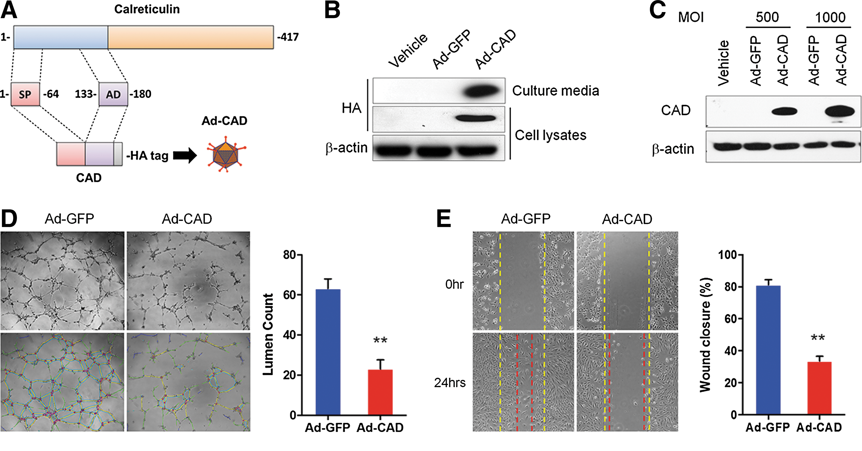
The effect of adenovirus-mediated calreticulin anti-angiogenic domain (CAD) gene delivery on in vitro angiogenic activities. (
To evaluate the anti-angiogenic activity of adenovirus-mediated CAD, tube formation and cell migration assay were performed. Compared with Ad-GFP-infected HUVECs, cells infected with Ad-CAD showed a significant decrease in their capacity to form tube-like networks on Matrigel (lumen count in Ad-GFP 63.2 ± 4.8% compared with A-CAD 23.0 ± 4.6%; p < 0.001, n = 4; Fig. 1D). Ad-CAD infected cells also showed poorer migration in the scratch migration assay (wound closure in Ad-GFP 78.5 ± 2.9% compared with A-CAD 33.7 ± 2.9%; p < 0.001, n = 8; Fig. 1E).
Duration of in vivo adenoviral transgene expression in the eye following subconjunctival injection
To evaluate the duration of transgene expression following subconjunctival injection, Ad-Luci were injected subconjunctivally in rat eyes and monitored using in vivo bioluminescence images. Twenty-four hours after injection, luciferase expression was significantly increased in the periocular area of the injected eye. This expression was sustained for up to 112 days (Fig. 2). Importantly, there was no bioluminescence detected in other organs, including the liver, of rats that had received Ad-Luci via subconjunctiva injection. This result is consistent with the known expression profile of an adenoviral-mediated transgene given subconjunctivally. 15
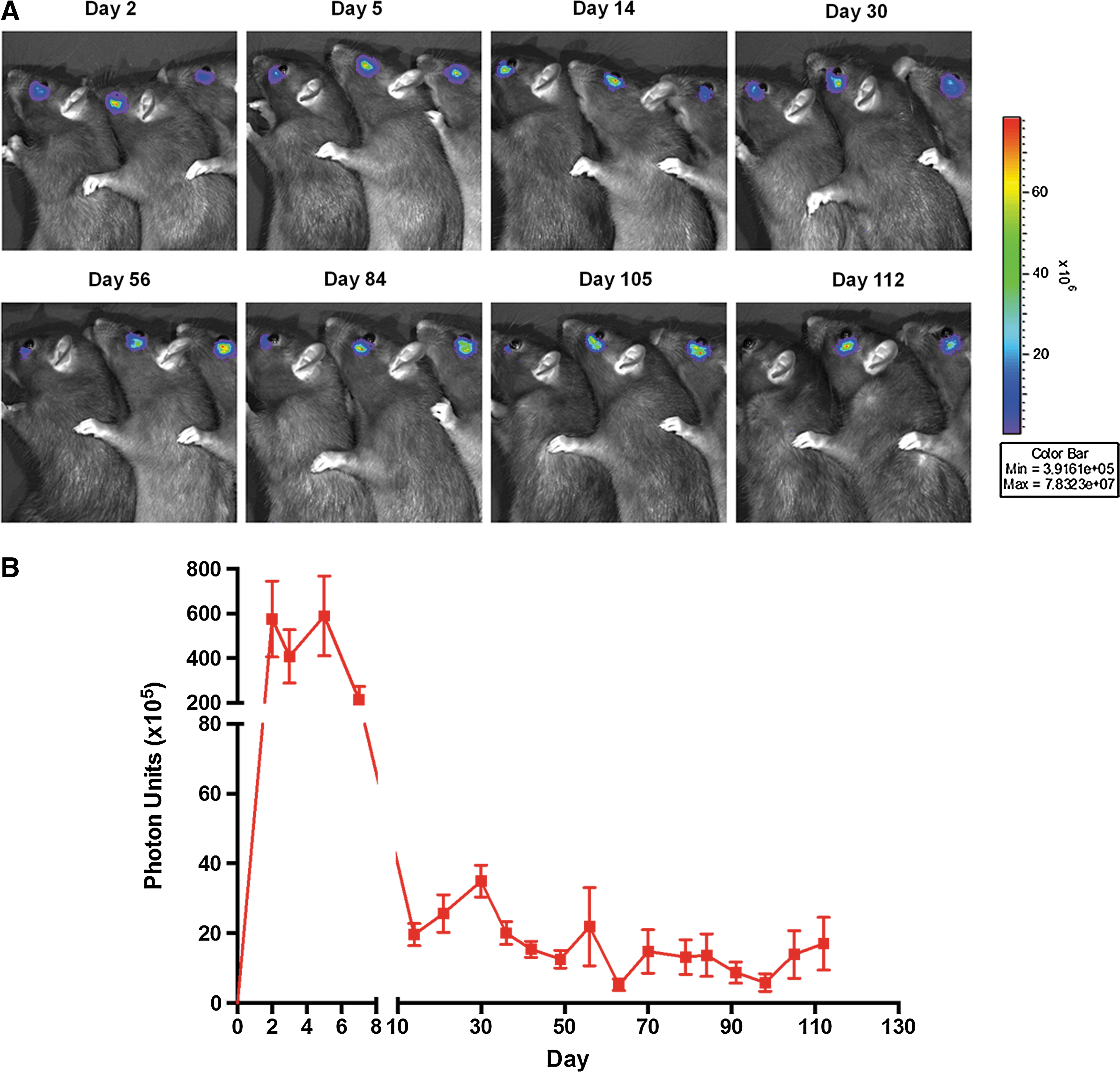
Duration of adenovirus-mediated gene delivery after a subconjunctival injection in rats. (
Evaluation of ocular expression of CAD after a subconjunctival Ad-CAD injection in the rats
To validate CAD expression in various ocular tissues after subconjunctival Ad-CAD injection, at day 5 after uniocular injection of Ad-LacZ or Ad-CAD, eyes were enucleated and dissected to isolate periocular tissue, sclera, choroid, and retina. CAD mRNA quantification with qPCR showed an increase in CAD expression in periocular tissue (8,062.1-fold; n = 3). Increased expression was also detected in the sclera (157.1-fold; n = 3), retina (88.5.1-fold; n = 3), and choroid (15.5-fold; n = 3) in Ad-CAD-injected eyes compared with the Ad-LacZ-injected eyes (Fig. 3). At day 5 after Ad-CAD injection, Western blot using the calreticulin antibody confirmed CAD protein expression in the periocular tissues (Fig. 3).
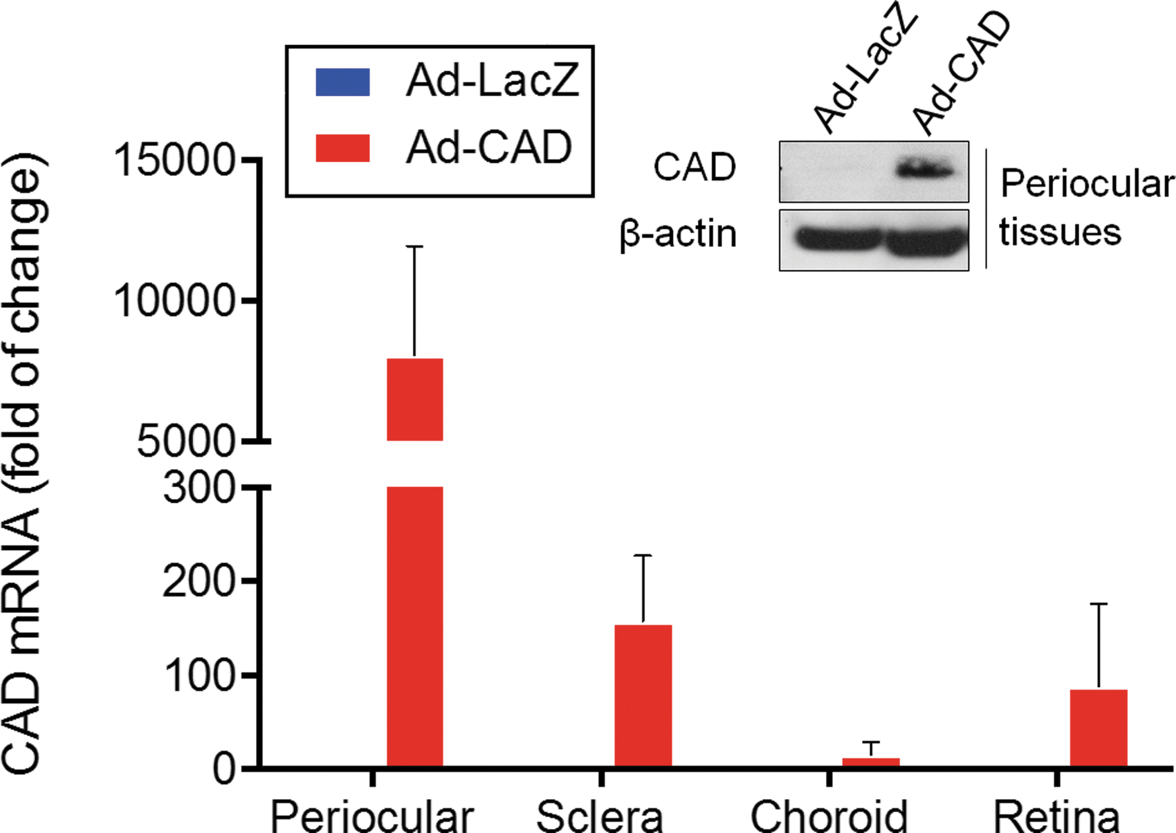
Evaluation of ocular expression of CAD after a subconjunctival Ad-CAD injection in rats. Five days after rats received Ad-LacZ (n = 3) or Ad-CAD (n = 3) injection in one eye, the eyes were enucleated and dissected into periocular tissue, sclera, choroid, and retina for evaluation of CAD expression by quantitative polymerase chain reaction and Western blot analysis. Representative Western blot of CAD protein expression in periocular tissues at day 5 after Ad-CAD injection. Color images are available online at
Retinal function and structure analysis by ERG and OCT
To evaluate the safety of Ad-CAD gene delivery via subconjunctival injection, the ERG was employed to evaluate retinal function in rats. Figure 4 summarizes the effect of Ad-LacZ (Fig. 4A and B) and Ad-CAD (Fig. 4C and D) on retinal function. Six weeks after subconjunctival injection, there was no significant difference in outer (t9 = 0.52; p = 0.60, n = 5), middle (t9 = 0.60; p = 0.56, n = 5), or inner retinal function (t9 = 0.64; p = 0.53, n = 5) in Ad-CAD-treated eyes compared with their contralateral controls. Likewise, subconjunctional injection of Ad-LacZ did not significantly affect retinal function (outer: t9 = 0.58, p = 0.57; middle: t9 = 0.28, p = 0.79; inner: t9 = 0.53, p = 0.604).

Effect of subconjunctival Ad-CAD injection on retinal function. (
OCT was performed to validate the effect of Ad-LacZ and Ad-CAD gene delivery on retinal structure. Figure 5A–C shows a representative retinal OCT image from an animal in each of the three groups (vehicle, Ad-LacZ, and Ad-CAD). Six weeks after subconjunctival injection, there was no significant difference in RNFL (Fig. 5D; t9 = 0.27; p = 0.79, n = 5), ganglion cell layer (GCC; Fig. 5E; t9 = 1.66; p = 0.13, n = 5), or TRT (Fig. 5F; t9 = 2.12; p = 0.07, n = 5) in Ad-CAD-treated eyes compared with their contralateral controls. Likewise, Ad-LacZ did not significantly affect retinal structure function (RNFL: t9 = 1.84, p = 0.10; GCC: t9 = 1.80, p = 0.11; TRT: t9 = 1.11, p = 0.30). Thus, Ad-CAD gene delivery via subconjunctival injection does not lead to any adverse effect on retinal function and structure.
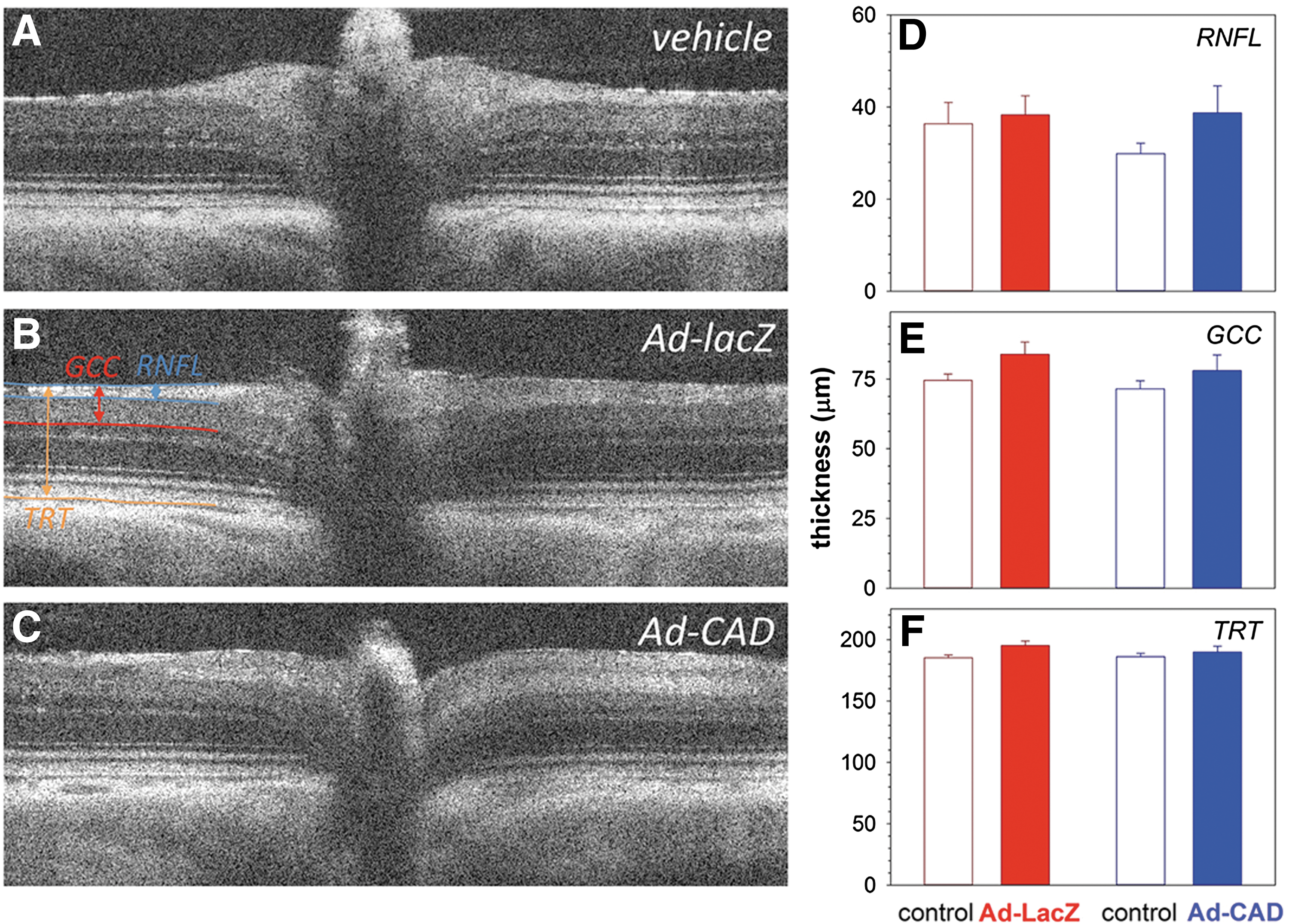
Effect of subconjunctival Ad-CAD injection on retinal structure. Representative optical coherence tomography images from a vehicle- (
Subconjunctival injection of Ad-CAD alleviates laser-induced CNV lesions
To evaluate the therapeutic potential of Ad-CAD, the rat model of laser-induced CNV was employed. After laser injury, rat eyes that had received Ad-CAD showed a significant reduction of CNV lesions (8,985 ± 608 μm2; p < 0.001, n = 20 from eight eyes) compared with vehicle- (16,910 ± 715 μm2; n = 20 from eight eyes) or Ad-LacZ-injected eyes (18,325 ± 1,325 μm2; n = 20 from eight eyes; Fig. 6A and B). Similarly, FFA analysis revealed that eyes that had received Ad-CAD had significantly smaller CNV lesion area (17,452 ± 614 μm2 at day 21; 15,947 ± 617 μm2 at day 28; 14,672 ± 516 μm2 at day 35; 15,910 ± 544 μm2 at day 42; p < 0.05 for each a comparison, n = 42–48 from eight eyes) compared with eyes that had received PBS (22,112 ± 1,044 μm2 at day 21, 21,779 ± 875 μm2 at day 28, 20,789 ± 819 μm2 at day 35, 22,004 ± 1,072 μm2 at day 42) or Ad-LacZ (21,618 ± 1,088 μm2 at day 21, 22,220 ± 1,065 μm2 at day 28, 21,264 ± 800 μm2 at day 35, 21,935 ± 1,014 μm2 at day 42; n = 42–48 from eight eyes) on days 21, 28, 35 and 42 (Fig. 7A and B).
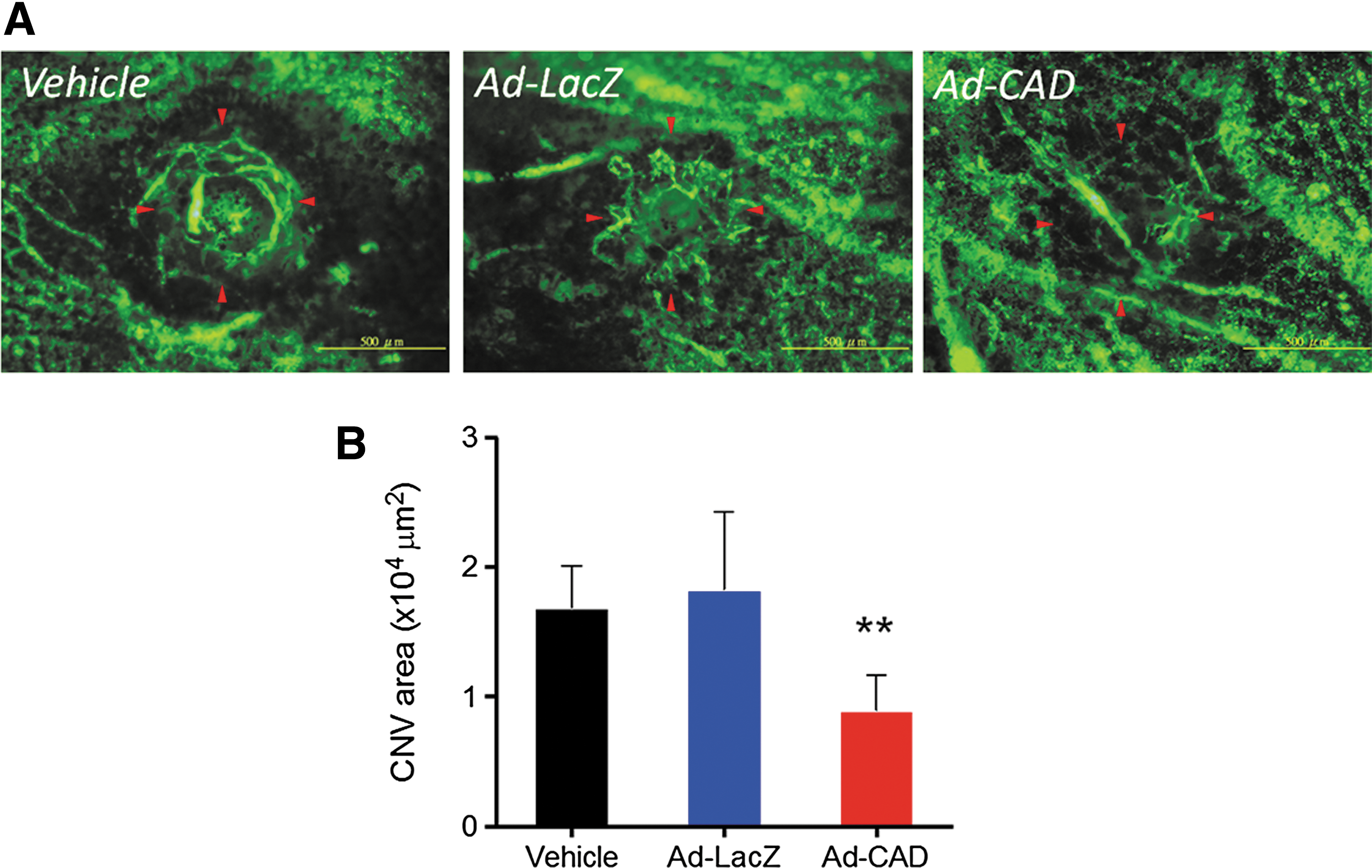
Flat-mount analysis of choroidal vascularity after a subconjunctival Ad-CAD injection. Choroidal vascularity of laser-induced choroidal neovascularization (CNV) lesions was examined by labeling using fluorescein isothiocyanate (FITC)-dextran. (

Fluorescein angiographic analysis of CNV lesions after a subconjunctival Ad-CAD injection. (
Discussion
This study shows that subconjunctival delivery of CAD via an adenoviral vector is an effective approach for attenuating the development of CNV. Gene delivery of CAD by adenoviral vector specifically inhibited the angiogenic activity of primary endothelial cells in vitro, and subconjunctival injection of Ad-CAD alleviated the laser-induced CNV lesions in vivo. The most widely used route of administration in registered clinical trials of anti-VEGF therapeutic agents is via intravitreal injection. 24 This route is invasive and has significant risks. An alternative is intravenous delivery of gene therapies, which may be less invasive but produces increased risk of systemic side effects. 25 Compared with these traditional gene delivery routes, subconjunctival injection is less invasive and has been shown to be a safe and efficient route of gene therapy for ocular diseases in previous animal studies. 26 –28 It has recently been demonstrated that transgene expression mediated by a single subconjunctival injection of adenoviral vector could extend to a number of ocular tissues. Specifically, higher expression was found in the sclera, followed by the choroid, retina, and cornea. 15 This distribution is similar to the current findings for CAD expression in rat eyes. The data also suggest that subconjunctival injection is an efficient and safe route for gene delivery in the short term, with no effect on retinal function or structure, and no apparent systemic toxicity. 15
Recent experimental work and clinical trials in gene therapy have gained momentum, with successes in treating both anterior and posterior eye disease. 29,30 It is easier and less costly to manufacture gene therapy vectors than it is to produce large quantities of purified protein. Recent preclinical 31,32 and clinical studies 33 provide strong support that ocular gene therapies targeting VEGF, a central mediator of pathological ocular neovascularization, are a viable long-term treatment option for neovascular AMD. However, chronic VEGF suppression in the eye may have deleterious side effects, including retinal neuronal toxicity. Gene transfer of endogenous antiangiogenic proteins is an alternative approach that has the potential to provide long-term suppression of neovascularization and/or excessive vascular leakage in the eye, 34 with potentially fewer side effects associated with systemic suppression of VEGF. Preclinical studies of gene transfer in animal models and clinical trial provided impressive preliminary results with a number of transgenes. 35 –38 Vasostatin is the N-terminal domain of calreticulin, which is inclusive of amino acids 1–180. This endogenous protein has been shown to have potent endogenous antiangiogenic properties and could be useful for the treatment of disease resulting from aberrant growth of blood vessels. 19,39 –43 Recently, the functional domain of vasostatin was identified as a 48 amino acid fragment, vasostatin48, and topical administration in rat eyes attenuates the development of laser-induced CNV. 11 The CAD used in the current study is the 112 residues of vasostatin, combining a secretory domain (residues 1–64) along with vasostatin48 (residues 133–180). Similar to vasostatin48, CAD has anti-angiogenic potency, but does not lead to accumulation in infected cells if it is delivered by a viral vector. Moreover, CAD has a low molecular weight, which provides an additional benefit by way of improving penetrance for subconjunctival delivery. While encouraging, the therapeutic efficacy of CAD against ocular neovascularization in comparison to its parent protein, vasostatin, and anti-VEGF treatments already in clinical use needs further evaluation.
Success in targeting angiogenesis is highly dependent on the delivery method to produce sustained expression of the therapeutic molecule in vivo. Gene therapy is one option that provides an emerging and powerful modality in the treatment of diseases resulting from pathological neovascularization. An adenovirus is the preferred viral vector owing to its ability to infect dividing and non-dividing cells and high efficiency for transgene expression. 44 However, as adenoviral vectors can trigger cytotoxic T-lymphocyte-mediated immunogenicity against adenoviral-transduced cells, 45 transgene expression can be short-lived, with a duration of a month or less. 46 The present study showed that subconjunctival injection of Ad-Luci resulted in expression of luciferase in injected eyes within 24 h. Importantly, luciferase expression persisted for at least 112 days after a single dose. It was previously reported that acute inflammatory responses induced by subconjunctival injection of an adenovirus was confined to the conjunctival injection site and gradually diminished within 2 weeks. 15,47 Therefore, it is believed that the subconjunctival injection of an adenovirus induces only a mild and temporary local immune response, leading to transgene expression for up to 112 days. Nevertheless, to achieve long-term transgene expression, adeno-associated virus (AAV) remains the preferred vector used in gene therapies for ocular disorders. AAV vectors are able to induce a moderate immune response and yield high-level, stable gene expression that is sustained for several years. 24 However, the issue with AAV vectors is that onset of gene expression is delayed and slower to reach the peak levels. The slower expression of transgene via AAV delivery can be attributed to the time needed for synthesis of the complementary strand of the single-stranded viral DNA genome before transcription. 48 A self-complementary AAV (scAAV) vector has been developed to overcome this limitation. 49 Compared with the single-strand AAV (ssAAV) vector, the scAAV vector shows an accelerated onset of expression and improved transduction efficiency in the retina. 26 The present study provides support for further exploration of CAD gene therapy in the treatment of CNV, and AAV-mediated gene delivery might be a more viable option for CAD gene therapy than the adenoviral vector used here.
In conclusion, this study demonstrated that adenovirus-mediated CAD gene delivery is able to attenuate the development of laser-induced CNV in rat eyes. Although further investigation is required to assess the optimal dosage, therapeutic mechanisms, and pharmacokinetics, these data make a compelling case that gene delivery of CAD by subconjunctival injection may be used as a safer and less invasive therapeutic alternative to conventional therapies for management of CNV and reduce the risks associated with frequently intravitreal injections.
Footnotes
Acknowledgments
This work was supported by grants from Kaohsiung Veterans General Hospital (VGHKS 97-106, 98-062, 99-062, and 100-066), Kaohsiung Medical University (KMU-TP104G00, KMU-TP104G03 and KMU-TP104G04), NSYSU-MKU Joint Project (102-P035), the Ministry of Science and Technology of Taiwan (MOST 103-2325-B-110-002), and the National Health and Medical Research Council of Australia (NHMRC#1061912), the Ophthalmic Research Institute of Australia, and the Angior Family Foundation. G.J.D. receives a Principal Research Fellowship from NHMRC. The Centre for Eye Research Australia receives Operational Infrastructure Support from the Victorian Government.
Author Disclosure
No competing financial interests exist.