
Abstract
A number of previous studies have indicated that the genetic variation at the collage type I alpha 1 (COLIA1) gene locus influences susceptibility to osteoporosis. However, seldom have studies reported the effect of gene delivery using an adenovirus vector carrying human recombinant COLIA1 cDNA on stimulating osteogenic activity of osteoblasts and enhancing fracture healing of ovariectomized rats. The current study was performed to demonstrate whether direct gene delivery using an adenovirus vector carrying human recombinant COLIA1 cDNA could stimulate osteogenic activity of osteoblast in vitro and enhance fracture healing of ovariectomized rats in vivo. In vitro, the tet-on system regulated COLIA1 gene adenovirus was constructed and transfected to osteoblasts. COLIA1 mRNA and collagen type I levels were assessed by reverse transcription polymerase chain reaction and enzyme-linked immunosorbent assay to determine whether adenovirus transfected successfully. Osteogenic activity of the osteoblasts was assessed by alkaline phosphatase activity, immunohistochemical staining, immunofluorescent staining, mineralized matrix formation, and extracellular calcium levels. In vivo, adenovirus-delivered COLIA1 gene was injected into the fracture site of the tibia in an ovariectomized rat model of osteoporosis, and bone callus condition was assessed to determine whether the COLIA1 gene could accelerate osteoporotic fracture healing. In vitro, the results showed that COLIA1 gene adenovirus transfection could increase osteoblast COLIA1 gene expression and collagen type I protein synthesis, increase alkaline phosphatase activity, and stimulate calcium nodules formation, which exhibited a direct osteogenic effect on the osteoblasts. In vivo, local injection of COLIA1 gene adenovirus increased collagen type I expression, restored bone mineral density, and accelerated fracture healing in ovariectomized rats, without increasing serum collagen type I and liver COLIA1 mRNA levels. This study suggests direct gene delivery using an adenovirus carrying human COLIA1 cDNA can stimulate the osteogenic activity of osteoblasts in vitro and enhance bone fracture healing in vivo. The tet-on system is an ideal gene regulatory system for effective and safe regulation of the therapeutic gene.
Introduction
O
Gene therapy has the potential to provide sustained protein production and to deliver proteins in a more physiological manner than is possible by direct recombinant proteins. It has several advantages over protein delivery, including the flexibility to express the protein locally and focally or in a disseminated fashion, as needed. 7 As a common gene delivery vector, adenovirus is capable of delivering transgenes directly to different sites within the musculoskeletal system. It has been proven to possess several advantages, including its ease of preparation, high titers, and ability to infect cells in situ with high transduction efficiency. 8 Much previous research has demonstrated that direct local or general application of adenovirus vectors encoding osteogenic gene sequences, including bone morphogenetic protein-2 (BMP-2), osteoprotegerin (OPG), vascular endothelial growth factor (VEGF), insulin-like growth factor-1 (IGF-1), and transforming growth factor (TGF), was able to promote osteoblast activity, increase BMD, and enhance fracture healing in osteoporotic animal models. 9 –13
However, the overexpression of the targeted gene may induce a host immune response, side effects, and serious adverse events. 14,15 The ability to control transgene expression safely from virus vectors is a long-term goal, and a number of approaches have been considered. The tetracycline-regulated gene expression system (tet system), which was originally developed by Gossen and Bujard, has proved to be efficient and reliable in controlling transgene expression, including both the “tet-on” and “tet-off” systems. 16,17 The tet-on system is composed of two elements: a rtTA and a TRE. In this system, the rtTA binds to the TRE and induces transgene expression in the presence of tet or the tet analog dox. Gene expression is normally off in the absence of tet or dox. As the “opening key” of transgene expression, the tet-on system has been used in experimental models of musculoskeletal diseases, and its reliable ability to control transgene expression has been shown. 18,19
To the authors' knowledge, seldom have previous studies reported the use of COLIA1 gene adenovirus delivery or reported the usage of the tet-on system to control transgene expression in the field of osteoporosis treatment. On this basis, a pilot study was performed. An overexpression recombinant adenovirus vector carrying human COLIA1 cDNA was constructed, and its effect on osteoblasts was demonstrated after transfection. 20 A further investigation was performed to demonstrate whether direct gene delivery using an adenovirus vector carrying the human recombinant COLIA1 cDNA (Adv. COLIA1) could stimulate the osteogenic activity of osteoblasts in vitro and enhance fracture healing of osteoporotic rats in vivo. The study also aimed to prove whether the tet-on system could control COLIA1 gene expression in the presence or absence of dox.
Methods
Adenovirus vector construction
The tet-on system regulated adenovirus encoding human recombinant COLIA1 cDNA vector (tet-on Adv. COLIA1) was constructed according to the methods reported by previous studies and the adeno-X™ adenovirus system user manual (Clontech code no. PT5177-1). 21 The target COLIA1 gene fragment was obtained and amplified using polymerase chain reaction (PCR; PCR primers: forward 5′-GTAACTATAACGGTCATGTTCAGCTTTGTGGACC-3′; reverse 5′-ATTACCTCTTTCTCCCAGGAAGCAGACAGGGCCAACGTCGAAGCC-3′). The target COLIA1 cDNA fragment was connected to a pAdenoX-Tet-on 3G Vector set (Clontech cat. no. 631179) by using an In-Fusion HD cloning kit (Clontech cat. no. 639648). The transgene tet-on COLIA1 cDNA was cloned into the E1 region of the viral genome under the regulation of the human CMV early promoter to generate the vectors tet-on COLIA1. The recombinant tet-on COLIA1 virus was propagated in 293 cells. Cells were harvested, and adenovirus vector was released by freezing and thawing, after which the vector was purified using heparin sulfate chromatography. The adenovirus titer was determined using an anti-hexon antibody cell staining assay (Adeno-X Rapid Titer Kit; cat. no. 632250). Finally, the virus was stored at a concentration of 6 × 10⁹ ifu/mL at −80°C until use. To characterize gene expression, the final virus was used in a previous study, and satisfactory results were obtained. 20
In vitro experiments
Primary osteoblast culture and intervention
All procedures involving human subjects were approved by the Clinical Trials and Biomedical Ethics Committee of West China Hospital and were performed in accordance with relevant guidelines and regulations. Written informed consent was obtained from all participants. Primary human osteoblasts (H-OBs) were isolated and cultured using the collagenase digestion technique introduced by others. 10 Briefly, bone samples were obtained with informed consent from patients who had received total hip arthroplasty and after approval by the Clinical Trials and Biomedical Ethics Committee. The trabecular bones were cut into small matchstick-like pieces and washed several times with serum-free Dulbecco's modified Eagle's medium (DMEM) to remove the bone marrow. The bone chips were incubated twice with collagenase for 10 min at 37°C, and the supernatant was discarded. Then, the bone chips were digested in collagenase for 20 min, and the supernatant containing the released cells was recovered. Washing and recovering were repeated three times, and the enzymatic reaction was stopped by adding 10% fetal bovine serum (FBS). Cells were centrifuged for 5 min at 1,000 g, re-suspended in DMEM containing 10% fetal calf serum, seeded into a 25-cm3 flask, and cultured at 37°C in a humidified atmosphere of 5% CO2. Culture medium was replaced with fresh medium twice weekly. When cultures become almost confluent, cells were detached by mild treatment with trypsin and used for experiments.
H-OBs were divided into five groups: low-, medium-, and high-concentration experimental groups (L-E group, M-E group, and H-E group); control experimental group (C-E group); and blank control group (B-C group). H-OBs were plated, and different interventions were added according to the grouping. For experimental groups and the control experimental group, the virus, diluted in 1 mL of DMEM, was added to each well at different concentration: L-E group, virus multiplicity of infection (MOI) = 10 ifu/cell; M-E group, virus MOI = 50 ifu/cell; H-E group, virus MOI = 100 ifu/cell; and C-E group, virus MOI = 10 ifu/cell; 1 mL of DMEM without virus was added to the well of the B-C group. Cells were continued to culture at 37°C in a humidified atmosphere of 5% CO2 for 4 h. Then, medium was removed, and 100 ng dox, which diluted in 1 mL of DMEM, was added to each well of the L-E, M-E, and H-E groups. DMEM (1 mL) without dox was added to the well of the C-E and B-C groups. After continuing culture for 48 h, the cells and supernatant were collected from each well, and further analysis was performed.
Measurement of H-OBs COLIA1 mRNA expression
Quantitative real-time (RT) PCR was performed to measure the mRNA levels of COLIA1 in each group. β-actin (ACTB) was used as the endogenous control. Primers were designed and used as following: COLIA1, forward 5′-GTGCTAAGGGTGAAGCTGGT-3′, reverse 5′-CATCAGCACCAGGGTTTCCAG-3′; ACTB, forward 5′-GAAGATCAAGATCATTGCTCCT-3′, reverse 5′-TACTCCTGCTTGCTGATCCACA-3′. Total RNA was isolated from cells using TRIzol reagent (Invitrogen, Carlsbad, CA). cDNA synthesis was performed, and all RT-PCRs were conducted in a final volume of 20 μL of power SYBR green PCR master mix (Applied Biosystems, Foster City, CA) containing fast-start Taq DNA polymerase for a “hot start” and DNA-intercalating SYBR green 1 dye for the detection. The reaction program was 50°C for 2 min, 95°C for 10 min, and 40 cycles of 95°C for 15 s and 60°C for 1 min, followed by one cycle of 95°C for 15 s, 60°C for 1 min, and 95°C for 15 s. All experiments were carried out in triplicate on the same plate. The expression levels of the COLIA1 gene were calculated relative to the expression of the ACTB gene, for which relative quantification was calculated by normalizing the test crossing thresholds (Ct) with the ACTB amplified control Ct. Data are presented as the means of three independent experiments.
Measurement of H-OBs collagen type I expression
The levels of collagen type I were detected by performing an enzyme-linked immunosorbent assay (ELISA) using a commercial kit according to the manufacturer's instructions (Hermes Criterion Biotechnology; Elixir Canada Medicine Company Ltd., Vancouver, Canada).
Immunohistochemical and immunofluorescent staining
Immunohistochemical staining (IHS) and immunofluorescent staining (IFS) were performed to determine the collagen type I levels of H-OBs in each group. Briefly, cells were plated in six-well plates with 20,000 cells per well and were fixed in 10% formalin for 10 min. After fixation, the cells were washed twice in PBS, and 0.1% Trition-100 was added for 15 min at room temperature. Samples were then incubated with the primary antibody (rabbit anti-rat collagen type-I; 1:100 dilution with PBS; Boster Biological Technology, Wuhan, China) and stored overnight at 4°C. The samples were washed three times with PBS and incubated with secondary antibody (for IHS, goat anti-rabbit IgG, 1:100 dilution with PBS; for IFS, goat anti-rabbit IgG-FITC, 1:32; Boster Biological Technology) for 30 min at 37°C. The digital images were captured by an optical microscope with or without fluorescence attachments (Nikon Eclipse Ti, Tokyo, Japan). Cell positive for collagen type I expression appeared brown in IHS and fluorescent green in IFS.
Measurement of alkaline phosphatase activity and staining
To assess alkaline phosphatase (ALP) activity, the H-OBs were washed in PBS once and lysed with 150 μL of 1 × passive lysis buffer (PLB). The substrate solution for ALP consisted of six-tablet AP substrate, AMP buffer, and dH2O. PLB cell lysate (10 μL) was added to 90 μL of ALP substrate in a microplate, and the mixture was incubated for 30 min. The reaction was stopped with 0.3 M of Na3PO4, and ALP activity was assessed at 405 nm using a microplate reader. The results were normalized against total protein. To perform ALP staining, the cells were fixed in 70% ethanol for 15 min and washed with dH2O. Then, the cells were stained at room temperature following the manufacturer's instructions using the tetrazolium method. Digital images were captured by a microscope with 100 × magnification (Nikon Eclipse Ti). Cells positive for ALP appeared blue.
Measurement of mineralized matrix formation and calcium levels
After transfection with virus, H-OBs were cultured in α-MEM medium containing 10% FBS, 300 ng/mL of BMP2, 50 μg/mL of ascorbic acid, and 5 mM of β-glycerolphosphate for 18 days. Mineralized matrix was revealed by Alizarin Red S staining. Extracellular matrix calcium deposits for mineralized nodule formation were stained with Alizarin red S dye, which combines with calcium ions, and the calcified nodules appeared bright red with Alizarin red staining. The supernatant was collected, and the optical density (OD) value was read on a microplate spectrophotometer reader (Multiskan GO; Thermo Fisher Scientific, Waltham, MA) at 422.7 nm. Total calcium was calculated by comparing to standard solutions prepared in parallel.
In vivo experiments
Animals and intervention
All procedures involving rats were approved by the Clinical Trials and Biomedical Ethics Committee of West China Hospital, and all experiments were performed in accordance with relevant guidelines and regulations. Female SD rats were used in experiments and were housed under specific pathogen-free conditions (20–26°C, 12 h/12 h light-dark cycle, and 50–55% humidity) with free access to food pellets and tap water. The bilateral ovariectomy operations performed to create the osteoporotic model in rats were conducted as described in the reported literature. 22 Then, a mid-diaphyseal tibial osteotomy was created in the right tibia of each rat to conduct the fracture model, and the fracture was fixed with 1.0 mm Kirschner wire intramedullary. The rats were divided into three groups. In group A (dox + adv group), the rats were feed with 100 mg/kg/day of dox for 7 days. Then, 1 × 10⁸ ifu/kg of adenovirus suspended in 50 μL of saline was slowly injected into the fracture site of the tibia. In group B (adv group), the rats were feed with normal food pellets for 7 days and injected with the same dose of adenovirus into the tibial fracture site. In group C (control group), the rats were feed with normal food pellets, and 50 μL of saline was injected into the fracture site. Samples of blood were collected with a capillary tube from the tail vein under anesthesia. The serum was separated from the blood and stored at −70°C until analysis. Samples of the right tibia and livers of each rat were also removed for further analysis. All samples were collected every 2 weeks after adenovirus administration for a total of 8 weeks.
Measurement of bone callus collagen type I
Bone callus lysates were collected for determination of collagen type I expression levels using commercial ELISA kits (Hermes Criterion Biotechnology; Elixir Canada Medicine Company Ltd.). The assays were performed according to the manufacturer's instructions.
IFS assessment of collagen type I
To investigate the level of collagen type I expression, an IFS assessment was conducted on the bone callus from the rats. Tissues were prepared in 5 μm sections. The commercial kits for IFS assessment were purchased from Boster Biological Technology, and the assessment was performed following the manufacturer's instructions. The digital images were captured by a fluorescence microscope (Nikon Eclipse Ti) with 100 × magnification. Cells positive for collagen type I expression appeared fluorescent green.
Measurement of bone callus BMD
The bone callus segment of each rat in the three groups was scanned by dual-energy X-ray absorptiometry using an iDEXA densitometer (DPX-L; Lunar, Madison, WI) to determine BMD.
Histological analysis
Callus tissue was prepared by staining 5 μm sections with hematoxylin and eosin (H&E) and Masson staining. The sections were mounted with neutral balsam, and the morphology of the tissue was observed by light microscopy.
Measurement of serum collagen type I
The levels of serum collagen type I were detected by performing an ELISA using kits according to manufacturer's instructions (Hermes Criterion Biotechnology; Elixir Canada Medicine Company Ltd.).
Measurement of liver COLIA1 mRNA expression
Hepatic lysates of each rat were collected for determination of COLIA1 mRNA levels using RT-PCR. The steps of RT-PCR are introduced in the cell part.
Statistical analysis
All data were recorded as the mean and standard deviation (SD). One-way analysis of variance was used to compare the differences among the groups. All statistical analysis was performed using SPSS for Windows v13.0 (SPSS, Inc., Chicago, IL), and p-value of <0.05 was considered statistically significant.
Results
Tet-on Adv. COLIA1 stimulates H-OBs expression of COLIA1 mRNA and collagen type I
As shown in Fig. 1a–c, H-OBs expression levels of COLIA1 mRNA in the L-E, M-E, and H-E groups were elevated significantly compared to those in the C-E and B-C groups (p < 0.05). COLIA1 mRNA elevated significantly with increasing virus concentration, which means adenovirus stimulates COLIA1 mRNA expression in a dose-dependent manner. H-OBs collagen type I expression levels in the L-E, M-E, and H-E groups were significantly elevated compared to those in the C-E and B-C groups (p < 0.05). Results of IHS and IFS showed different optical staining density of collagen type I among five groups: H-OBs treated with adenovirus and dox exhibited higher staining density and fluorescence emission in the L-E, M-E, and H-E groups. In comparison, H-OBs in the C-E and B-C groups exhibited lower staining density and fluorescence emission, which demonstrated adenovirus stimulated COLIA1 mRNA and collagen type I expression in H-OBs.

The results of real-time polymerase chain reaction (RT-PCR), enzyme-linked immunosorbent assay (ELISA), immunohistochemical staining (IHS), and immunofluorescent staining (IFS).
Tet-on Adv. COLIA1 stimulates ALP activity of H-OBs
Figure 2a–c shows the results of ALP activity and staining of H-OBs. ALP activity of H-OBs in the L-E, M-E, and H-E groups was increased significantly compared to that of the C-E and B-C groups, which demonstrated H-OBs treated with adenovirus and dox exhibited higher osteogenic activity. The results of ALP staining were coincident with ALP activity. Microscopic images of ALP staining revealed that higher OD was found in the L-E, M-E, and H-E groups compared to a lower OD in the C-E and B-C groups. The results demonstrated adenovirus carrying COLIA1 could stimulate the osteogenic activity of H-OBs in vitro.

Alkaline phosphatase (ALP) activity and ALP staining of H-OBs.
Tet-on Adv. COLIA1 stimulates mineralized matrix formation and increases calcium levels
Figure 3a–c shows the results of Alizarin Red S staining and calcium levels. H-OBs treated with adenovirus and dox exhibited higher ability of mineralized matrix formation, and the number of mineralized nodules in the L-E, M-E, and H-E groups was larger than that of the C-E and B-C groups. Calcium levels in the L-E, M-E and H-E groups were increased significantly compared to those of the C-E and B-C groups, which was coincident with Alizarin Red S staining. The results of Alizarin Red S staining and calcium levels indicate that H-OBs treated with adenovirus have the tendency to stimulate mineralized matrix formation and increase matrix calcium accumulation.
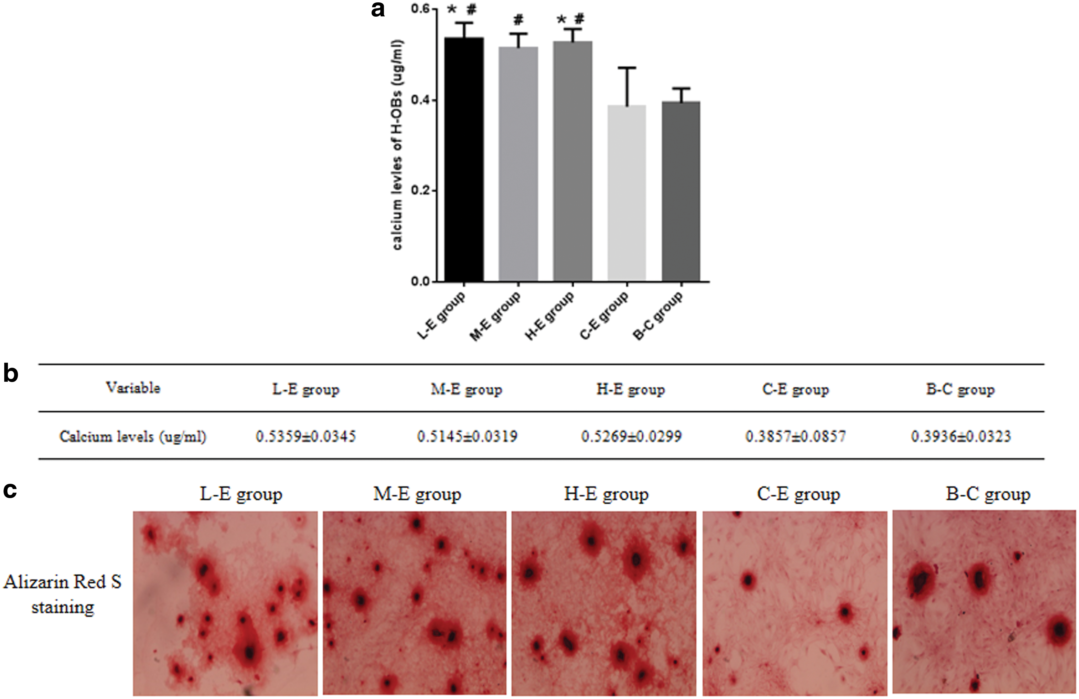
The results of Alizarin Red S staining and extracellular matrix calcium levels.
Tet-on Adv. COLIA1 stimulates bone callus collagen type I expression
Figure 4a and b shows the results of collagen type I expression in the bone callus. The collagen type I levels in group A peaked 2 weeks after adenovirus injection and then declined progressively for the subsequent 6 weeks. As time went on, no significant change in collagen type I was found in groups B and C after adenovirus injection. The collagen type I levels in group A were elevated significantly compared to those in groups B and C at 2 and 4 weeks. There was no significant difference among the three groups at 6 and 8 weeks.
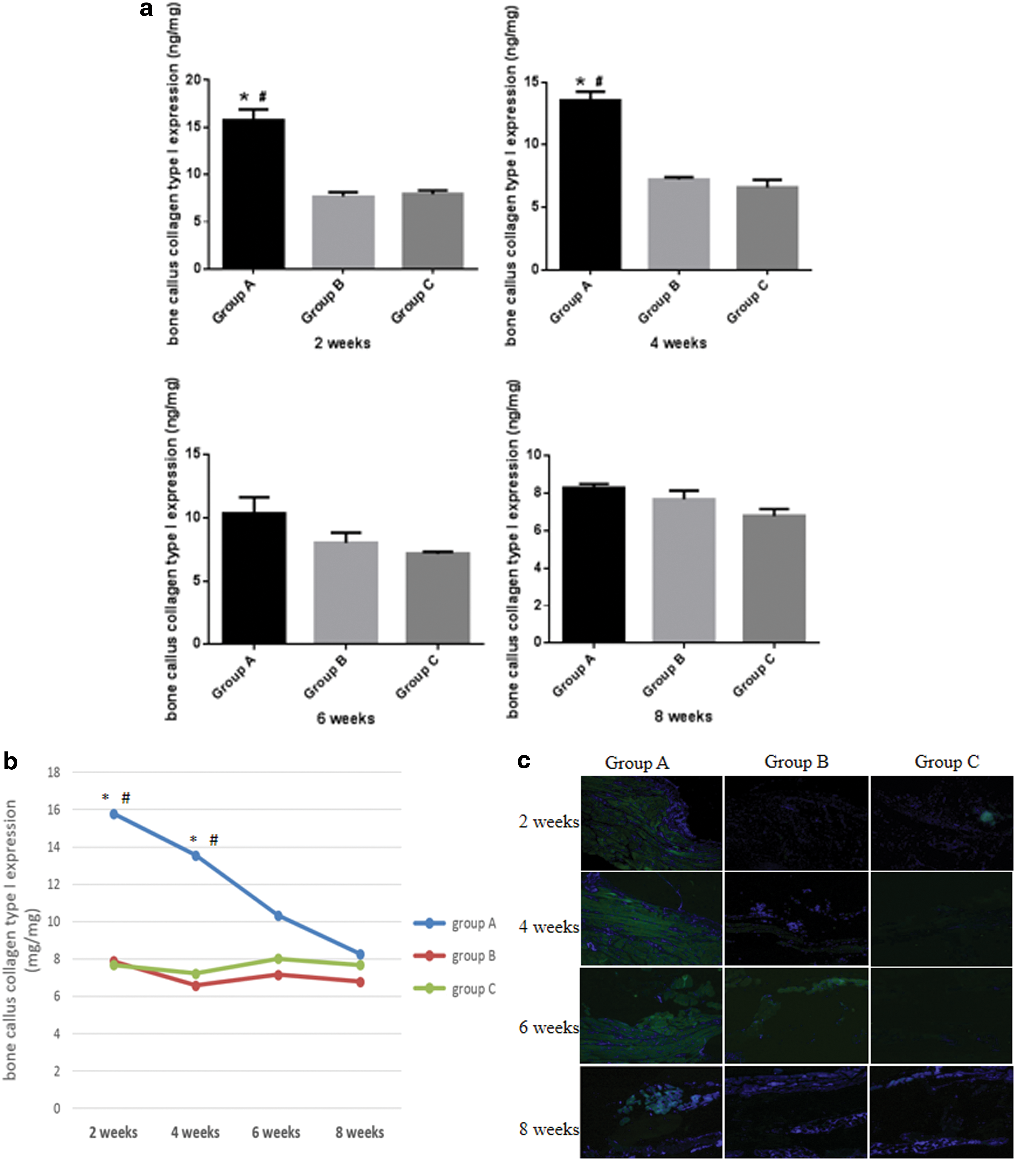
The results of bone callus collagen type I expression and fluorescent staining.
Figure 4c shows the representative microscopic views of IFS of the bone callus. Rats treated with adenovirus injection and a dox diet exhibited higher fluorescence emission than those injected with adenovirus only or saline, which indicates the collagen type I expression was elevated in group A.
Tet-on Adv. COLIA1 increases the BMD of the bone callus and stimulates bone fracture healing
The results of bone callus BMD are shown in Fig. 5a. In group A, callus BMD increased progressively after adenovirus injection and peaked at 8 weeks. No significant change in BMD was found in groups B and C. Callus BMD in group A was increased significantly compared to that in groups B and C at 8 weeks, and no significant difference was found among the three groups at 2, 4, and 6 weeks after virus injection. Figure 5b shows the results of bone callus histological analysis at 8 weeks. Eight weeks after adenovirus injection, all bone fractures in each rat were nearly healed. In group A, H&E and Masson staining showed active bone-cell differentiation and proliferation, regular arrangement of collagen fibers and bone trabecula, and no obvious inflammatory cell infiltration. In groups B and C, H&E and Masson staining showed a lower expression of bone cell, collagen fibers, and bone trabecula; inter-trabecula lacunas existed, with mild inflammatory cell infiltration. The results revealed that better fracture healing was obtained in group A compared to groups B and C.
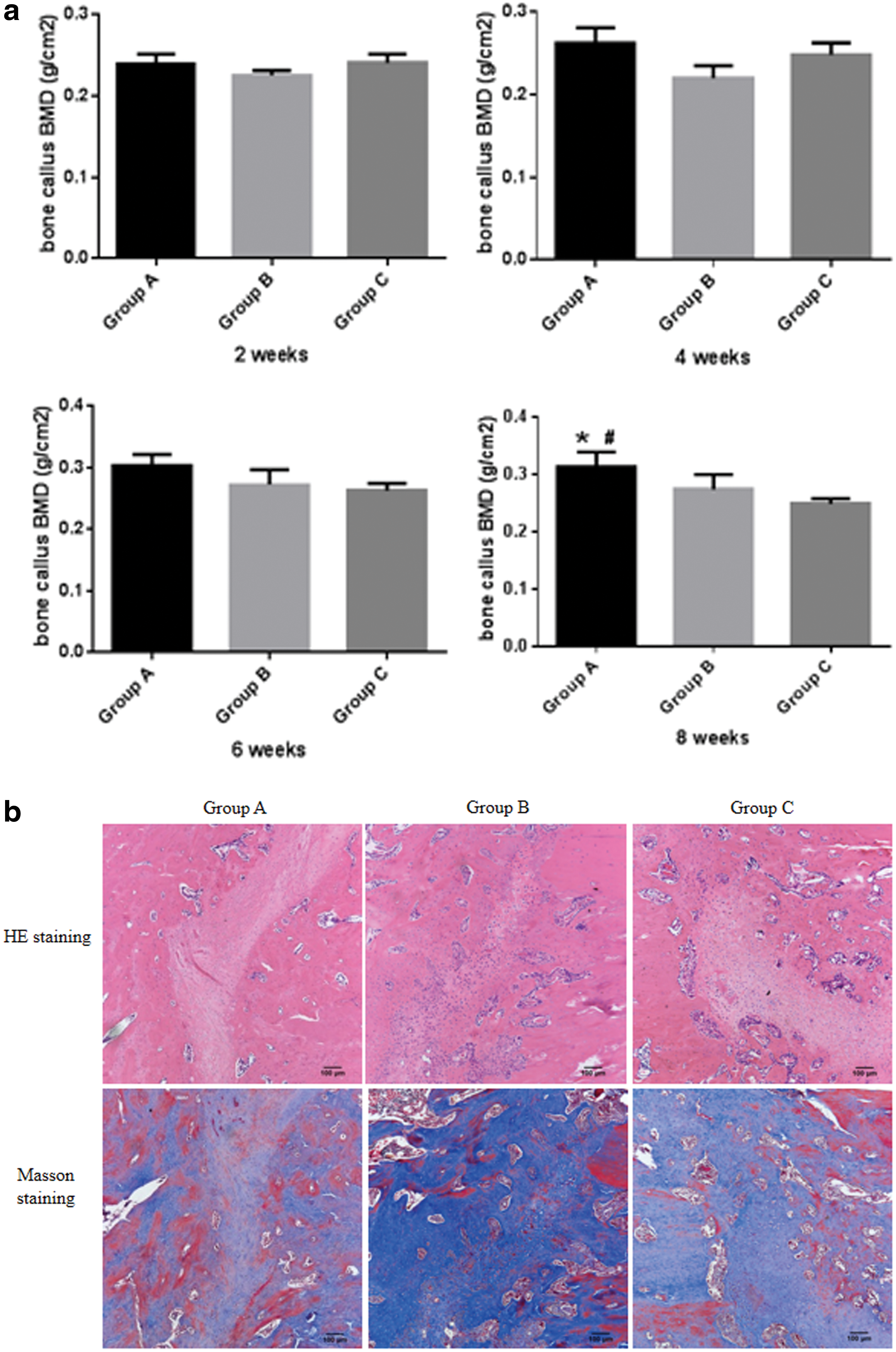
The results of bone callus bone mineral density (BMD) and histological analysis.
No overexpression of COLIA1 mRNA in extra bone tissue
Figure 6 shows the results of serum collagen type I and liver COLIA1 mRNA levels. The results show that there was no significant difference among the three groups in terms of serum collagen type I and liver COLIA1 mRNA levels at any weeks after adenovirus injection.

The results of serum collagen type I and liver COLIA1 mRNA levels. Serum collagen type I and liver COLIA1 mRNA levels were determined by ELISA and RT-PCR, respectively. The results showed there was no significantly difference among the three groups in terms of serum collagen type I and liver COLIA1 mRNA levels at any weeks after adenovirus, which indicated the COLIA1 gene from the adenovirus was not overexpressed in the blood and liver tissue.
Discussion
The present study demonstrated that direct gene delivery using tet-on Adv. COLIA1 could increase COLIA1 gene expression and collagen type I synthesis, which exhibited a direct osteogenic effect on H-OBs in vitro and enhanced osteoporotic fracture healing in vivo. Furthermore, the study also proved that the tet-on system could be an ideal system for tightly regulated gene expression mediated by the presence or absence of dox.
Osteoporosis is a common age-related systemic bone disorder characterized by low bone mass and micro-architectural changes, leading to an increased risk of low-impact fractures. 1,23 Several factors such as smoking, male sex, nutrition, sex-hormone status, and exercise are known to exert a significant influence on bone mass, but genetic factors have also been shown to be of great importance. 24,25 Several genes have been examined during the last 10–15 years, and those genes encoding the vitamin D receptor, estrogen receptor alpha, BMP, TGF-beta, and collagen protein are among the genes that have been shown to be associated with low BMD and increased fracture risk. 26 –29 For these genes variants, the COLIA1 gene is the most extensively studied candidate gene in the field of osteoporosis. 29 –33 This gene encodes the alpha 1 chain of collagen type I, the most abundant of all bone matrix proteins, and two alpha 1 chains interact with one alpha 2 chain to form the characteristic triple-helix collagen type I fibrils. It is evident that this protein is vital to bone strength, since coding mutations in the COLIA1 gene cause osteoporosis and osteogenesis imperfecta (a rare genetic disorder causing severe bone fragility). 34 A previous study on rats proved that the area of collagen type I in the callus is essential during the healing process of an osteoporotic fracture, and the combination of collagen type I and microarchitecture may act as a factor to determine the biomechanical strength of callus independent of BMD. 35 Two large-scale meta-analyses also confirmed the previous results and demonstrated that genetic variation at the COLIA1 locus influences susceptibility to osteoporosis. 5,6
Gene therapy is a technique whereby new genes are introduced into cells in order to treat disease by restoring or adding gene expression. 7,36,37 It has been widely used in the orthopedics field. Various cytokines and growth factors, including BMP, OPG, VEGF, IGF-1, TGF-alpha/beta, and omentin-1, have been found to reverse osteoporotic bone loss and enhance the healing process of osteoporotic fractures. 8 Using gene therapy, previous research has demonstrated that direct local or general application of virus encoding the gene sequence of the cytokine or growth factors above were able to promote osteoblast activity, increase BMD, and enhance fracture healing. 9 –13 Xie et al. 9 investigated the effects of omentin-1 on bone metabolism using virus gene transfection. The results showed adenovirus-mediated overexpression of omentin-1 reduced osteoclast formation through stimulating OPG and inhibiting RANKL production in vitro and partially resorted BMD and bone strength in ovariectomized mice in vivo. Egermann et al. 10 generated recombinant adenovirus vectors carrying human BMP-2 cDNA and tested their ability to transduce and stimulate mineralization and differentiation in primary ovine osteoblasts and mesenchymal stem cells. Large increases in ALP and mineralization confirmed the ability of local adenovirus delivery of BMP-2 to simulate osteoblastic differentiation. Bolon and Kostenuik performed animal studies on mice and found that virus vector carrying OPG cDNA reverses established osteopenia in ovariectomized mice without evidence of liver toxicity, which demonstrated virus delivery appears to be a safe and effective method for producing sustained systemic exposure to the target gene sequence. 13,38
Previously, an in vitro study was performed to determine the influence of −1997 G/T promoter polymorphism of the COLIA1 gene on the biochemical function of human osteoblasts. 39 The results showed that human osteoblasts with the TT genotype have low expression of COLIA1 mRNA and collagen type I. The lower amount of collagen type I synthesized by the TT genotype osteoblasts can decrease the matrix outside the bone cells and results in an insufficient site for calcium deposition, which explain why people with the TT genotype have lower BMD and a predisposition to osteoporosis. In the pilot study, an overexpression recombinant adenovirus vector carrying human COLIA1 cDNA was constructed, and its effect on osteoblasts after transfection was determined. 20 The results showed the osteoblast expression level of COLIA1 mRNA increases after transfection by adenovirus, leading to an improvement in collagen type I content and calcium nodule amount in the extracellular matrix. The current study incorporated the tet-on system into the upstream region of the COLIA1 gene and constructed the tet-on system regulated COLIA1 gene adenovirus. The results revealed, in the present of dox, the virus could increase COLIA1 mRNA expression and collagen type I levels, increase ALP expression, and stimulate extracellular calcium nodule formation, which exhibited an osteogenic effect on osteoblasts in vitro. The study also found that a single adenovirus local injection carrying COLIA1 cDNA could increase collagen type I expression and BMD in the callus, which might enhance osteoporotic fracture healing in vivo. No overexpression of serum collagen type I and liver COLIA1 mRNA was found. So, the series of studies demonstrated a strong correlation between the COLIA1 gene and osteoporosis, and the promising future of using genetic approaches to treat osteoporosis-associated disease.
In the in vitro study, the results showed there were no significant differences among the L-E, M-E, and H-E groups in terms of COLIA mRNA expression, H-OBs collagen type I expression, ALP activity, and mineralized matrix formation and calcium levels. The virus MOIs were 10 ifu/cell, 50 ifu/cell, and 100 ifu/cell in the L-E, M-E, and H-E groups, respectively. The same dose of 100 ng of dox, diluted in 1 mL of DMEM, was added to each group. The results demonstrate that dox could induce tet-on tet-on Adv. COLIA1 over expression of COLIA1 mRNA and collagen type I protein, which further promote ALP activity, stimulate mineralized matrix formation, and elevate calcium levels. However, this effect was not in a concentration-dependent manner. The optimal virus concentration and effect time need to be determined in the next step study.
The tet-on system is ideal for tightly regulated therapeutic gene expression mediated by adenovirus vector, which consists of two components: the rtTA and the TRE. In this system, tet or its analog dox controls the transgene expression: in the present of dox, rtTA binds to TRE and induces transgene expression; conversely, the gene expression is normally off unless dox is present. 16,17 The tet-on system was employed in several studies to regulate target gene expression, either following viral-mediated gene transfer or using plasmid transfection. 18,19 Tsai et al. 40 performed a study on mice, and the results showed that the tet-on system regulates IL-3 gene expression and enhances the immune response to prostate cancer. Wubbenhorst et al. 19 constructed an efficient tet-on system for the expression of BMP-2 in primary rabbit chondrocytes, which allow for the induction and termination of growth factor gene expression once cartilage regeneration is complete. The results showed the tet-on system is an effective strategy for efficient, repeatedly inducible expression of BMP-2 in primary rabbit chondrocytes. Xiong et al. 41 demonstrated that the regulatory tet-on system appears to be safe to use and capable of sustaining transgene expression in the brain, even in the presence of an immune response against its components. In addition, other studies that were performed in mice also demonstrated that the use of the tet-on system may be a promising approach to regulate transgene expression. 18,42 The current study showed, after being transfected by tet-on-regulated adenovirus, human osteoblasts expressed a low level of COLIA1 mRNA and collagen type I in the absence of dox (C-E group in the in vitro study and group B in the in vivo study). However, by adding an additional dose of dox, expression could be enhanced significantly, with the COLIA1 mRNA and collagen type I expression increasing significantly in the three experimental groups in the in vitro study and in group A in the in vivo study. The results demonstrate that the tet-on system is an ideal gene regulatory tool for effective and safe regulation of the therapeutic gene.
In conclusion, this study demonstrates that direct gene delivery using an adenovirus vector carrying human recombinant COLIA1 cDNA (Adv. COLIA1) can stimulate the osteogenic effect of osteoblasts in vitro and enhance bone fracture healing in vivo. It also proved that the tet-on system can control COLIA1 gene expression in the presence or absence of dox. These findings provide an important proof-of-concept that gene therapy with COLIA1 may be beneficial for preserving bone mass and strength in osteoporosis patients.
Footnotes
Acknowledgments
This research was funded by the National Natural Science Foundation of China Program (81601936 and 30600622) and Health and Family Planning Commission of Sichuan Province Program (17ZJ018 and 17PJ124).
Author Disclosure
The authors declare that they have no competing financial interests.