
Abstract
Oncolytic virotherapy is a new and safe therapeutic strategy based on the inherent cytotoxicity of oncolytic viruses and their ability to replicate and spread within tumors in a selective manner. In a previous study, a new type of oncolytic herpes simplex virus type 2 (oHSV-2, named OH2) was constructed to treat human cancers. That study demonstrated that OH2 is genetically and biologically stable. Its antitumor activity was maintained, even after passaging the virus for >20 generations. To advance OH2 into a clinical trial, a systematic preclinical safety evaluation was performed, which included: an acute toxicity test of OH2 in BALB/c mice; repeated dose toxicity tests of OH2 in BALB/c mice and cynomolgus monkeys; and biodistribution assays of OH2 in BALB/c mice, tumor-bearing mice, tumor-bearing nude mice, and cynomolgus monkeys. The results of this preclinical safety evaluation of OH2 indicate that OH2 is safe and suitable for clinical trials.
Introduction
Cancer is a type of malignant disease that threatens human health. Although medical science and technology have advanced quickly and the molecular mechanisms underlying carcinogenesis are being increasingly revealed, the total 5-year survival rate of human cancers remains low. 1,2 Surgical resection of primary tumors combined with chemotherapeutics and various types of radiotherapy remain the mainstream treatments. 3,4 In view of the shortcomings of the existing therapies, researchers have sought to exploit new therapeutic modes to conquer this deadly disease. Oncolytic virus (OV) therapy is an anticancer approach that utilizes replication-competent viruses specifically to kill tumor cells. 5 OV therapy with several viruses, including adenoviruses, herpesviruses and reoviruses, has recently shown promise in many tumor models. The herpes simplex virus (HSV) is an enveloped, double-stranded DNA virus that causes cold sores in its wild-type form. 6,7 There are eight human herpes viruses, which can be subdivided into the following three subfamilies: (i) Alphaherpesvirinae; (ii) Betaherpesvirinae; and (iii) Gammaherpesvirinae. 8 HSV type I (HSV-1) was one of the first viruses to be developed into a recombinant oncolytic virotherapeutic vector. 6 Modified HSV is utilized to diagnose and treat cancers. To date, several HSV mutants, including OncovexGM-CSF(talimogene laherparepvec [T-Vec]), 1716, G207, NV1020, HF10, and G47Δ, have either completed or entered Phase I, II, and III clinical trials to treat cancers of various degrees of malignancy from tissue origins, including melanoma, breast cancer, and glioma. 9 –14 However, determining of the optimal tumor treatment indications requires further investigation. In a previous study, a novel oncolytic HSV type 2 (HSV-2; HG52/ICP34.5-/ICP47-) was constructed that expressed human granulocyte-macrophage colony-stimulating factor (GM-CSF; OH2). The study demonstrated that the morphology, genome, and antitumor activity of OH2 remained stable, even after passaging the virus for >20 generations. 5 In the present study, a preclinical safety evaluation of OH2 was performed, including a single-dose acute toxicity test of OH2 in BALB/c mice, a repeated dose toxicity test of OH2 in BALB/c mice and cynomolgus monkeys, and a biodistribution test of OH2 in various murine models and cynomolgus monkeys. Before any novel medicines are approved for the initiation of human clinical trials, their safety must be established to support the clinical development and licensure of the product. Therefore, to advance OH2 into a human Phase I clinical trial, the present study comprised a comprehensive preclinical safety assessment of OH2, including single and repeated-dose toxicity studies conducted in mice and cynomolgus monkeys. The results of the preclinical safety evaluation of OH2 indicate that OH2 is safe and suitable for clinical trials.
Methods
Ethical statement
All animal tissue collection procedures were performed according to protocols approved by the Hubei Province, P.R. China, Biological Studies Animal Care and Use Committee, or the Institutional Animal Care and Use Committee of the National Centre for Safety Evaluation of Drugs, Beijing, P.R. China.
Cells and virus
The Vero cell line from Africa green monkey kidney cells and the HT-29 cell line from human colorectal cancer cells were purchased from the American Type Culture Collection (ATCC, Manassas, VA). Cells were cultured in Dulbecco's modified Eagle's medium (DMEM)/F12 medium supplemented with 5% newborn bovine serum and incubated at 37°C with 5% CO2 and saturated humidity. The CT26 mouse colon cancer cell line was purchased from the Cell Resource Center of Peking Union Medical College and was cultured in DMEM/F12 medium supplemented with 10% fetal bovine serum. OH2 oncolytic HSV-2 was constructed from the HG52 strain.
Mice and reagents
Male and female cynomolgus monkeys were purchased from Guangxi Guidong Quadrumana Development Laboratory Co. Ltd. (Guangxi, P.R. China). Seven-week-old male and female BALB/c and BALB/c nude mice were purchased from the Beijing Vital River Laboratory Animal Technology Co. Ltd (Beijing, P.R. China). All animals were housed under specific pathogen-free conditions.
Hemolysis test and red blood cell agglutination/flocculent test of OH2
Fresh rabbit blood (10 mL) was taken from the artery of the ear of a healthy rabbit, and the blood was shaken vigorously in a triangular flask with glass beads. Later, defibrinated blood with the fibrinogen removed was generated using an NR-150 cradle to shake the flask at room temperature at 10 g for 30 min. Next, physiological saline was added at a ratio of 10:1 (saline:defibrinated blood), vigorously shaken, and centrifuged at 400 g for 5 min. After the supernatant was removed, the precipitated red blood cells (RBCs) were then washed with physiological saline several times until the supernatant was clear. Next, 1 mL of precipitated RBCs plus 49 mL of normal saline was used to make the 2% rabbit erythrocyte suspension. For the experiment, seven clean tubes (nos. 1–5 for OH2, no. 6 for the 0.9% sodium chloride negative control, and no. 7 for the sterilized water positive control) were used (Supplementary Table S1). After adding the 2% RBC suspension, all tubes were immediately incubated at 37 ± 0.5°C. The tubes were monitored every 15 min for the first hour and once every hour for an additional 2 h.
Anaphylaxis test of OH2
Hartley male guinea pigs (n = 42) weighing 316.4–381.8 g were used. Four groups were established, including the formulation buffer group (the negative control), the bovine serum albumin group (BSA; positive control), and two OH2 test groups, including 6.25E5 and 6.25E6 CCID50/kg (drug concentration of 5E5 and 5E6 CCID50/mL, respectively). The sensitization to the back of the neck consisted of three subcutaneous injections made every 2 days at a 0.5 mL volume. On the 14th and 21st days after the last injection, animals 1–6 and animals 7–12 in each group were stimulated intravenously, respectively (1.0 mL/stimulation) (Supplementary Table S2). Allergy symptoms were monitored after the stimulation.
Acute toxicity test of OH2 in BALB/c mice
The following four groups were established, each containing 10 male and 10 female mice: the vehicle control (formulation buffer) group, the low-dose group, the high-dose group, and the negative control (saline solution, 0.9% NaCI) group. Animals were intravenously injected with OH2 (low-dose group: 2E6 CCID50/mouse; high-dose group: 4E6 CCID50/mouse; Supplementary Table S3). On day 15 after the injection, general toxicity was examined.
The following three groups were established, each containing 10 male and 10 female mice: the vehicle control (formulation buffer) group, the OH2 group, and the negative control (saline solution, 0.9% NaCI) group. Animals were subcutaneously injected with OH2 (4E6 CCID50/mouse; Supplementary Table S3). On day 15 after the injection, general toxicity was examined.
Repeated dose toxicity test of OH2 in BALB/c mice
The following four groups were established, each containing 72 male and 72 female mice: the vehicle control (formulation buffer) group, the low-dose group, the medium-dose group, and the high-dose group. Animals were subcutaneously injected with OH2 once a week for a total of six injections with the indicated dose (low-dose group: 1E5 CCID50/mouse; middle-dose group: 1E6 CCID50/mouse; high-dose group: 2E6 CCID50/mouse; Supplementary Table S4). On days 37 and 92, examinations of general toxicity, hematology, serum biochemistry, and pathological were carried out. The general toxicity assay included observations of clinical symptoms and the injection site, as well as measurements of body weight, organ weight, and food consumption. The blood test assay included analyses of the following: white blood cells (WBC), lymphocytes, granulocytes, monocytes, RBC, hemoglobin (Hgb), hematocrit, mean corpuscular volume, mean corpuscular Hgb (MCH), MCH concentration, platelet count, mean platelet, CD4+ lymphocytes, CD8+ lymphocytes, and CD4+/CD8+ ratio. The serum biochemistry assay included analyses of the following: alanine aminotransferase, aspartate aminotransferase, alkaline phosphatase, creatine kinase, lactate dehydrogenase, total bilirubin, blood urea nitrogen, creatinine, glucose, cholesterol, triglycerides, total protein, albumin (ALB), ALB/globulin (A/G), K+, Na+, and Cl−. The pathological examinations included the brain, spleen, heart, kidney, liver, lung, thymus, adrenal gland, ovary (only females), uterus (only females), testis (only males), and epididymis (only males).
The following four groups were established, each containing 36 male and 36 female mice: the control group (formulation buffer), low-dose group, middle-dose group, and high-dose group. Animals were subcutaneously injected with OH2 once per week for a total of six injections with the indicated dose (low-dose group: 1E5 CCID50/mouse; middle-dose group: 1E6 CCID50/mouse; high-dose group: 2E6 CCID50/mouse; Supplementary Table S5). Immunological tests were carried out on days 21, 42, and 92, including the following analyses: GM-CSF content in serum, antibodies against GM-CSF in serum, and antibodies against HSV-2.
Repeated dose toxicity test of OH2 in cynomolgus monkeys
The following three groups were established, each containing five male and five female cynomolgus monkeys: the vehicle control (formulation buffer) group, low-dose group, and high-dose group. Animals were subcutaneously injected with OH2 once per week for a total of six injections with the indicated dose (low-dose group: 1E7 CCID50/mL; high-dose group: 2E7 CCID50/mL). The repeated dose toxicity test of OH2 was carried out on days 37 and 92, including general toxicity, blood test, serum biochemistry, and pathological examination. The general toxicity assay includes observations of clinical symptoms and the injection site, as well as measurements of body weight, organ weight, and food consumption. The blood test assay included analyses of the following: WBC, lymphocytes, granulocytes, monocytes, RBC, Hgb, hematocrit, mean corpuscular volume, MCH, MCH concentration, platelet count, mean platelet, CD4+ lymphocyte, CD8+ lymphocyte, and the CD4+/CD8+ ratio. The serum biochemistry assay included the following: alanine aminotransferase, aspartate aminotransferase, alkaline phosphatase, creatine kinase, lactate dehydrogenase, total bilirubin, blood urea nitrogen, creatinine, glucose, cholesterol, triglycerides, total protein, ALB, A/G, K+, Na+, and Cl−. The pathological examinations included the brain, spleen, heart, kidney, liver, lung, thymus, adrenal gland, ovary (only females), uterus (only females), testis (only males), and epididymis (only males).
Biodistribution of OH2 in BALB/c mice
The following four groups were established, containing six or nine male and six or nine female mice per group: the negative control (saline solution, 0.9% NaCI) group, low-dose group, middle-dose group, and high-dose group. Animals were subcutaneously injected with OH2 once a week for a total of six injections with the indicated dose (low-dose group: 1E5 CCID50/per mouse; middle-dose group: 1E6 CCID50/per mouse; high-dose group: 2E6 CCID50/per mouse). On days 16, 37, and 92, the biodistribution of OH2 in indicated tissues was measured using HSV-2 gD sequence-based real-time polymerase chain reaction (PCR). Briefly, DNA was extracted from each frozen tissue type (approximately 25 mg) using a DNA Purification Kit (Tiangen, Beijing, P.R. China) according to the manufacturer's instructions. The concentration of eluted DNA was determined by UV spectrophotometry and adjusted to a final concentration suitable for quantitative PCR (qPCR). The primers (PF: 5′-CACGGATACGATCCGTAAGGA-3′; PR: 5′-CGAAAACGACGAGGTCTTGGT-3′) and probe (5′-FAM-CCTGCGGCTGTCGCTGGC-BHQ1-3′) were designed using Primer Express software (Applied Biosystems, Foster City, CA). The qPCR protocol consisted of an initial heating step at 94°C for 3 min, and then 40 cycles of incubation at 94°C for 30 s and 60°C for 1 min. A standard curve of plasmid DNA (101–106 copies), no template controls, and background DNA controls were all run in duplicate reactions. Furthermore, to monitor qPCR inhibition, tissue from a naive animal separately spiked with plasmid DNA (102–104 copies) was analyzed, and no inhibition was detected. The results were expressed as copy number per microgram of tissue, and the limit of detection was 10 copies of the target sequence.
Biodistribution of OH2 in tumor-bearing mice
The following two groups were established, each containing 18 male and 18 female mice per group: the negative control group (saline solution, 0.9% NaCI) and the OH2 group. Animals were subcutaneously injected with CT-26 cells. Five days after the injection, when tumors reached a volume of 300 mm 3 , mice were intratumorally injected with OH2 (2E7 CCID50/mL) on days 1, 4, and 7. The biodistribution was investigated on days 8, 14, and 28 using HSV-2 gD sequence-based real-time PCR as described above.
Biodistribution of OH2 in tumor-bearing nude mice
The following two groups were established, each containing 18 male and 18 female mice: the negative control group (saline solution, 0.9% NaCI) and the OH2 group. Animals were subcutaneously injected with HT-29 cells. Eight days after the injection, when tumors reached a volume of 300 mm 3 , mice were intratumorally injected with OH2 (2E7 CCID50/mL) on days 1, 4, and 7. The biodistribution was investigated on days 8, 14, and 28 using HSV-2 gD sequence-based real-time PCR as described above.
Biodistribution of OH2 in cynomolgus monkeys
The following three groups were established, each containing five male and five female cynomolgus monkeys: the negative control (saline solution, 0.9% NaCI) group, low-dose group, and high-dose group. Animals were subcutaneously injected with OH2 once a week for a total of six injections with the indicated dose (low-dose group: 1E7 CCID50/mL; high-dose group: 2E7 CCID50/mL). On days 37 and 92, the biodistribution of OH2 in indicated tissues was investigated using HSV-2 gD sequence-based real-time PCR as described above.
Statistical analysis
The data are presented as the mean ± standard deviation. Statistical analysis was performed using SPSS Statistics for Windows v17.0 (SPSS, Inc., Chicago, IL). A p-value of <0.05 was considered significant, and a p-value of <0.01 was considered highly significant.
Results
Hemolysis test and RBC flocculent test of OH2
To investigate whether OH2 leads to hemolysis and RBC flocculants, OH2 with 5E6 CCID50/mL and a 2% RBC suspension were used for the hemolysis and RBC flocculent test (Supplementary Table S1). The results indicated that OH2 did not cause hemolysis or RBC flocculants.
Anaphylaxis test of OH2
To investigate whether OH2 causes allergic reactions in Hartley guinea pigs, 42 Hartley guinea pigs were used for the anaphylaxis test (Supplementary Table S2). Twelve Hartley guinea pigs were in formulation buffer negative control group, six animals were in the BSA positive control group, and 12 animals were in the high-dose OH2 group and the low-dose OH2 group. These Hartley guinea pigs received a subcutaneous injection of OH2 once a day for a total of three sensitizations, followed by an intravenous injection of OH2 on days 14 and 21 after the third sensitization. Allergy symptoms were monitored after the stimulation. None of the OH2-tested animals that received both sensitization and stimulation exhibited allergy symptoms. The incidence of OH2-caused anaphylaxis was 0%.
Acute toxicity test of OH2 in BALB/c mice
To investigate the acute toxicity of OH2 in BALB/c mice, BALB/c mice were subcutaneously or intravenously injected with OH2. In the subcutaneous groups, 10 female BALB/c mice and 10 male BALB/c mice were subcutaneously injected with a single dose of OH2 at 4E6 CCID50 (Supplementary Table S3). In the intravenous groups, 10 female BALB/c and 10 male BALB/c mice were intravenously injected with a single dose of OH2 at 2E6 CCID50 or 4E6 CCID50 (Supplementary Table S3). No animals died during the test. Drug administration was not associated with abnormal clinical signs or abnormal body weight changes. In addition, there were no abnormal necropsy findings. All doses were well tolerated, with no signs of toxicity. The no observed adverse effect level (NOAEL) for the subcutaneous and intravenous administration of OH2 in mice was considered to be higher than 2E8 CCID50/kg in both sexes.
Repeated dose toxicity test of oncolytic HSV-2 in BALB/c mice
To investigate the repeated dose toxicity of OH2 in BALB/c mice, 216 BALB/c mice were used for the assay (Supplementary Tables S4 and S5). In this assay, 7- to 8-week-old BALB/c mice were subcutaneously injected with 1E5, 1E6, or 2E6 CCID50 OH2 once a week for a total of six injections. There were no treatment-related effects on body weight for males or females (Fig. 1A and B and Supplementary Table S6). There were no treatment-related gross necropsy findings for either scheduled euthanasia or early death animals in the study (Supplementary Table S7). There were no changes in hematology observed in this study (Supplementary Tables S8 and S9). No histopathological changes were observed between control group and OH2 administration group (Supplementary Figs. S1–S4). The concentration of GM-CSF in serum was not changed in the study. Anti-GM-CSF antibodies in the serum were not detected in the low-, middle-, or high-dose groups (Table 1). Anti-HSV-2 antibodies in the serum were detected in the low-, middle-, and high-dose groups (Table 2). There were no dose-dependent changes in serum biochemistry (Supplementary Tables S10 and S11) or organ weight (Supplementary Tables S12 and S13). The NOAEL for the subcutaneous repeated administration of OH2 in mice was considered to be higher than 1E8 CCID50/kg in both sexes.

Group mean body weight of mice and cynomolgus monkeys following repeated administration of OH2.
Anti-GM-CSF antibody in the serum of mice
GM-CSF, granulocyte-macrophage colony-stimulating factor; −, negative result.
Anti-HSV-2 antibody in the serum of mice
+, positive result.
Repeated dose toxicity test of oncolytic HSV-2 in cynomolgus monkeys
Thirty cynomolgus monkeys (15 female and 15 male) were randomly divided into three groups (Supplementary Table S14). The monkeys were subcutaneously injected with 3E6 CCID50/kg or 6E6 CCID50/kg OH2 separately once a week for a total of six injections. On days 37 or 92 post injection, the cynomolgus monkeys were sacrificed by lethal injection with an overdose of pentobarbitone by intracardiac injection after anesthesia. No animals died during the 5-week repeated dose toxicity study. There were no treatment-related changes in body weight (Fig. 1C and Supplementary Table S15), body temperature (Supplementary Table S16), electrocardiography (Supplementary Table S17), blood pressure (Supplementary Table S18), eyes (Supplementary Table S19), urinalysis (Supplementary Table S20), hematology (Supplementary Table S21), serum biochemistry (Supplementary Table S22), or serum protein electrophoresis (Supplementary Table S23). No histopathological changes were observed between the control group and the OH2 administration group (Supplementary Figs. S5–S8). The concentration of GM-CSF in the serum was not changed in the study. Anti-GM-CSF antibodies in the serum were not detected in the low-, middle-, and high-dose groups (Table 3). Anti-HSV-2 antibodies in the serum were not detected in the low- and high-dose groups (Table 4). Significant changes in the classification of bone-marrow cells between the vehicle control group and the high-dose group were not detected (Supplementary Table S24), and there were no dose-dependent changes in organ weight (Supplementary Table S25). The NOAEL for the subcutaneous administration of OH2 in cynomolgus monkey was considered to be higher than 6E6 CCID50/kg in both sexes.
Anti-GM-CSF antibodies in the serum of monkeys
Anti-HSV-2 antibodies in the serum of monkeys
Biodistribution of OH2 in BALB/c mice, tumor-bearing mice, tumor-bearing nude mice, and cynomolgus monkeys
To investigate the biodistribution of OH2, 7-week-old BALB/c mice were subcutaneously injected with OH2 once per week for a total of six injections (Supplementary Table S26). The biodistribution of OH2 DNA was monitored, and the results showed that there was no obvious replication or widespread distribution of OH2 in the mice during the experiment (Fig. 2A).
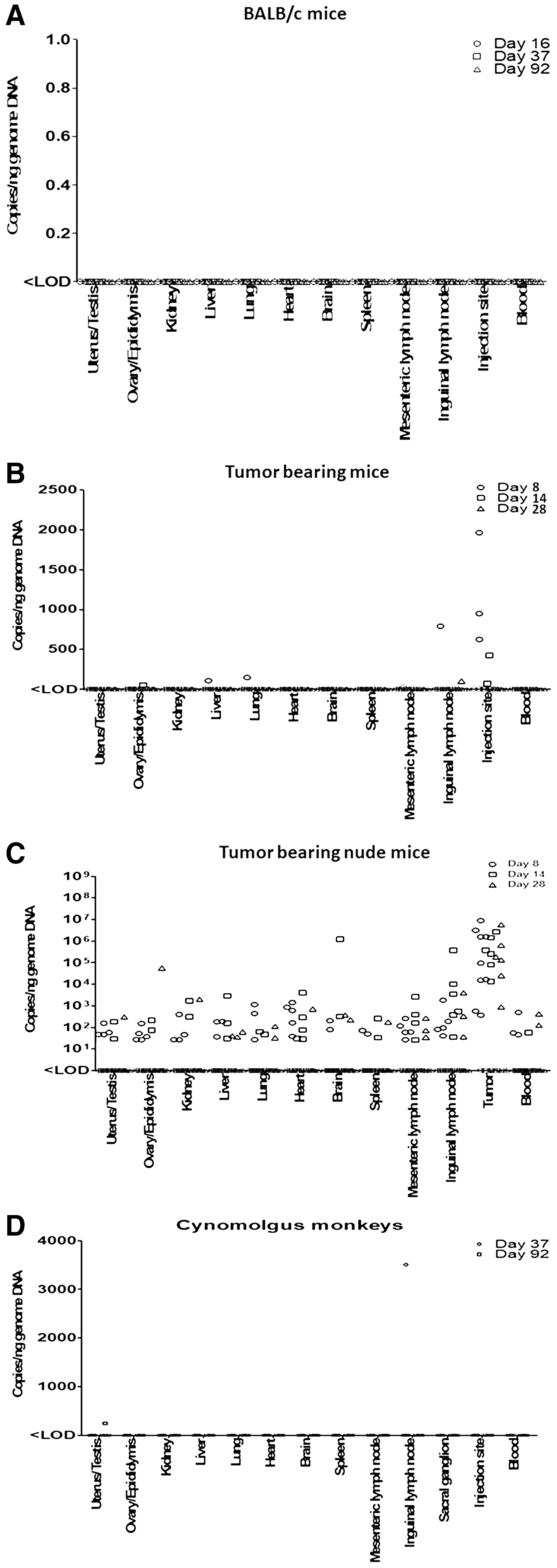
Biodistribution of OH2 DNA in various tissues of BALB/c mice
The biodistribution of OH2 was investigated using a tumor-bearing mouse model. Seven-week-old BALB/c mice were subcutaneously injected with CT-26 cells. Five days post injection, when tumors reached a volume of 300 mm 3 , mice were intratumorally injected with OH2 on days 1, 4, and 7. The biodistribution of OH2 DNA was detected on days 8, 14, and 28 (Supplementary Table S26). The results indicated no obvious replication or widespread distribution of OH2 in the mice during the experiment, except at the injection site and the inguinal lymph nodes. Additionally, OH2 was completely cleared in mice on day 28 (Fig. 2B).
To compare the biodistribution of OH2, a tumor-bearing nude mouse model was established. Nude mice were subcutaneously injected with HT-29 cells. Eight days post injection, when tumors reached a volume of 300 mm 3 , mice were intratumorally injected with OH2 on days 1, 4, and 7. The biodistribution of OH2 DNA was monitored on days 8, 14, and 28 (Supplementary Table S26). The results indicated that the test viral DNA was localized primarily in the tumor. Additionally, OH2 DNA was also widely distributed in other tissues, but the abundance of OH2 DNA in these tissues was much lower than that in the tumor and exhibited a decreasing trend over time (Fig. 2C).
To compare the biodistribution of OH2, a cynomolgus monkey model was established. Cynomolgus monkeys were subcutaneously injected with OH2 once per week for a total of six injections (Supplementary Table S26). The biodistribution of OH2 was monitored. Viral DNA >3,000 copies was detected in one animal in the inguinal lymph node near the injection site on day 32. The results indicated that OH2 did not replicate, and viral DNA was not detected in the animal tissues during the administration period or the recovery period (Fig. 2D).
Discussion
Oncolytic viral therapy is emerging as a novel cancer treatment. In previous work, a new type of oncolytic HSV-2 (OH2) was constructed to treat human cancers, and it was demonstrated that OH2 is stable in its genomic and biological characteristics. 5 Moreover, the antitumor activity persists, even after the passage of the virus for >20 generations. This study provides preclinical safety data, including an acute toxicity test of OH2 in BALB/c mice, a repeated dose toxicity test of OH2 in BALB/c mice and in cynomolgus monkeys, and an analysis of the biodistribution of OH2 in BALB/c mice, tumor-bearing mice, tumor-bearing nude mice, and cynomolgus monkeys. Taken together, these tests support OH2 as a potential therapeutic agent for cancer treatment.
Preclinical safety evaluations of OVs have been widely carried out because the systematic evaluation of OV toxicology in the preclinical stage is a pivotal step for OVs to enter clinical study. Fang et al. reported that oncolytic adenovirus was safe for treating bladder cancer in a subcutaneous xenograft tumor model in nude mice. 15 Nathan carried out toxicology studies of an oncolytic vesicular stomatitis virus (VSV) expressing human interferon-β in rhesus macaques and demonstrated that no neurological signs or abnormalities were observed, and all macaques developed neutralizing anti-VSV antibodies. 16 Preclinical safety evaluations were also carried out in HSV-1 viruses, of which 1716 (Δ34.5), G207 (ΔICP6 and 34.5 with expression of LacZ), and T-Vec (Δ34.5 and ICP47 with expression of GM-CSF) were shown to be safe in preclinical safety evaluations. 17 T-Vec was recently approved as the first oncolytic virus drug in the United States and Europe. 18
In preclinical pharmacology and toxicology research, OH2 has exhibited good efficacy and safety. In a series of toxicity assays under good laboratory practice (GLP) or non-GLP conditions, animals tolerated OH2 well, and no organs were damaged. These results suggest that OH2 does not propagate in normal tissues and organs. In addition, the following biological distribution assays showed that OH2 selectively propagated in tumor cells. (1) The biological distribution assay of BALB/c mice that received six repeated doses revealed that during the administration and recovery period, OH2 was not widely detected in the mice. (2) The biological distribution assay of tumor-bearing mice that received six intra-tumoural injections of OH2 revealed that OH2 was primarily localized in the tumor, and OH2 was only detected in a few animals. As time progressed, no OH2 was detected in the normal organs, and the OH2 in the tumors decreased significantly. (3) The biological distribution assay of tumor-bearing nude mice revealed that OH2 was primarily distributed in tumors after OH2 injection, and there was obvious viral replication. Although OH2 was also detected in normal tissues, the copy numbers of OH2 in normal tissues were much lower than those in the tumors, and the OH2 copies decreased over time. Cases in which viral DNA persisted in nude mice for a long time could be caused by the immune deficiency of nude mice. (4) The biological distribution assay of cynomolgus monkeys that received six doses injection revealed that there was no obvious replication of the OH2 virus in the cynomolgus monkeys during the administration period, and viral DNA was only detected in the inguinal lymph nodes near the injection site in certain animals at the end of the administration period. In contrast, OH2 viral DNA was not detected during convalescence. Interestingly, the OH2 sequence was detected in tested tissues, especially at the injection site and the inguinal lymph node of the tumor-bearing nude mice. Nude mice lacking a thymus presumably lacked the mechanism to clear OH2. Not surprisingly, oncolytic virus was found at the tumor injection site in nude mice in the preclinical safety evaluation, and Fang reported that the tumor injection site exhibited extremely high expression of viral genes compared with the control mice. 15
One purpose of the toxicology study is to determine the dose range that should be used in the subsequent clinical trials. The G207 Phase I trial included 21 patients who received doses ranging from 1 × 10⁶ to 3 × 10⁸ pfu in 0.1 mL and 1 × 10⁹ pfu in 0.3 mL at a single site, with a final cohort receiving 3 × 10⁹ pfu inoculated at five sites (0.2 mL each) 11 . A Phase I study of OncoVEXGM-CSF (T-Vec) included 13 patients in a single-dose group who received doses of 106–108 pfu/mL and 17 patients in a multi-dose group testing a number of dose regimens. 19 The results demonstrated that Onco VEXGM-CSF is well tolerated and can be safely administered. The initial and maximum doses of OH2 in patients are proposed based upon the preclinical safety data obtained from animal models. Determination of the initial dose of OH2 was based on the following: in the long-term toxicity study of subcutaneous injection of OH2 in cynomolgus monkeys, the doses of the low- and high-dose groups were 3E6 and 6E6 CCID50/kg, respectively. The animals were observed continuously for 3 months after administration of the treatment. During the experiment, the clinical symptoms, body weight, food intake, ophthalmologic examination, electrocardiogram, urine biochemical examination, hematology, hemagglutination, serum biochemistry, and peripheral blood T lymphocyte distribution showed no significant changes related to the test article. The organ weights of the animals did not show any significant changes associated with the test article. Therefore, taking the low dose 3E6 CCID50/kg to calculate the human equivalent dose (HED), HED = 3E6 CCID50/kg × 0.32 (conversion factor) = 0.96E6 CCID50/kg. The HED was further divided by the safety factor (usually set to 10) to get the maximum recommended starting dose (MRSD) of 0.96E5 CCID50/kg. Assuming a human body weight of 50 kg, the MRSD would be 0.96E5 × 50 = 4.8E6 CCID50. For further safety consideration, the starting dose of OH2 was set to 1E6 CCID50. Determination of the maximum dose of OH2 was based upon the following: the preclinical safety evaluation showed that the NOAEL of subcutaneous/intravenous OH2 virus in mice was 4E6 CCID50/mouse (20 g), equivalent to 2E8 CCID50/kg. Based on mouse body weight of 20 g, the HED was calculated as follows: HED = 2E8 CCID50/kg × 0.08 (conversion factor) = 1.6E7 CCID50/kg. Assuming a human body weight of 50 kg, the human equivalent dose was 1.6E7 × 50 = 8E8 CCID50/dose.
In conclusion, the systematic preclinical safety data presented here are sufficient to approve OH2 for clinical trials.
Footnotes
Acknowledgments
This work was supported by General Program of Hubei Province Basic Science Foundation (2015CFB470); National Major Scientific and Technological Special Project for “Significant New Drugs Development” (2015ZX09501007-004); National Major Scientific and Technological Special Project for Significant New Drugs Development (2018ZX09733002); and National Science and Technology Major Project of China (2018ZX09201017-001).
Author Disclosure
Y.D., J.Z., Z.M., X.S., and Z.F are employed by Wuhan Binhui Biotechnology Co. Ltd. The authors would like to state clearly that there are no commercial conflicts with this work. There are no patents, products in development, or marketed products to declare. Interpretation of the results was completely independent from any company's opinion. This affiliation does not alter the authors' adherence to all policies on sharing data and materials.
References
Supplementary Material
Please find the following supplemental material available below.
For Open Access articles published under a Creative Commons License, all supplemental material carries the same license as the article it is associated with.
For non-Open Access articles published, all supplemental material carries a non-exclusive license, and permission requests for re-use of supplemental material or any part of supplemental material shall be sent directly to the copyright owner as specified in the copyright notice associated with the article.