
Abstract
Inefficient autologous tissue recovery in skin wounds increases the susceptibility of patients to infections caused by multidrug resistant microorganisms, resulting in a high mortality rate. Genetic modification of skin cells has become an important field of study because it could lead to the construction of more functional skin grafts, through the overexpression of antimicrobial peptides that would prevent early contamination and infection with bacteria. In this study, we produce and evaluate human skin equivalents (HSEs) containing transfected human primary fibroblasts and keratinocytes by polyplexes to express the antimicrobial peptide LL-37. The effect of LL-37 on the metabolic activity of normal HSEs was evaluated before the construction of the transfected HSEs, and the antimicrobial efficacy against Pseudomonas aeruginosa and Staphylococcus aureus was evaluated. Subsequently, the levels of LL-37 in the culture supernatants of transfected HSEs, as well as the local expression, were determined. It was found that LL-37 treatment significantly promoted the cellular proliferation of HSEs. Furthermore, HSEs that express elevated levels of LL-37 were shown to possess histological characteristics close to the normal skin and display enhanced antimicrobial activity against S. aureus in vitro. These findings demonstrate that HSEs expressing LL-37 through nonviral modification of skin cells are a promising approach for the prevention of bacterial colonization in wounds.
Introduction
Healthy and intact skin acts as a barrier that prevents the entrance of microorganisms from the environment. 1 Wounds caused by mechanical trauma, surgical interventions, chronic wounds, burns, or the natural aging compromise the integrity of this barrier and favor bacterial contamination, which can complicate wound closure and patient recovery, increasing morbidity and mortality, as well as the socioeconomic impacts worldwide. 2,3
Opportunistic pathogens such as Pseudomonas aeruginosa and Staphylococcus aureus are able to infect open wounds and produce biofilms that, once established, protect the bacteria against the effects of commonly used antibiotics. Consequently, chronic wounds infected with populations of resistant bacteria can develop that compromise the lives of patients. 4 –6 Multiple dressing formats have been commonly implemented to inhibit microbial proliferation or treat local infections. Broad-spectrum antimicrobial agents can be combined with dressing materials such as foams, hydrogels, alginates, collagens, or films. 7,8 However, because of increasing antimicrobial resistance, these agents cannot be used for extended periods of time. Autologous skin grafts are also used for the treatment of these lesions, nevertheless, they are limited by the availability of healthy donor tissue in cases of large skin loss. 9 Therefore, the development of innovative strategies to prevent and combat infected cutaneous wounds is still needed. 10,11
The physical barrier provided by the skin is augmented by the innate immune system and the expression of the host defense peptides (HDPs), which act as a chemical barrier to reinforce the protection of the tissue. However, the wound environments are often depleted of HDPs, which can decrease the early response against microorganisms. HDPs are produced mainly by keratinocytes of the epidermis. In mammalian species, the two major classes of HDPs are the defensins and cathelicidins. The linear peptide LL-37 is the only member of the cathelicidins family identified in humans. 12,13 The expression of LL-37 is regulated by the transcription of the CAMP (cathelicidin antimicrobial peptide) gene, which translates to an 18 kDa proprotein known as hCAP18 (human cationic antimicrobial protein), that releases the active C-terminus 37 amino acid peptide after post-translational processing.
In addition to its antimicrobial activity against a variety of bacteria, 14,15 LL-37 is involved in the chemotaxis of immune and endothelial cells, as well as keratinocyte proliferation during the wound healing processes. 16 For these reasons, LL-37 has been a promising tool for wound treatment development as an alternative to conventional antibiotics. However, the therapeutic application of synthetic LL-37 in clinical practice has been hindered by the high cost of peptide synthesis and biological disadvantages, such as their susceptibility to proteolysis in the wound bed and their poor activity under physiological conditions. 17
Currently, human skin equivalents (HSEs) offer another alternative for the treatment of skin wounds. HSEs are three-dimensional (3D) models based on biopolymers, in which different cell populations can be incorporated to favor the covering of the lesion and provide a bed for new cells to regenerate the lost tissue. Biopolymers such as fibrin have been used successfully for the construction of HSE, as it can stimulate the production of extracellular matrix by fibroblasts, providing an environment that promotes tissue regeneration. 18,19 Moreover, gene therapy has allowed to genetically modify the skin cells to increase the production of antimicrobial peptides such as the LL-37. 20 These modified cells can be incorporated in the HSEs to function as “bioreactors” in vivo. The gene transfer to epidermal cells has been commonly performed through retroviral methods, which, despite its high efficiency, have disadvantages such as immunogenicity, oncogenicity, and development of mutagenic processes. 21,22
Consequently, the use of synthetic nonviral vectors has generated a great interest in protocols for clinical use in gene therapy. In particular, polyethylenimine (PEI) polyplexes have been widely used for nonviral transfection in mammalian cells due to their ability to protect nucleic acids against premature degradation, and thus facilitate their delivery through the plasma membranes. 23,24 This system, together with the use of scaffolds such as HSEs, allows maintaining the in vitro expression of the protein of interest and reducing the cytotoxicity that some of these systems present against the target cells. In addition, the development of HSEs with modified autologous cells could provide new and improved properties by targeting the excretion of those proteins of interest specifically to the site of action, preventing any systemic side effects such as immune recognition and rejection. 25
In this report, we first investigate the antimicrobial efficacy of the synthetic LL-37 peptide and its effects on human primary fibroblasts and keratinocytes. Then, we demonstrate that HSEs generated from skin cells genetically modified by linear polyethyleneimine (LPEI) polyplexes express elevated levels of the LL-37 and possess enhanced antimicrobial activity in vitro. This approach could support the coverage of cutaneous wounds and prevent bacterial growth, which could keep the wound bed in more sterile conditions favoring the closure and recovery of the tissue.
Materials and Methods
Materials
Synthetic LL-37 (LLGDFFRKSKEKIGKEFKRIVQRIKDFLRNLVPRTES)was purchased from PolyPeptide group (Malmö, Sweden) at ≥90% purity, confirmed by high-performance liquid chromatography. LPEI (25 kDa) was purchased from Polysciences and the recombinant plasmid containing the CAMP sequence coding to the hCap18/LL-37 expression was purified from existing Escherichia coli stocks, using a plasmid maxi kit (Qiagen EndoFree®). The plasmid (hereafter called pDNA) allows the expression of both the reporter gene (red fluorescent protein [RFP]) and the gene of interest (CAMP). Normal human dermal fibroblasts (NHDF) and normal human epidermal keratinocytes (NHEK) tested and certified as mycoplasma free and virus free (HIV-1, hepatitis B, and hepatitis C) were purchased from Lonza, The Netherlands. The reagents for cell culture such as Dulbecco's modified Eagle's medium (DMEM) high glucose, the nutrient mixture F-12 Ham (Ham's F-12), and fetal bovine serum (FBS) were purchased from Gibco, Thermo Fisher Scientific, USA.
Bacterial strains and culture conditions
S. aureus ATCC 25923 and P. aeruginosa PAO1 were grown from stock solutions (7% dimethylsulfoxide [DMSO], at −80°C) on blood agar plates at 37°C for 24 h. Bacteria were cultured to late-stationary phase in tryptic soy broth (TSB; OXOID, Basingstoke, United Kingdom) as described hereunder. Single colonies were inoculated into 10 mL TSB and incubated overnight at 37°C. Then, this preculture was inoculated into 200 mL TSB in the absence or presence of 50 mM sodium bicarbonate (NaHCO3, pH 7.4) to evaluate the impact of bicarbonate on the antimicrobial potency of the LL-37 peptide. Bacterial cultures were grown for 24 h at 37°C with continuous rotation (200 rpm) and harvested by centrifugation for 5 min at 5,000g. After removal of the broth, bacterial pellets were washed twice in phosphate-buffered saline (PBS; 5 mM K2HPO4, 5 mM KH2PO4, 150 mM NaCl, pH 7.4), and sonicated three times for 10 s (Vibra cell model 375; Sonics and Material, Inc.). The final concentrations were adjusted to 1 × 10⁵ bacteria/mL using a Bürker–Türk counting chamber.
Minimum inhibitory concentration and minimum bactericidal concentration assays
The minimum inhibitory concentrations (MICs) of the synthetic LL-37 peptide were determined using the broth microdilution method. In brief, twofold dilution series of the peptide starting from 256 to 0.25 μM were prepared in 96-well plates in PBS. In each well, 100 μL of a bacterial suspension containing 1 × 10⁵ bacteria/mL prepared in absence or presence of 50 mM NaHCO3 (pH 7.4) was added. The 96-well plates were incubated at 37°C for 24 h and the MIC was determined as the lowest peptide concentration at which bacterial growth was not visually observed. Subsequently, the minimum bactericidal concentrations (MBCs) were determined from the broth dilution of MIC assays (starting from the MIC values with no visual growth) by inoculating 10 μL on TSB agar plates. The agar plates were incubated for 24 h at 37°C. The lowest concentrations of LL-37 at which no colonies were seen on the plates were defined as the MBC.
Also, the absorbances from the wells were recorded after 24 h of incubation using a spectrophotometer (Shimadzu, Japan) at 600 nm to compare bacterial growth in the absence and presence of NaHCO3 with the respective negative controls, which consist of the bacterial suspension without peptide exposition or 0 μM LL-37. Three independent experiments with separately cultured bacteria were performed, each with three replicates.
Cell cultures and generation of HSEs
Fibroblasts were cultured in DMEM high glucose supplemented with 10% (v/v) FBS and 1% (v/v) penicillin–streptomycin. Keratinocytes were cultured on a mitomycin C-treated 3T3 feeder layer and DMEM: Ham's F-12 (3:1) supplemented with 10 (v/v) % FBS and 10 ng/mL epidermal growth factor (EGF; Austral Biologicals, USA). Only passages between 1 and 5 were used for all experiments.
The generation of the HSEs was performed according to the protocol established by the Tissue Engineering and Cellular Therapy Group, University of Antioquia, Colombia although with some modifications. 26 In brief (Fig. 1), a mixture of pooled human plasma (blood derived from donors), 150 mM NaCl, 5 μL of 5 mg/mL tranexamic acid (CAS Number 1197-18-8; Sigma), and a fibroblast suspension (2 × 10⁵ cells/mL) was prepared. Then, 1% CaCl2 (Sigma) was added to promote fibrin polymerization, and after gently mixing the solution, it was poured into cell culture inserts (Transwell®; Corning 3460) with 0.4-μm porous membranes, and incubated at 37°C, 5% CO2 for 30 min. Subsequently, each fibrin gel was covered with DMEM, high glucose supplemented with 10% FBS, and incubated overnight at 37°C. Afterward, a keratinocyte suspension (2 × 10⁵ cells/1.2 cm2) was seeded on top of each gel in the transwell, to obtain keratinocytes versus fibroblasts ratio of 3:1. The cultures were kept immersed in the cell culture medium for 7 days, refreshing the medium every 3 days. Next, the medium from the “epidermal” zone in the transwell was removed and the keratinocytes were exposed to the air–liquid interface for 14 days at 37°C, and 5% CO2 to stimulate the stratification.
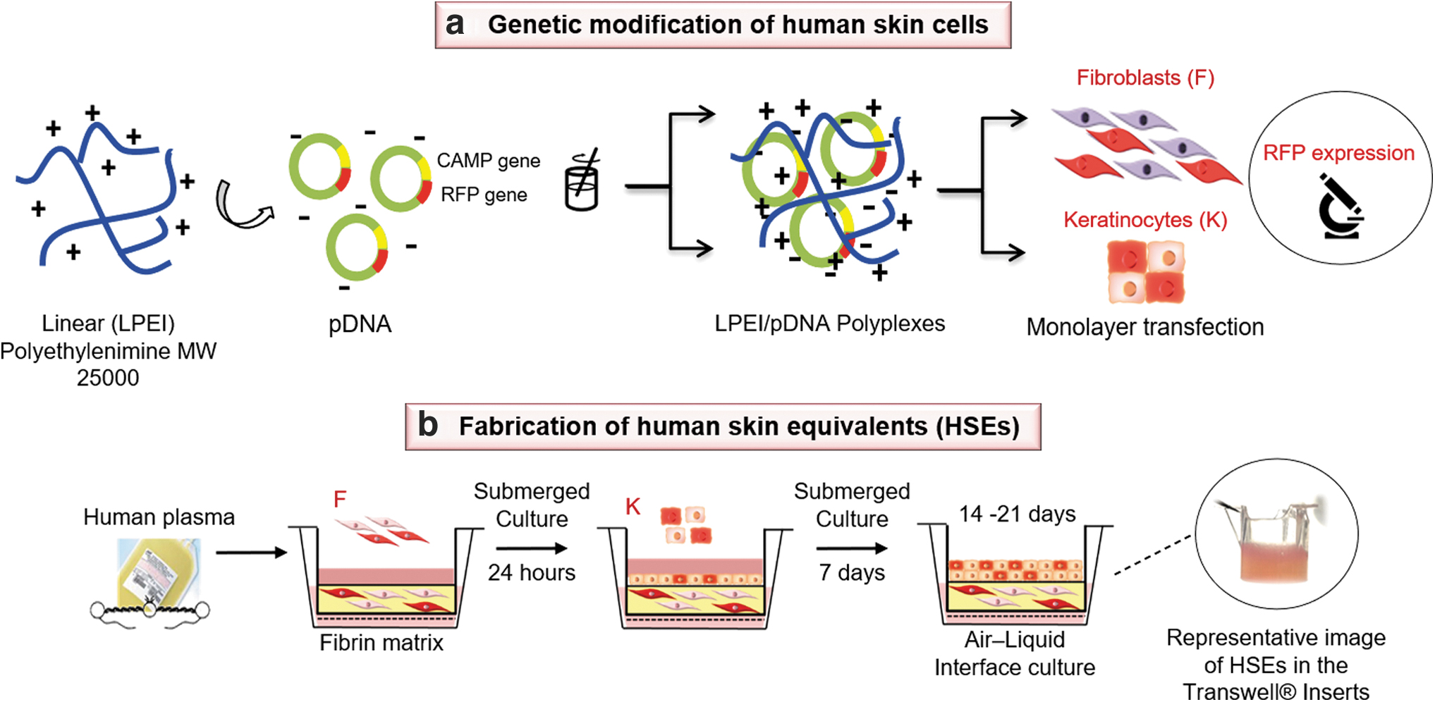
Schematic representation of the process for fabrication of HSEs with transfected cells. In
Generation of HSEs modified by LPEI/pDNA polyplexes
Figure 1 shows the culture protocol overview of HSEs with transfected skin cells. In brief, keratinocytes and fibroblasts were seeded separately 24 h before transfection into 24-well plates (5 × 10³ cells/cm2) and incubated at 37°C in 5% CO2. The transfection procedure was carried out using a protocol previously published. 27,28 LPEI/pDNA polyplexes were initially prepared in 4-(2-hydroxyethyl) piperazine-1-ethane-sulfonic acid (HEPES)-buffered saline (150 mM NaCl, 20 mM HEPES, pH 7.4) with a N/P ratio of 19 (molar ratio of nitrogen in LPEI [6.3 μg] to phosphorus in pDNA [2.5 μg]). Next 50 μL of this polyplexes solution was mixed with 0.5 mL DMEM:Ham's F-12 (3:1) low in serum (1% FBS) without antibiotics. Subsequently, the plates were centrifuged at 210g for 5 min and the cells were incubated with the polyplexes for 6 h at 37°C, and 5% CO2. Finally, the medium was replaced with fresh medium to remove the remaining polyplexes, and the cells were incubated for an additional 24 h.
The cells were visualized using fluorescence microscopy (Leica DMR Leica, Wetzlar, Germany) to confirm the expression of the RFP reporter gene and the HSEs were constructed as previously described with a final volume of 0.5 mL.
Effects of LL-37 on HSEs
To examine the effect of LL-37 peptide, the metabolic activity of HSEs at different stages of the cell culture was determined. Samples of HSEs were prepared as already described and cultured in cell growth medium [DMEM: Ham's F-12 (3:1) supplemented with 10% FBS] at 37°C and 5% CO2 atmosphere. The growth medium was mixed with decreasing concentrations of LL-37 (twofold dilutions from 256 to 2 μM) and then added to the HSEs for 1, 7, and 14 days. The medium was replaced with fresh medium supplemented with LL-37 every 3 days.
Subsequently, a solution of 500 μL of XTT [(2,3-bis (2-methoxy-4-nitro-5-sulfophenyl)-2H-tetrazo-lium-5-carboxanilide salt); A8088 AppliChem, The Netherlands] was added to the growth medium present on top of each sample. After 6 h of incubation at 37°C, 100 μL of the mixture was taken from each sample and absorbances A460nm and A690nm were measured using a spectrophotometer (Shimadzu, Japan). The metabolic activity of the LL-37-treated HSEs was calculated relative to the negative control samples (HSEs exposed to growth medium in the absence of LL-37) according to Equation (1).
The absorbance of all samples was corrected by subtracting the culture medium background from samples that do not contain cells and treated identically as the HSEs exposed to LL-37 or negative controls.
To study the effect of LL-37 on cell proliferation of HSEs, a group of samples exposed to various concentrations of the peptide was fixed with a 3.7% paraformaldehyde (Sigma) solution for 1 h and subsequently washed with PBS three times. Then, the samples were permeated using a 0.5% Triton X-100 (Sigma) solution for 3 min and blocked for nonspecific binding with 5% bovine serum albumin (BSA; Sigma) in PBS (PBSA) for 30 min. Afterward, the samples were stained using 4′,6-diamidino-2-phenylindole (DAPI) (Sigma; D9564, ratio: 1:500), and tetramethylrhodamine isothiocyanate-phalloidin for visualization of the nucleus and cytoskeleton of the cells, respectively. Cells were visualized with a fluorescence microscope (Leica DMR, Wetzlar, Germany) after 14 days in culture and the number of cells was reported as the number of nuclei in 0.5 mL of gel.
Enzyme-linked immunosorbent assay
To test whether microbial stimulation increases the expression of the LL-37 peptide, culture supernatants were collected from monolayers of fibroblasts and keratinocytes transfected and nontransfected, cultured for 7 days. For the stimulation, a bacterial culture suspension was prepared by inoculating 1 × 10⁵ bacteria/mL S. aureus ATCC 25923 in TSB overnight, then, bacteria were harvested by centrifugation for 5 min at 5,000g and the supernatant was carefully removed, and filter sterilized using a standard PTFE filter (Millipore; PTFE, 0.22 μm). The bacterial spent medium was mixed in DMEM–TSB (9:1) and poured on top of the cell monolayers. Then, the cultures were incubated for 24 h, at 37° C and 5% CO2.
In addition, cell culture supernatants from transfected and nontransfected HSEs, cultured for 14 and 21 days, were collected.
Concentrations of LL-37 in the culture supernatants from both cell monolayers and HSEs were quantified with a commercially available hCAP-18/LL-37 ELISA kit (Hycult Biotech, The Netherlands) using a plate reader (Shimadzu, Japan) according to the manufacturer's protocol. Cell supernatants from three independent experiments were analyzed.
Histology and immunohistochemistry
HSEs and transfected HSEs cultured for 21 days were fixed in 3.7% (v/v) paraformaldehyde. The fixed tissues were paraffin embedded or frozen in Tissue-Tek® O.C.T. Compound (Sakura Finetek Europe B.V., Alphen aan den Rijn, The Netherlands) for histological and immunofluorescent analysis, respectively. Paraffin-embedded blocks were sectioned into 5-μm thickness samples and stained with hematoxylin and eosin (H&E).
Frozen HSEs were cryosectioned to 7 μm thick slices in a Slee MNT cryotome, fixed in acetone at −20°C and air dried. Next, the sections were incubated with a blocking solution (2% BSA in PBS) for 30 min and then incubated for 1 h at 37°C with rabbit polyclonal antibodies against LL-37 (1:150) (Innovagen, Sweden). Afterward, samples were carefully washed in PBS and exposed to polyclonal swine antirabbit immunoglobulins/horseradish peroxidase (1:100) (Dako) (1% BSA in PBS) for 45 min at room temperature. Sections were counterstained with a H&E solution, and examined using an inverted microscope (Leica DMR, Wetzlar, Germany). Sections incubated only with the secondary antibody were included as negative controls (Supplementary Fig. S1).
Antimicrobial activity of HSEs
To test the antimicrobial activity of the transfected and nontransfected HSEs, an in vitro bacterial inoculation and growth assay were performed. Under sterile conditions, HSEs with a final volume of 0.5 mL cultured for 21 days were inoculated with 1 mL of a solution of 1 × 10⁵ bacteria/mL (S. aureus ATCC 25923) in DMEM supplemented with 10% TSB medium. Only sterile medium was used as a negative sterility control. The cocultures were incubated for 24 h at 37°C and 5% CO2. Subsequently, the culture supernatants and the HSEs were collected and separately processed to determine the number of CFU/mL.
After removing the HSEs from the wells, they were weighed and transferred to sterile tubes (Falcon™). The HSEs were put in 0.5 mL PBS and homogenized by sonication (Transonic TP 690; ELMA, Germany, 160 W, 35 kHz) for 30 s at room temperature to achieve the dispersion of the bacteria. Then, 10 μL from the resultant solution was plated on TSB agar plates and incubated for 24 h at 37°C.
Results
Synthetic LL-37 possesses in vitro antibacterial activity against planktonic S. aureus and P. aeruginosa
Table 1 gives MIC and MBC values determined for the synthetic LL-37 against S. aureus ATCC 25923 and P. aeruginosa PAO1 planktonic bacterial strains in PBS with and without NaHCO3 (50 mM, pH 7.4). NaHCO3 is the dominant buffer in the human body and it has been shown to enhance the in vitro activity of various mediators of host defenses, including cathelicidins. 29 For S. aureus, the MIC of LL-37 was determined to be 32 μM in the presence of bicarbonate, however, the displayed MBC was higher than the highest concentration evaluated (>256 μM). Interestingly, the MIC and MBC values for P. aeruginosa did not differ regardless of the addition of NaHCO3. These results confirm those reported in previous studies in which NaHCO3 was shown to have an effect on the susceptibility of S. aureus due to changes in the bacterial membrane that enhance the permeabilization by cationic compounds such as LL-37. 30
The minimum inhibitory concentration and minimum bactericidal concentration of LL-37 against planktonic strains of Staphylococcus aureus ATCC 25923 and Pseudomonas aeruginosa PAO1
MBC, minimum bactericidal concentration; MIC, minimum inhibitory concentration.
In addition, these findings also coincided with the dose–response curves (Supplementary Fig. S2) wherein the bacterial growth was evaluated related to the optical density values at OD600. The absorbances from S. aureus decreased significantly in the presence of NaHCO3 from 32 μM, whereas for P. aeruginosa, LL-37 demonstrated a stimulated growth at low LL-37 concentrations and showed to be completely bactericidal from 64 μM even in the absence of NaHCO3.
Increasing concentrations of LL-37 enhance cell proliferation in HSEs
Fibroblasts and keratinocytes are the major cell types that respond to the inflammatory phase during the cutaneous regeneration process. The response of these cells to the inflammatory signals activates cell proliferation in the wound bed, which is a critical factor to facilitate wound closure. To evaluate the ability of the synthetic LL-37 peptide to induce cell proliferation in HSEs, the HSEs were constructed and cultured in the presence of increasing concentrations of the peptide for 1, 7, and 14 days.
Figure 2a shows that compared with the control (0 μM LL-37), the metabolic activity of the HSEs increased when exposed to concentrations up to 64 μM of LL-37 after 7 and 14 days in culture. No differences were observed at higher concentrations (128 and 256 μM), however, the values remained close to or greater than 100%. DAPI nuclear staining was used to quantify number of cells (Fig. 2b) in HSEs exposed to the MICs of LL-37 against planktonic S. aureus ATCC 25923 and P. aeruginosa PAO1 (32 and 64 μM, respectively). After 14 days in culture, the number of cells displayed significant difference among controls and treated HSEs. These results were confirmed by the fluorescence images (Fig. 2c), which demonstrated an increased number of nuclei and a greater number of actin filaments. Taken together, these results suggest that cells cultured in HSEs proliferated better in the presence of LL-37.

Effect of LL-37 on cell proliferation and metabolic activity in HSEs.
LPEI/pDNA polyplexes increase LL-37 secretion in skin cells and HSEs in response to bacterial stimulation
LL-37 peptide levels were first determined by enzyme-linked immunosorbent assay (ELISA) in cell supernatants from both transfected and nontransfected monolayers of fibroblasts and keratinocytes. In the absence of bacterial stimulation, significant differences in the expression of LL-37 between transfected and nontransfected cells were only observed for keratinocytes (Fig. 3a, b), possibly because LL-37 is endogenously synthesized by keratinocytes and its secretion was increased due to the transfection.
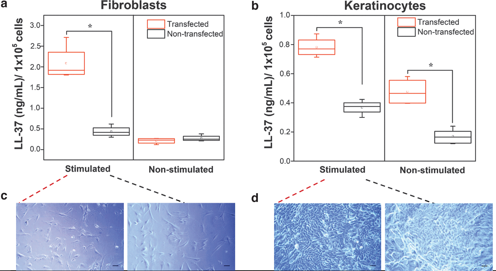
LL-37 peptide expression in cell culture supernatants of genetically modified
The effect of stimulation with S. aureus ATCC 25923 metabolites (spent medium) for 24 h on the secretion of LL-37 in transfected and nontransfected fibroblasts and keratinocytes showed that the secretion of LL-37 by transfected cells was significantly increased compared with nontransfected cells (Fig. 3a, b). Figure 3c and d shows that transfected and nontransfected fibroblasts and keratinocytes morphology was not altered when cultured under bacterial stimulation. This indicates that the increase in LL-37 levels is due to the metabolic response to stress by the cells in contact with bacterial metabolites, and not to the modification of the culture conditions or proliferation. To increase the level of LL-37 peptide in HSEs, monolayer cultures of fibroblasts and keratinocytes were genetically modified with LPEI/pDNA polyplexes, after which the transfected cells were included in the HSE protocol.
After 14 and 21 days in culture, transfected HSEs were exposed to bacterial metabolites for 24 h and cell supernatants were collected and analyzed by ELISA. Figure 4 shows that metabolites from bacteria were able to induce the secretion of significant levels of the LL-37 peptide in HSEs constructed with transfected cells compared with the nontransfected cells after 14 days in culture. Cell culture supernatants collected at day 21 did not show significant differences between the groups; however, the LL-37 expression in HSEs with transfected cells is higher than those of nontransfected cells by ∼10%.
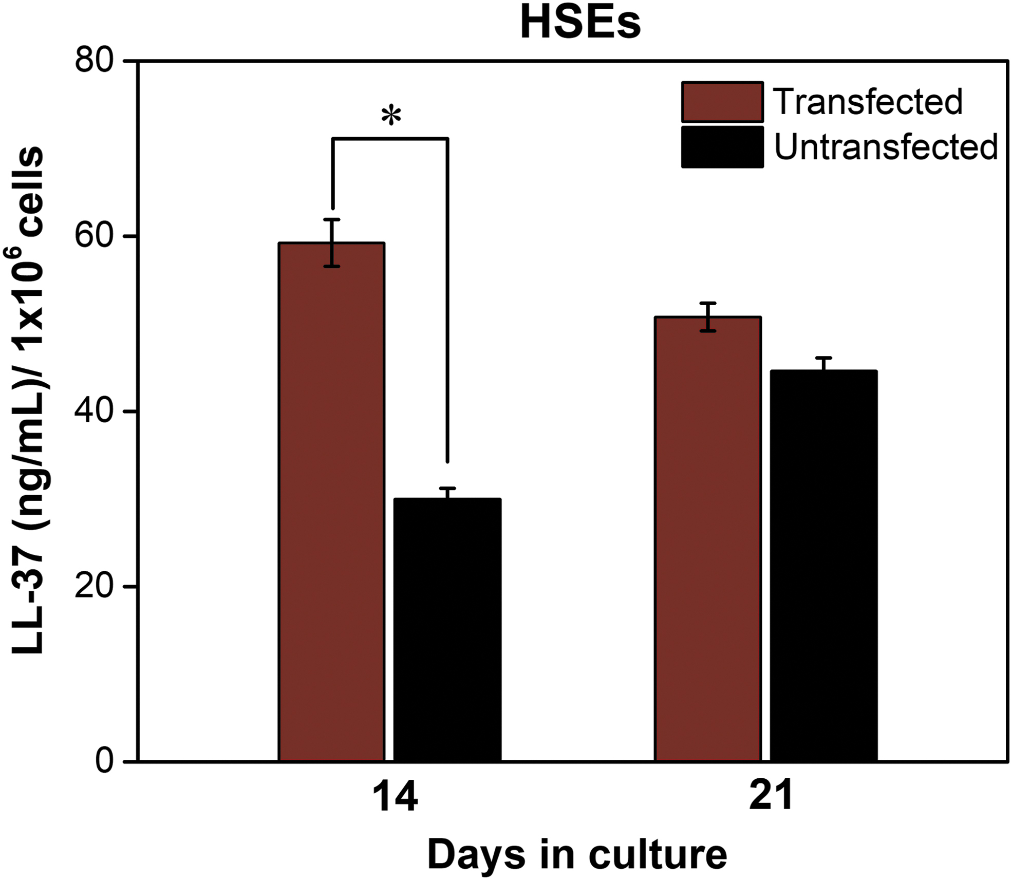
LL-37 peptide excretion by HSEs with transfected cells and controls (nontransfected) stimulated with Staphylococcus aureus ATCC 25923 metabolites detected by ELISA after 14 and 21 days in culture. Note that the number of cells on the y-axis represents combined keratinocytes and fibroblasts. The means ± standard deviation are shown (n = 3, independent experiments). Mann–Whitney U-test. *p < 0.05. Color images are available online.
HSEs expressing LL-37 resemble human native skin
After 21 days of culture at the air–liquid interface, the localization of LL-37 in HSEs constructed with transfected and nontransfected cells was explored. Cryosections from both tissues were immunostained using an antibody against the LL-37 peptide (Fig. 5). Figure 5b shows that the transfected HSEs displayed staining in the basal layer of the epidermis, as well as in some areas of the dermis, whereas the nontransfected HSEs were stained only in the epidermis (Fig. 5a), which could be related to the endogenous expression of LL-37 in the keratinocytes from the human skin. 31
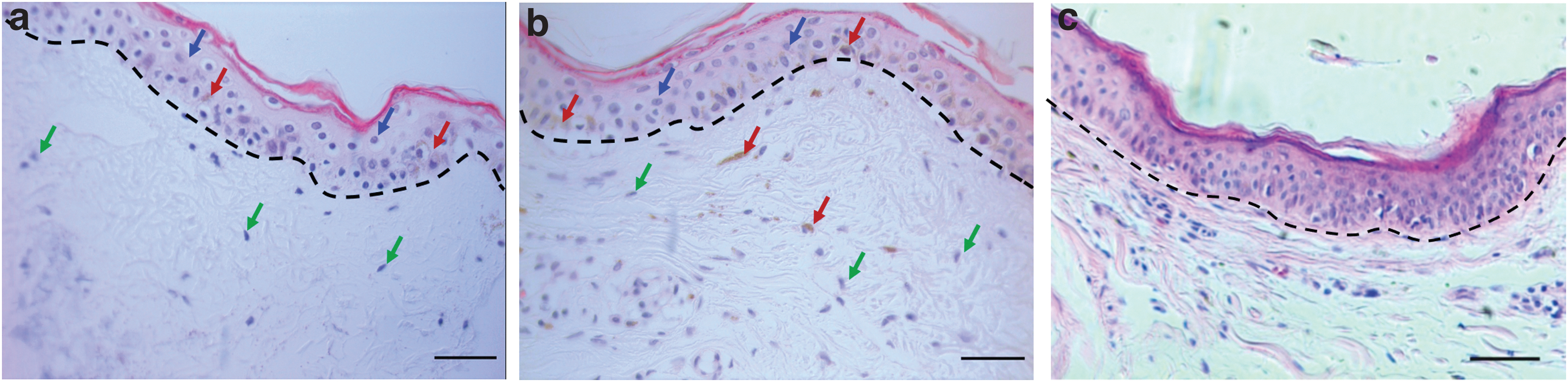
HSEs expressing LL-37 exhibit an architecture similar to native skin and produce LL-37 in both the epidermis and dermis.
Furthermore, histological evaluation by H&E staining showed that HSEs constructed with transfected cells have an architecture similar to those with nontransfected cells, and both types of HSEs showed characteristics close to the native human skin (Fig. 5c), with a dermal matrix populated by fibroblasts, and a partially stratified epidermis. These results suggest that there are no alterations in the formation of the dermal and epidermal compartments of the HSEs as a result of the increased LL-37 transgene expression. In addition, the localization of LL-37 in both transfected and nontransfected samples is consistent with the pattern displayed by normal skin tissues, in which LL-37 is expressed mainly in the basal layer. 32
Higher levels of LL-37 in HSEs enhance antimicrobial activity in vitro
To assess the antimicrobial activity in vitro of HSEs generated with transfected and nontransfected fibroblasts and keratinocytes, HSEs were cultured for 21 days. Then, the cell culture medium was challenged with S. aureus ATCC 25923 in growth medium containing 1 × 10⁵ bacteria/mL. The bacteria were cultured together with the HSEs for 24 h. Figure 6a shows that HSEs constructed with transfected cells significantly decreased bacterial growth by ∼1 log compared with the HSEs with nontransfected cells. In addition, the CFU/mL recovered from the cell culture supernatants from HSEs with transfected cells was significantly lower (50%) (Fig. 6b) as compared with the HSEs with nontransfected cells. These findings confirm that the increased expression of LL-37 in the HSEs could be advantageous to prevent early bacterial colonization.
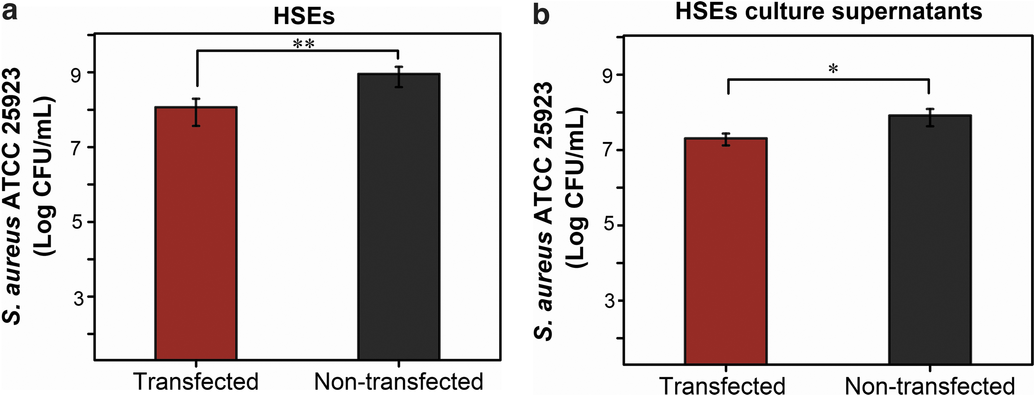
In vitro antibacterial efficacy of HSEs expressing LL-37 against planktonic Staphylococcus aureus ATCC 25923.
Discussion
As an alternative to the traditional antibiotics, gene therapy has allowed to genetically modify skin cells. Incorporated into biopolymer-based matrices they can secrete in a sustained manner, proteins of interest such as LL-37. 33 In this study, we describe a nonviral approach to increase the expression of the LL-37 peptide in HSEs generated with genetically modified human fibroblasts and keratinocytes by LPEI/pDNA polyplexes. Transfected HSEs express higher concentrations of LL-37 and possess characteristics close to native skin and exhibit improved antimicrobial activity against S. aureus strain in vitro.
Despite the fact that several studies have investigated the in vitro activity of LL-37 against two of the most common bacterial isolates in human wounds, S. aureus and P. aeruginosa, there are still discrepancies in the reported values for MICs and MBCs. Some of these concentrations previously reported in the literature are very high so that the combined use of antimicrobial peptides together with standard antibiotics has been proposed to achieve the killing efficacy. 34,35 However, the synergistic effect of LL-37 and an antibiotic can be explained by the amphipathic conformation of LL-37 that could mediate membrane permeabilization and facilitate the entry of the antibiotic into bacterial cells to achieve their goals. 36 In addition, the possible cytotoxicity of the combinations between antibiotics and antimicrobial peptides on human cells is not clear, whereas the use of LL-37 has shown positive effects on the proliferation and differentiation of various cell types, which could be a possible adjunct for regeneration. 37 –39 Therefore, the application of LL-37 alone remains an attractive strategy, as it is less likely to induce resistance and mutagenesis in bacteria in the natural environment compared with antibiotics. 40
In previous studies, 36,37 the attenuation of the antimicrobial activity of antimicrobial peptides has been observed when tested under conditions in which existing traditional antibiotics are active. This has been attributed to the inactivation of the structures of the cationic peptides by physiological concentrations of NaCl and divalent metal cations, among other variables. 41 For that reason, we have explored the manipulation of the bacterial culture medium including the addition of NaHCO3, making them physiologically more relevant, and therewith decrease the MICs for the treatment of skin infections in vivo.
Our results demonstrated that at sub-MICs, starting at 16 μM, LL-37 was effective against S. aureus in the presence of NaHCO3 (pH 7.4) (Supplementary Fig. S2a). Moreover, LL-37 demonstrated to be bactericidal against P. aeruginosa at concentrations >64 μM, regardless of the presence of NaHCO3 (Supplementary Fig. S2b). Moreover, LL-37 demonstrated to be bactericidal against P. aeruginosa at concentrations >64 μM, regardless of the presence of NaHCO3 (Supplementary Fig. S2b). This concentration is higher compared with that of S. aureus possibly due to the activity of the extracellular DNA (eDNA) that is actively released or secreted by bacteria. 42 eDNA has been shown to bind metal cations such as magnesium (Mg2+). In P. aeruginosa, this is especially important, since it is known that the two-component regulatory systems, called PmrA–PmrB and PhoP–PhoQ, respond to the presence of limiting concentrations of Mg2+, which regulates the expression of virulence genes that modifies lipopolysaccharides and contributes to survival against the activity of antimicrobial peptides. 43,44 Results for S. aureus seem to be in line with those presented in the literature where LL-37 was found to be less effective toward the Gram-positive strains cultured in normal bacterial medium (without NaHCO3). 45,46 NaHCO3 can contribute to the activity of antimicrobial peptides such as LL-37 through the alteration of the electrochemical gradient of protons, known as the proton motive force (PMF), generated through the bacterial membrane. 30 The PMF is necessary for the synthesis of ATP and active transport of molecules across the bacterial membrane.
It has been shown that LL-37 peptide is capable of interacting with several human cell types and influencing their behavior, improving cell migration and proliferation processes, and even promoting differentiation. 47,48 Significant advances have been achieved regarding the elucidation of the LL-37 effects over human cells such as neutrophil and monocytes. However, current knowledge regarding its effects on cells incorporated in biological matrices such as HSEs is largely unknown. In this study, we demonstrate that concentrations of LL-37 ranging from 2 to 256 μM were safe for the HSEs, and that concentrations up to 64 μM increased the metabolic activity after 7 and 14 days in culture (Fig. 2a). These findings coincide with various literature reports showing that LL-37 is cytocompatible toward keratinocytes and fibroblasts from the skin and that it could also stimulate cell proliferation, mainly through its association with cell receptors such as fibroblast growth factor receptor and EGF receptor. 13,49,50 In fact, we evaluated the number of cells in HSEs exposed to different concentrations of LL-37, specifically those previously established as MICs for S. aureus and P. aeruginosa (32 and 64 μM, respectively). The number of cells in the treated HSEs was higher than the negative controls (HSEs exposed to 0 μM LL-37) (Fig. 3b, c). These findings suggest that possibly an increase in the production of LL-37 by transfected keratinocytes and fibroblasts could positively regulate their proliferation when cultured in HSE.
The use of matrices constructed with biomaterials such as fibrin, in conjunction with delivery systems such as polyplexes, has allowed the expression of the transgene to be maintained for a longer period of time and a more localized release of the protein. 51 –53 In a previous study, the production of LPEI polyplexes for nonviral transfection of human keratinocytes and fibroblasts was optimized, and resulted in a higher number of copies of the CAMP gene (encoding LL-37 peptide). 27 Using this optimized system gave a higher expression of LL-37 in transfected cells when stimulated with bacterial culture supernatants compared with nontransfected cells (Fig. 3). The stimulation could be explained due to the presence of products derived from the S. aureus cell wall such as peptidoglycans, polysaccharides, and lipoteichoic acids as well as proteins (e.g., the staphylococcal protein A) that are anchored to the cell wall envelope, and constitute critical factors for bacterial adherence to and invasion of host tissue. 54
In addition, transfected HSEs expressed higher levels of LL-37 for up to 14 days in culture (Fig. 4), although on day 21, LL-37 release was similar to controls. This is corresponding to the endogenous LL-37 expression by the keratinocytes of the human skin. 31,55 In fact, LL-37 immunostaining was detected in the epidermal and dermal layers of the transfected HSEs, but this pattern of staining was only observed in the epidermal layer of nontransfected HSEs (Fig. 5). Furthermore, transfected HSEs possess histological characteristics close to native human skin, demonstrating that the expression of LL-37 did not alter the architecture of the model.
LL-37 expression decreases considerably in chronic skin wounds, which could explain why these wounds are more predisposed to bacterial colonization and less efficient in healing. 56 Enhanced production of LL-37 in fibrin-rbased HSEs constructed with fibroblasts and keratinocytes transfected by polyplexes system could favor wound healing through its antimicrobial properties. A report showed that enhancing LL-37 production through its adenoviral expression in burn wounds in rats that genetically modified cells was more efficient in controlling infection than direct application of the peptide. 57 Moreover, there is evidence that LL-37 exhibits antimicrobial properties through its nonviral expression in the human keratinocyte progenitor cell line (NIKS) cultured in a full-thickness human skin substitute generated with type 1 collagen. 20 In this study, HSEs containing transfected cells were able to decrease bacterial growth of a planktonic strain of S. aureus after 24 h of coculture, suggesting that the levels of LL-37 were sufficient to control bacterial colonization in the HSEs. Furthermore, as 3D models, HSEs can be produced in different sizes to increase the population of cells expressing the protein of interest, which could more easily prevent bacterial attachment or early biofilm formation in the wound environment.
Therapeutic use of these genetically engineered skin models with improved antimicrobial activity could have important advantages compared with the use of antibiotics, due to the natural difficulty that microorganisms have to acquire resistance to HDPs. 13,58 In addition, LL-37 has been shown to enhance wound healing and vascularization by promoting endothelial proliferation and the chemotaxis of immune cells. 59 Thus, future studies could focus on the evaluation of processes such as angiogenesis and re-epithelialization of the wound.
In conclusion, our study demonstrates that HSEs built with cells that express higher levels of the LL-37 antimicrobial peptide through the linear polyplexes system could be an efficient way to prevent bacterial colonization and therewith prevent infections in skin wounds.
Footnotes
Author Disclosure
No competing financial interests exist.
Funding Information
This study was supported by the Abel Tasman Talent Program (ATTP) of the University of Groningen and COLCIENCIAS National Doctoral program (code 727-2015).
Supplementary Material
Supplementary Figure S1
Supplementary Figure S2
References
Supplementary Material
Please find the following supplemental material available below.
For Open Access articles published under a Creative Commons License, all supplemental material carries the same license as the article it is associated with.
For non-Open Access articles published, all supplemental material carries a non-exclusive license, and permission requests for re-use of supplemental material or any part of supplemental material shall be sent directly to the copyright owner as specified in the copyright notice associated with the article.