




Abstract
Tenascin-C (TN-C), a key component of extracellular matrix (ECM), is strongly expressed in fetal and cancer tissues. Large-molecular-weight variants of TN-C, including different combinations of its alternative spliced FNIII repeats, are specifically expressed in tissues under certain pathological conditions. Here we report the production of monoclonal antibodies (MAbs) against FNIII domain D (FNIII D) of human TN-C. Complementary DNA encoding the FNIII D region was generated by RT-PCR from human osteosarcoma (OS) cell line, and the recombinant FNIII D-GST fusion protein was expressed and purified. Two hybridoma cell lines secreting monoclonal antibodies (MAbs) against FNIII D were obtained by routine murine hybridoma technique. The MAbs were identified by indirect enzyme-linked immunosorbent assay (ELISA), Western blot, and immunohistochemistry (IHC). Both of them were applicable in Western blot and IHC. With our MAbs, we found TN-C was positive in OS and most of it was among the tumor stroma. To conclude, these MAbs to human FNIII D domain of TN-C may be useful for exploring OS pathogenesis and potential clinical application.
Introduction

Diagram of human TN-C monomer.
Osteosarcoma (OS) is a kind of malignancy and the tumor cells can form malignant bone-like or bone tissues. OS is most often seen within primary bone malignant neoplasm and makes up one third of all the bone-originated malignancies.(7) A previous study proved that TN-C promotes distant metastases of osteosarcoma expression.(8) However, no further observations were taken to identify what kind of spliced variants got involved in OS pathogenesis. In this study, we isolated cDNA encoding FNIII D from an OS cell line and expressed the recombinant protein containing the FNIII D domain. Finally, two strains of MAbs specific for FNIII D were obtained and characterized.
Materials and Methods
Cell culture
Murine myeloma cell line Sp2/0 and human osteosarcoma cell line MG-63 were incubated in RPMI 1640 (Sigma-Aldrich, St. Louis, MO) containing 100 mL/L fetal calf serum (FCS, Gibco-Invitrogen, San Diego, CA). Hybridomas were incubated in RPMI 1640 supplemented with 20 mL/L FCS.
Molecular cloning of FNIII D domain cDNA
A total of 106 MG-63 cells were solubilized in RNA extraction solution (TRIzol, Invitrogen). Total RNA was isolated using chloroform, and precipitated with isopropanol. The resulting 5 μg of RNA was used as template for single-strand cDNA synthesis with 20 U avian myeloblastosis virus (AMV) reverse transcriptase (Takara Biotechnology, Dalian, China) according to the manufacturer's instructions. Primers specific for human FNIII D of TN-C and β-actin were synthesized by Shanghai Sangon Biological Engineering and Technology Service (Shanghai, China). Primers were as follows: sense, 5′-gaagccgaaccggaagttga-3′ (20 nt at position 4851–4870 relative to the start codon of human tenascin-C,
Expression of FNIII D-GST fusion protein in Escherichia coli
The cDNA of FNIII-D was subcloned with pyrobest polymerase (Takara, China) and inserted into a pGEX-4T-1 vector (Amersham Pharmacia Biotech, Piscataway, NJ). The corresponding protein was expressed in E. coli as glutathione S-transferase (GST) fusion protein induced by isopropyl-b-D-thiogalactopyranoside (IPTG, Sigma) and purified with glutathione-sepharose.
Preparation of hybridomas secreting MAb to FNIII D of TN-C
Female Balb/c mice (4 weeks old) were immunized with 40 mg of purified FNII D-GST fusion protein in complete Freund's adjuvant (Sigma) by subcutaneous (sc) injection. Subsequently, immunizations were carried out twice with 40 mg of antigen in incomplete Freund's adjuvant (Sigma) by sc and intraperitoneal (ip) injection, respectively, at 3-week intervals. Ten days after the third immunization, mice were bled from caudal vein and the serum titer was determined by ELISA. The immunized mice were boosted with 20 mg of FNII D-GST fusion protein by ip injection 3 days before fusion. Splenocytes from immunized mice and Sp2/0 myeloma cells were fused according to the general procedure in the presence of polyethylene glycol (PEG, MW 4000, Merck, Darmstadt, Germany).(9) Positive hybrids were selected by indirect ELISA and subcloned three times using the limiting dilution method. MAbs were produced either from supernatants of the hybridoma culture or from ascitic fluid of Balb/c mice intraperitoneally injected with hybridoma. Immunoglobulin (Ig) isotypes were identified by using an isotyping kit (Pierce, Rockford, IL).
ELISA
ELISA was performed on 96-well plates (Nunc, Nagel Inc., Roskilde, Denmark), which were coated with 5 mg/L FNIII D-GST or GST, respectively, in coating buffer (0.05 mol/L carbonate/bicarbonate buffer [pH 9.6]) and incubated overnight at 4°C. After washing three times with PBS/0.5 mL/L Tween-20, the supernatants of hybridoma culture or various dilutions of corresponding murine ascites were added and the plates were incubated for 1 h at 37°C. The plates were washed three times and the working dilution (1:2000) of horseradish peroxidase (HRP)-conjugated sheep anti-mouse IgG (Dako, Glostrup, Denmark) was added. After incubation at 37°C for 1 h and three more washes, the substrate 2,2’-azino-bis (3-ethylbenzo thiasoline-6-sulfonic acid [ABTS], Sigma) was added to each well and incubated for 15 min; then the absorbance at 410 nm (A410 nm) was detected with a microplate reader.
Western blot analysis
Samples containing 50 μg of purified recombinant protein were separated on a polyacrylamide gel with Tris-glycine-SDS running buffer and were subsequently transferred onto a polyvinylidene difluoride membrane (Bio-Rad, Hercules, CA) for 1 h. Membranes were blocked with 5% milk in TBS-Tween-20 and incubated with the primary antibody overnight. Membranes were washed and incubated with a secondary peroxidase linked antibody, and the reactive bands were detected by enhanced chemiluminescence (ECL) according to the manufacturer's protocol (Amersham Biosciences).
Immunohistochemistry
Formalin-fixed and paraffin-embedded malignant human osteosarcoma tissue sections were maintained in our department. All tissues were sampled from surgical specimens within 2 h of resection. Three-micrometer thick sections were mounted on poly-L-lysine-coated slides, followed by deparaffinization in xylene and dehydration in graded alcohol. The endogenous peroxidase activity was blocked by incubation with 3 mL/L H2O2 in methanol for 30 min. Tissue sections were subjected to antigen retrieval by boiling in 0.01 mol/L sodium-citrate (pH 6.0) for 10 min in a microwave oven. After blocking with 15 mL/L normal goat serum for 1 h, immunohistochemistry assay was carried out with an Elivision Plus staining kit (Maixub-bio, Fuzhou, China) according to the manufacturer's manual. In brief, tissue sections were stained with MAb against FNIII D of TN-C with a concentration of 50 mg/L for 1 h at room temperature and washed three times; after that an enhancer was added for 30 min, followed by three washes, before HRP-conjugated goat anti-mouse was added and washed another three times. Finally, diaminobenzidine (DAB) was added for coloring.
Results
FNIII D domain of TN-C cDNA cloning and fusion protein expression
FNIII D domain of TN-C cDNA was successfully cloned from human osteosarcoma cell line MG-63 (Fig. 2), and the sequence was identical to the data in GenBank. The optimal condition inducing the soluble FNIII D-GST fusion protein to the greatest extent was at 30°C for 8 h with 80 nmol/L IPTG. The SDS-PAGE results showed the FNIII D-GST fusion protein had a molecular weight (MW) of ∼38 kDa (Fig. 3).

Representative RT-PCR results of FNIII D domain of TN-C from osteosarcoma cells. Lane 1, FNIII D cDNA; lane 2, marker of DL2000.
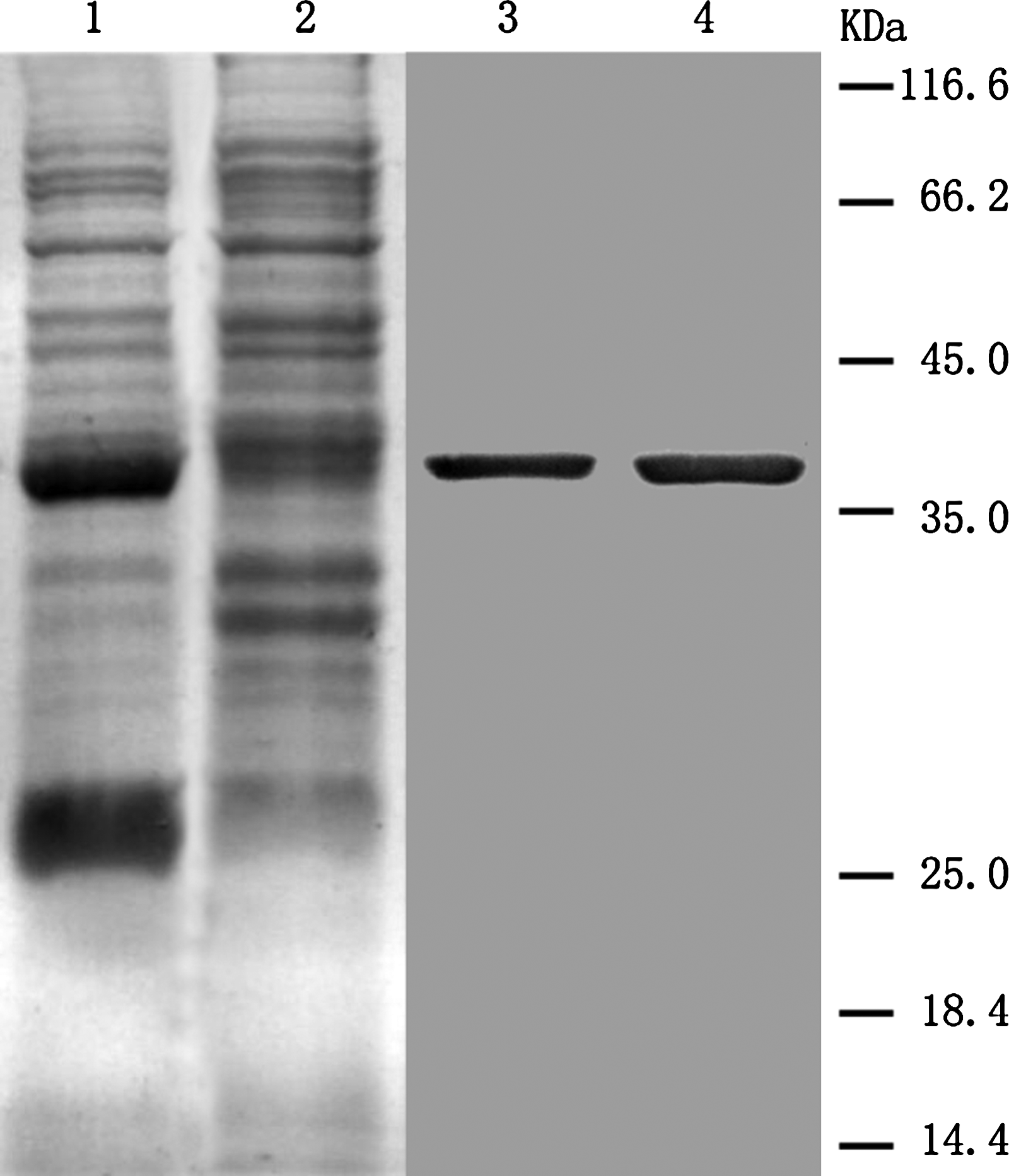
SDS-PAGE and Western blot of FNIII D-GST fusion protein. Lane 1, bacteria culture with IPTG induction; lane 2, bacteria culture without IPTG induction; lane 3, Western blot of MAb 3A8; lane 4, Western blot of MAb 4E2.
Characterization of MAbs against FNIII D domain of TN-C
Altogether two hybridoma clones secreting MAbs to FNIII D were obtained by the routine hybridoma technology and designated as 3A8 and 4E2, respectively. The clone name, isotype, and ascitic titers are listed in Table 1.
The isotypes of MAbs were identified by an isotyping kit (Pierce).
The titers of MAbs in ascites were measured by indirect ELISA.
Both of the MAbs could be used for Western blot (Fig. 3) and immunohistochemical staining (Fig. 4) on tissue sections. In the Western blot assay, the MAbs stained the protein bands corresponding FNIII D. In the IHC results, TN-C was found distributed within the matrix around the malignant tissues.
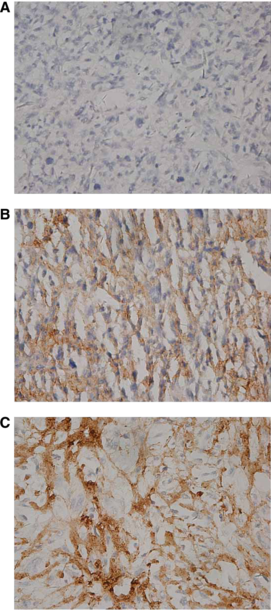
Tenascin-C staining with anti-FNIII D domain of TN-C MAb on osteosarcoma tissues (× 400). (
Discussion
Within the organism, cells interact with the ECM components. The communication between them determines the cellular behavior and morphology and thus influences cell growth and differentiation.(10) Among the components, members of tenascin family, composed of tenascin-C, tenascin-R, tenascin-X and tenascin-W, play important roles in tissue homeostasis.(11) TN-C, founding member of the family, acts as modulators of cell adhesion, migration, and growth. The spliced variants were always present during some disease pathogenesis but seldom seen under normal conditions.(12)
Although large variant mRNAs containing FNIII repeats D were identified in some diseases, such as chronic hepatitis C, no antibodies specific for FNIII D domain were prepared.(13) FNIII D domain of TN-C embraced 91 amino acids and, in this study, we isolated cDNA of FNIII D and expressed it as a GST-fusion protein, rather than chemical amino acid synthesis, to increase its immunogenicity.
Osteosarcoma occurred on the bones, which always burdens the stress of muscles and body weight. Interestingly, TN-C was among the few genes that were induced upon dynamic mechanical stress and its expression was related to metastasis.(14) In our study, we isolated cDNA from OS cell line and observed the corresponding FNIII D protein in the OS tissues, which informed us that our MAbs could supply more detailed and precise interpretation of OS physiology and may have potential diagnostic and therapeutic value for osteosarcoma in the future.
Footnotes
Acknowledgments
This work was supported by the Critical Project Foundation on Social Development of ShaanXi province (no. 2008K09-09).