




Abstract
The septin family of GTPase proteins has been shown to be important for cell division, cytoskeletal organization, and membrane-remodeling events. Septin 9 (SEPT9) is a member of the septin family (also designated MSF/eseptin/Sint1) and has been implicated in tumorigenesis. The present study reports on the preparation and properties of a monoclonal antibody (MAb) directed against SEPT9. The antibody was produced by hybridization of mouse myeloma cells with lymph node cells from an immunized rat. The MAb 7B5 specifically recognized SEPT9, as evidenced by immunoblotting using a variety of extracts from cultured cells. In immunostaining using MAb 7B5, a filamentous pattern near the plasma membrane was observed. The MAb 7B5 promises to be useful in immunoblotting and immunostaining experiments in various cells and tissues to determine the expression levels of SEPT9, as well as to further the analysis of the biological function of this protein.
Introduction
In the present study, we report on the production and characterization of a monoclonal antibody (MAb) against SEPT9. This MAb, referred to as 7B5, has the potential for use in ELISA, immunoblotting, and immunostaining analyses.
Materials and Methods
Design of peptide
The SEPT9 peptide MERDRITALKRC was synthesized by Sigma-Aldrich (Tokyo, Japan). The peptide corresponds to the 11 N-terminal amino acids of rat SEPT9. The peptide was coupled to Keyhole limpet hemocyanin (KLH) or BSA using 3-Maleimidobenzoic acid N-hydroxysuccinimide ester (MBS).
Immunization of rat and production of monoclonal antibody
An anti-SEPT9 rat monoclonal antibody was generated on the basis of the rat lymph node method established by Sado et al.(13) 200 μL of an emulsion containing 30 μg of SEPT9 peptide-coupled KLH and Freund's complete adjuvant were injected into a 10-week-old male WKY/NCrj rat (Charles River, Yokohama, Japan) via the hind footpads. After 3 weeks, cells from the medial iliac lymph nodes of the immunized rat with the antigen were fused with mouse myeloma SP2 cells at the ratio of 5:1 in a 50% polyethylene glycol (PEG4000, Merck, Darmstadt, Germany) solution. The resulting hybridoma cells were plated onto 96-well plates and cultured in HAT selection medium (Hybridoma-SFM [Invitrogen, Carlsbad, CA]; 5% fetal bovine serum [FBS]; 5% BM-condimed H1 [Roche, Indianapolis, IN]; 100 μM hypoxanthine; 0.4 μM aminopterin; 16 μM thymidine). After 10 days post-fusion, the hybridoma supernatants were screened by means of an enzyme-linked immunoadsorbent assay (ELISA) against the SEPT9 peptide-coupled BSA. Positive clones were subcloned and rescreened by ELISA and immunoblotting. The IgG subclass of antibody was determined using a Rat MonoAb ID/SP kit (Zymed, San Francisco, CA) according to the instructions of the manufacturer.
ELISA
SEPT9 peptide-coupled BSA (5 μg/mL) in 10 mM sodium phosphate buffer (pH 7.0) was adsorbed on the surface of 96-well Nunc-immunoplates (Nunc, Roskilde, Denmark) by overnight incubation at 4°C. The plates were blocked with 1% bovine serum albumin (BSA) in PBS (8.10 mM Na2HPO4 [pH 7.4], 137 mM NaCl, 2.68 mM KCl, and 1.47 mM KH2PO4) to avoid non-specific binding. The hybridoma supernatants were incubated for 1 h at room temperature, and then washed three times with TBS-T. Thereafter, plates were incubated for 30 min at room temperature with an alkaline phosphatase-conjugated anti-rat IgG antibody (Sigma, St. Louis, MO) diluted 1:10000. After washing three times with TBS-T, immunoreactivity was visualized using a pNPP phosphatase substrate system (KPL, Gaithersburg, MD).
Cell culture
Human cervical carcinoma HeLa cells, green monkey kidney epithelial COS-1 cells, murine fibroblast L-929 cells, murine fibroblast NIH3T3 cells, and normal rat kidney NRK cells were cultured in RPMI-1640 medium (for HeLa, L-929) or Dulbecco's modified Eagle's medium (for COS-1, NIH3T3, NRK) supplemented with 5% fetal bovine serum (FBS), penicillin (100 U/mL), and streptomycin (100 μg/mL) in a humidified atmosphere of 5% CO2 at 37°C.
Immunoblotting
Whole cell extracts of HeLa, COS-1, L929, NIH3T3, and NRK cells were separated by 10% SDS-PAGE and electrophoretically transferred to Immobilon-P polyvinylidene fluoride (PVDF) transfer membranes (Millipore, Bedford, MA). The membrane was blocked overnight at 4°C with a blocking solution containing 3% skim-milk in TBS-T (20 mM Tris-HCl [pH 7.5], 150 mM NaCl, and 0.05% Tween-20), and then incubated for 1 h at room temperature with anti-SEPT9 rat MAb 7B5. Following washing with TBS-T, the membrane was incubated for 30 min at room temperature with alkaline phosphatase-conjugated anti-rat IgG antibody (Sigma) and developed by treatment with nitroblue tetrazolium (NBT) and bromo-chloro-indolylphosphate (BCIP) after washing with TBS-T.
Immunofluorescence
HeLa cells were grown on coverslips in each culture medium. The cells were fixed with 3.7% formaldehyde in PBS for 15 min at room temperature followed by permeabilization with 0.5% Triton X-100 in PBS for 5 min at room temperature. After treatment with a blocking solution (PBS containing 50 mM glycine, 1% BSA, and 2% normal goat serum), the cells were incubated with anti-SEPT9 monoclonal antibody 7B5 and detected with Alexa488-conjugated goat anti-rat IgG (Invitrogen). Samples were examined using an IX-71 fluorescent microscope (Olympus, Tokyo, Japan).
Results and Discussion
A monoclonal antibody was generated against the N-terminal region of SEPT9 using a rat medial iliac lymph node method. The SEPT9 peptide was used as an antigen and immunized via the WKY/NCrj rat hind footpads with only a single injection. At three weeks post-immunization, lymphocytes were collected from the enlarged lymph nodes of the rat. The hybridomas obtained after fusing the lymphocytes with mouse myeloma SP2 cells were tested for the production of monoclonal antibodies that react with SEPT9 peptide in an ELISA experiment. 100 hybridoma supernatants, positive clones based on ELISA, were examined by immunofluorescence staining of HeLa cells. One, designated as MAb 7B5, was selected because it yielded the strongest signal in immunostaining. The specific immunoglobulin class of MAb 7B5 was determined using a rat isotyping kit. The analysis indicated that MAb 7B5 is the rat IgG2a (κ).
It is known that SEPT9 contains amino acid sequences that are highly conserved throughout the mammalian species. We examined whether the MAb 7B5 reacts with SEPT9 in cells from various species. As shown in Figure 1, MAb 7B5 recognizes a protein corresponding to about a 75 kDa single band, which is present in all cell extracts from HeLa (human), COS-1 (simian), L929 (mouse), NIH3T3 (mouse), and NRK (rat) cells. This suggests that, when used in conjunction with immunoblotting, this monoclonal antibody would be very useful for the analysis of expression levels of SEPT9 in various cell lines and tissues.
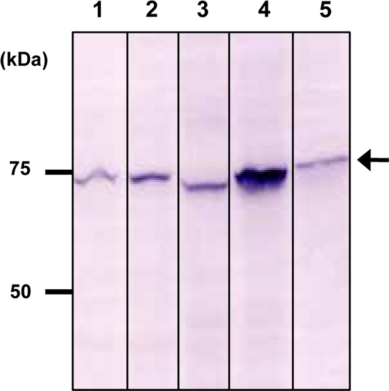
Species cross-reactivity of anti-SEPT9 MAb 7B5. Immunoblotting of various cell total extracts was performed using MAb 7B5: HeLa (human) (lane 1), COS-1 (simian) (lane 2); L929 (mouse) (lane 3); NIH3T3 (mouse) (lane 4); and NRK (rat) (lane 5). Molecular mass markers in kDa are indicated at the left of each panel.
This antibody was next characterized by the immunofluorescence staining of HeLa cells. In Figure 2, SEPT9 staining demonstrated a filamentous pattern as previously reported,(6) suggesting that it is involved in the cytoskeletal organization.
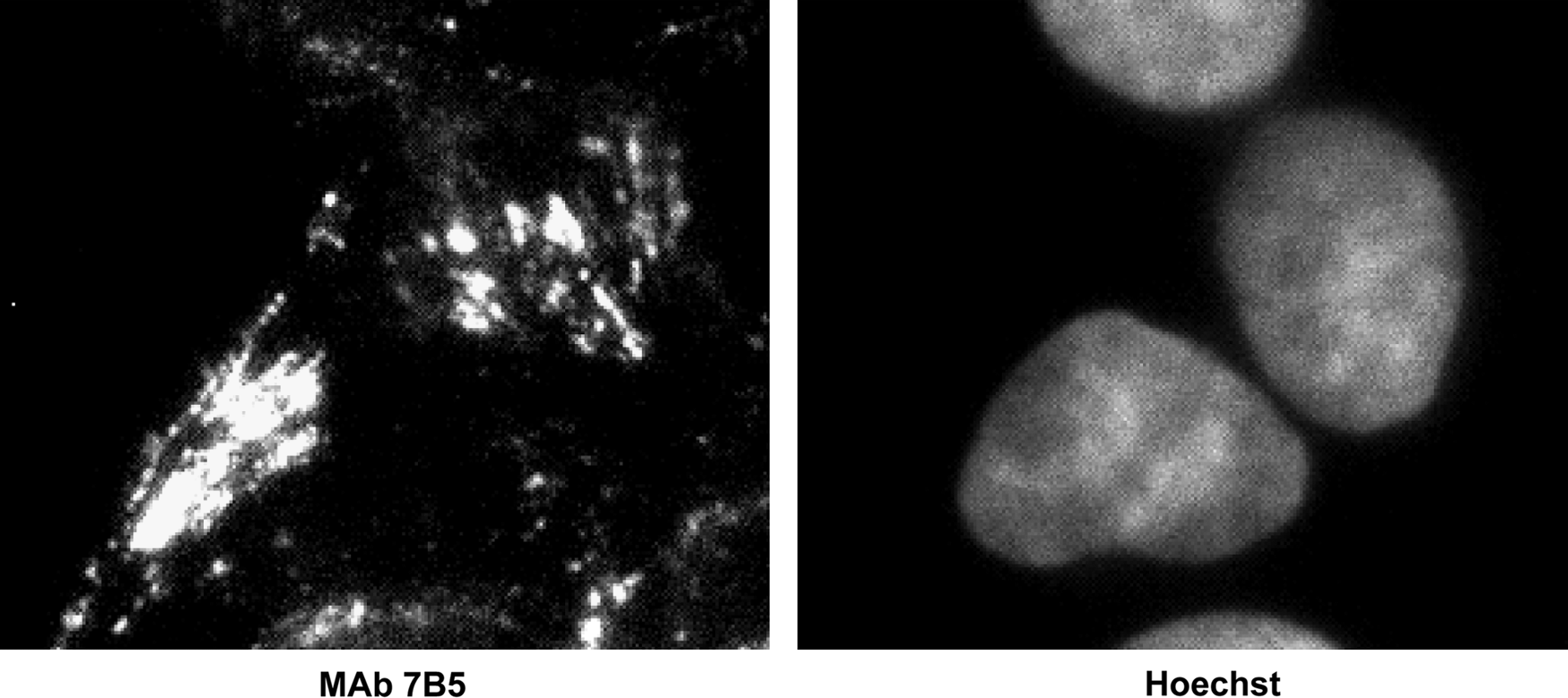
Indirect immunofluorescence of HeLa cell using anti-SEPT9 MAb 7B5. MAb was detected with Alexa488-conjugated anti-rat IgG and stained with Hoechst dye.
In summary, we report herein on the production and characterization of a monoclonal antibody that specifically recognizes SEPT9 in a variety of mammalian cells. It has recently been reported that SEPT9 is implicated in tumorigenesis and amyotrophy. Although the molecular mechanisms of SEPT9 biological action in the cell are not clear, our observations may be helpful in revealing its unknown functions. Application of the monoclonal antibody in a variety of methods is a powerful tool for investigation of biological molecules.(14–16) Therefore, MAb 7B5, a specific antibody for SEPT9, should aid in the elucidation of the biochemical and physiological functions of SEPT9.