




Abstract
RpfB, one of the five resuscitation-promoting factors (Rpfs) produced by Mycobacterium tuberculosis (MTB), plays an important role in the resuscitation and growth of the dormant MTB. RpfB is likely the target antigen recognized by the host immune system. Studies have shown that Rpf genes exist in many bacteria and their encoded proteins all contain Rpf-like domain. It is likely that this domain has biological characteristics and immunogenicity similar to that of the complete Rpf protein. Therefore, RpfB domain protein from M. tuberculosis was selected for this study. Mice were subcutaneously immunized three times over 2-week intervals. Mice splenocytes were then isolated and fused with SP20 cells. Hybridoma colonies were screened for monoclonal antibody (MAb) against RpfB domain. ELISA was used to examine the titer, specificity, and relative affinity of the antibody. The ability of produced anti-RpfB monoclonal antibody to recognize other proteins in the Rpf family and to inhibit the growth of Mycobacterium tuberculosis and Micrococcus luteus was examined. Our results showed that three anti-RpfB MAbs were successfully generated. All three MAbs can recognize RpfB domain specifically and can effectively inhibit the promoting effect of RpfB domain on the growth of MTB H37Ra strain and M. luteus at 1:1000 dilution, indicating that anti-RpfB domain MAbs may inhibit the reactivation of dormant or latent MTB in vivo. Therefore, they may be able to prevent the recurrence of the occult infection. The production of anti-RpfB domain MAbs provides a powerful experimental tool to further study the biological and immunological characteristics of the RpfB domain and to evaluate the possibility of using RpfB domain as a candidate component for tuberculosis subunit vaccine.
Introduction
Recent studies have found that five resuscitation-promoting factors (Rpfs) secreted by MTB, including RpfA, RpfB, RpfC, RpfD, and RpfE, play important roles in the resuscitation of the dormant MTB.(5) Rpf was originally found in Micrococcus luteus.(6) It is also the first secreted protein found so far that is able to restore the reproductivity of dormant bacteria. Rpf is also the target antigen recognized by the host immune system and induces strong immune response in vivo.(7) Specific antibody against Rpf produced by the host immune system can block the promoting effect of Rpf on MTB. Therefore, Rpf protein could provide a novel approach to preventing and controlling MTB infection. Homology analysis has revealed that Rfp-like proteins exist in a variety of G + C rich gram-positive bacteria. Proteins encoded by Rpf genes all contain Rpf-like domain.(8) Rpf domain is a 77-residue located between A42 and L118 in the full-length Rpf protein.(9) Studies have shown that Rpf domain has the same biological function as the Rpf protein. Rpf domain has the same ability in reactivation of dormant bacteria in vitro as the full-length Rpf protein. Rpf domain also shares similar immunogenicity as the full protein. Specific antibody induced by Rpf domain can possibly block all Rpf-like proteins of the Rpf family in MTB, and in turn inhibit the resuscitation and growth of bacteria. The five Rpf-like proteins in MTB have shown functional redundancy.(10) There was no obvious effect on bacterial growth when any one of the Rpf genes was knocked-out.(11) This suggests that the five Rpf-like proteins in MTB act in synergy to regulate MTB dormancy together. However, the synergistic effects of the five Rpf-like proteins and the optimized utilization among them have created many difficulties in the development of vaccine and the establishment of experimental detection methods. Among them, RpfB plays a more important role in MTB growth. Analysis of RpfB protein composition showed that it is a membrane protein with multiple T-cell and B-cell epitopes. Mutations of RpfB significantly reduced the growth rate of H37Rv.(12) Our previous study also found that RpfB has strong biological and immunological characteristics.(13) Hence, RpfB domain might be a starting point to study the immunological and biological functions of Rpf proteins in MTB.
In this study, RpfB domain peptide expressed and purified from the prokaryotic system was used as the antigen to produce monoclonal antibody (MAb), which provides a powerful tool to further study the biological and immunological characteristics of the RpfB domain.
Materials and Methods
Materials
MTB strain H37Rv was kindly provided by Shaanxi Provincial Institute for Tuberculosis Control (Xi'an, China); the Micrococcus luteus standard strain and Escherichia coli DH5α were kept in the bacteriology laboratory of the Department of Clinical Laboratory, Xijing Hospital (Xi'an, China); pPRO-EXHT-RpfB domain recombinant plasmid was constructed in the Department of Clinical Laboratory, Xijing Hospital(14); PEG, HT, and HAT were purchased from Promega (Madison, WI); Sp2/0 cell line was a gift from the Microbiology Department, the Fourth Military Medical University (Xi'an, China); BALB/c mice (female, 6∼8 week old) were from the Animal Center of Fourth Military Medical University; 6His MAb was purchased from the Immunology Department, the Fourth Military Medical University; HRP goat anti-mouse IgG antibody was purchased from Huamei Biotech (Luoyang, China); Ni2+ -NTA purification kit was purchased from Invitrogen (Carlsbad, CA).
Expression and purification of MTB RpfB domain peptide
E. coli DH5α harboring the prokaryotic expression plasmid pPRO-EXHT-RpfB was inoculated in the culture media, and 0.5 mM of IPTG was used to induce protein expression for 4 h. Expressed protein was resolved on a 160 g/L SDS-PAGE and confirmed by Western blotting. Fusion protein was purified by affinity chromatography using Ni2 + NTA purification kit under denaturing condition. By gradually decreasing the urea concentration, urea in the protein solution was removed while the denatured protein was spontaneously renatured during this course. The purified protein was first dialyzed against buffer containing 6 mol/L of urea at 4°C overnight. Buffers containing 4, 3, 2, 1, and 0.5 mol/L of urea were then used in order to dialyze the protein for more than 4 h, respectively. PBS buffer was last used for dialysis for over 4 h.
Establishment of MAb cell line
BALB/c mice were immunized with the purified target protein three times with 2-week intervals. Mice splenocytes were collected and fused with mouse myeloma Sp2/0 using PEG1500. Feeder cells were prepared 1 day before fusion. Fusion cells were cultured in HAT media for 5 days, followed by HT media until the eighth day. Purified recombinant RpfB domain protein was used to coat the microtiter plate and indirect ELISA was used to test the culture media. The supernatant from Sp2/0 culture and the serum from normal BALB/c mice were used as negative control; serum from immunized BALB/c mice was used as positive control. Monoclonal cell line was established by limited dilution of the colonies from the positive wells.
Preparation and purification of ascitic fluid containing MAb
The hybridoma cells were mixed well with serum-free media to adjust cell density to 1∼2 × 10⁹ cells/L. Each mouse was intraperitoneally injected with 0.5 mL of the cells. About 10∼15 days after the injection, the abdomen of the mice became apparently swollen. Ascitic fluid was collected and centrifuged at 3000 r/min for 15 min. Supernatant was collected. Ascitic fluid was subjected to purification using caprylic acid-ammonium sulfate precipitation.
Indirect sandwich ELISA to measure the titer of the purified antibody
ELISA plates were coated with 10 g/L of protein, and serially diluted purified antibody was added. HRP-conjugated goat anti-mouse antibody was used as the secondary antibody and color was developed with OPD. The supernatant from Sp2/0 cell culture and the serum from the immunized mice were used as negative and positive controls.
Western blot analysis to evaluate the specificity of the purified MAb
Expressed proteins were resolved on a 160 g/L of SDS-PAGE and then transferred onto nitrocellulose membrane. Fifty g/L of fat-free milk were used to block the membrane at 37°C for 1 h. Purified MAb was then added at 100 mg/L and incubated with the membrane in a 37°C shaker for 1 h. The membrane was washed with PBS three times; HRP-goat-anti-mouse IgG was added and incubated with the membrane in 37°C shaker for 1 h. The membrane was then washed with PBS three times and developed using OPD.
ELISA assay to evaluate MAb specificity
The plate was coated with both target protein and unrelated His-tag fusion protein. Purified MAb was used as the primary antibody for 1 h incubation at 37°C. The plate was washed and incubated with HRP-conjugated goat-anti-mouse antibody (1:1000 dilution) at 37°C for 1 h. The plate was washed again and developed with OPD, and the A value at 490 nm was measured.
ELISA assay to identify MAb type and sub-type
Serum-free supernatant from hybridoma cell culture was collected and tested with six type and sub-type specific goat-anti-mouse polyclonal antibodies. Type and sub-type can be determined by corresponding positive reaction.
Examination of MAb relative affinity
The plate was coated with 2 mg/L of RpfB domain at 4°C overnight, and rinsed and blocked with 10% fetal bovine serum for 1 h after washing. Serially diluted and purified MAb (from 200 to 10−7 μg/mL) was added and incubated at 37°C. The plate was rinsed and incubated with HRP-conjugated goat anti-mouse antibody (1:1000 dilution) at 37°C for 1 h. After washing, OPD was used for color development; OD value was measured at 490 nm and graphed to determine the relative affinity.
Antibody cross-reactivity of anti-RpfB domain MAb
M. luteus Rfp, Rpf domain, RpfB domain, MTB RpfA (expressed and purified in our lab; expression vector was kindly provided by Prof. Mike Young, University of Wales, Cardiff, United Kingdom), and H37Ra strains were used as antigens to coat ELISA plates. The purified MAb (B8G11 with the highest titer was used) served as the primary antibody in the ELISA assay to detect the cross-reactivity of anti-RpfB domain MAb with Rpf family proteins, as well as MTB.
Inhibition assay of anti-RpfB domain MAb
Dormant MTB strain H37Ra (100 μL) was inoculated into 5 mL of 7H9 culture medium, and 100 μL of M. luteus was inoculated into 5 mL of glucose broth culture medium. Purified RpfB domain protein and anti-RpfB domain MAb were added respectively at different concentrations. PBS (0.01 M) was added as the negative control. Assays were quintuplicated in five tubes at each concentration. Bacteria were cultured at 37°C. Small amounts of M. luteus culture were collected every 3 h (and every 6 h after 27 h) to test OD600. Small amounts of MTB H37Ra culture were collected every 5 days to test OD600. Growth curves for each group of M. luteus and MTB H37Ra were graphed based on the OD measurements.
Results
Expression and purification of RpfB domain protein
The expressed RpfB domain protein is insoluble. SDS-PAGE analysis showed a protein band with an Mr of 12,000 Dalton, which is consistent with the expected size. The target protein was purified under denaturing condition. The eluates were collected and subjected to 160 g/L SDS-PAGE analysis. Result showed a clear protein band with an Mr of 12,000 Dalton. The fusion protein has an achieved purity of up to 85% based on TLC analysis (Fig. 1).
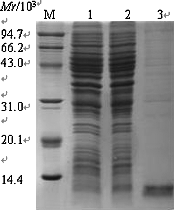
SDS-PAGE analysis of MTB RpfB domain fusion protein expression. SDS-PAGE analysis of the expressed MTB RpfB domain fusion protein in E. coli. Samples were electrophoresed on 160 g/L SDS-PAGE. Proteins were visualized by Coomassie blue R-250 staining. Fusion protein was purified by affinity chromatography using Ni2+ NTA purification kit under denaturing condition. M, protein marker; 1, pPro-EXHT-RpfB domain uninduced; 2, pPro-EXHT-RpfB domain induced; 3, purified RpfB domain.
Production and characterization of anti-Rpf domain MAb
Splenocytes from the immunized mice were fused with myeloma cells Sp2/0. After 8 days, indirect ELISA assay was performed and 20 wells were positive. After three rounds of cloning by limited dilution, three hybridoma cell lines that stably secrete MAb were obtained, namely, D3A5, B8G11, and A9C8, among which, D3A5 and B8G11 belong to IgG1 subtype, while A9C8 belongs to IgM subtype. The titer of the three monoclonal antibodies was significantly increased after purification from ascitic fluid (Table 1), and Western blot analysis showed that the MAb can specifically recognize the 12 kDa Rpf domain peptide (Fig. 2). Results of ELISA assay showed that the purified MAb did not recognize unrelated His-tagged protein, but recognizes RpfB domain specifically. This indirectly confirms that the produced MAb is specific to RpfB domain, but not to His tag. By using indirect ELISA assay, the OD values of reactions between serially diluted MAb and RpfB domain were plotted. The relative affinity of MAb was represented as the antibody concentration at an OD value corresponding to 50% of OD value at the plateau stage of antigen-antibody binding. The results showed that the relative affinities of these three MAbs are D3A5 0.1 μg/mL, B8G11 1.0 μg/mL, and A9C8 0.05 μg/mL, with the order of relative affinity being A9C8 > D3A5 > B8G11 (Fig. 3).
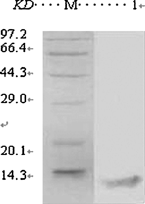
Western blot analysis of purified MAb. MTB RpfB domain fusion protein expressed in E. coli could be recognized by anti-Rpf domain MAb at 12 kDa. M, protein marker; 1, RpfB domain Western blot.
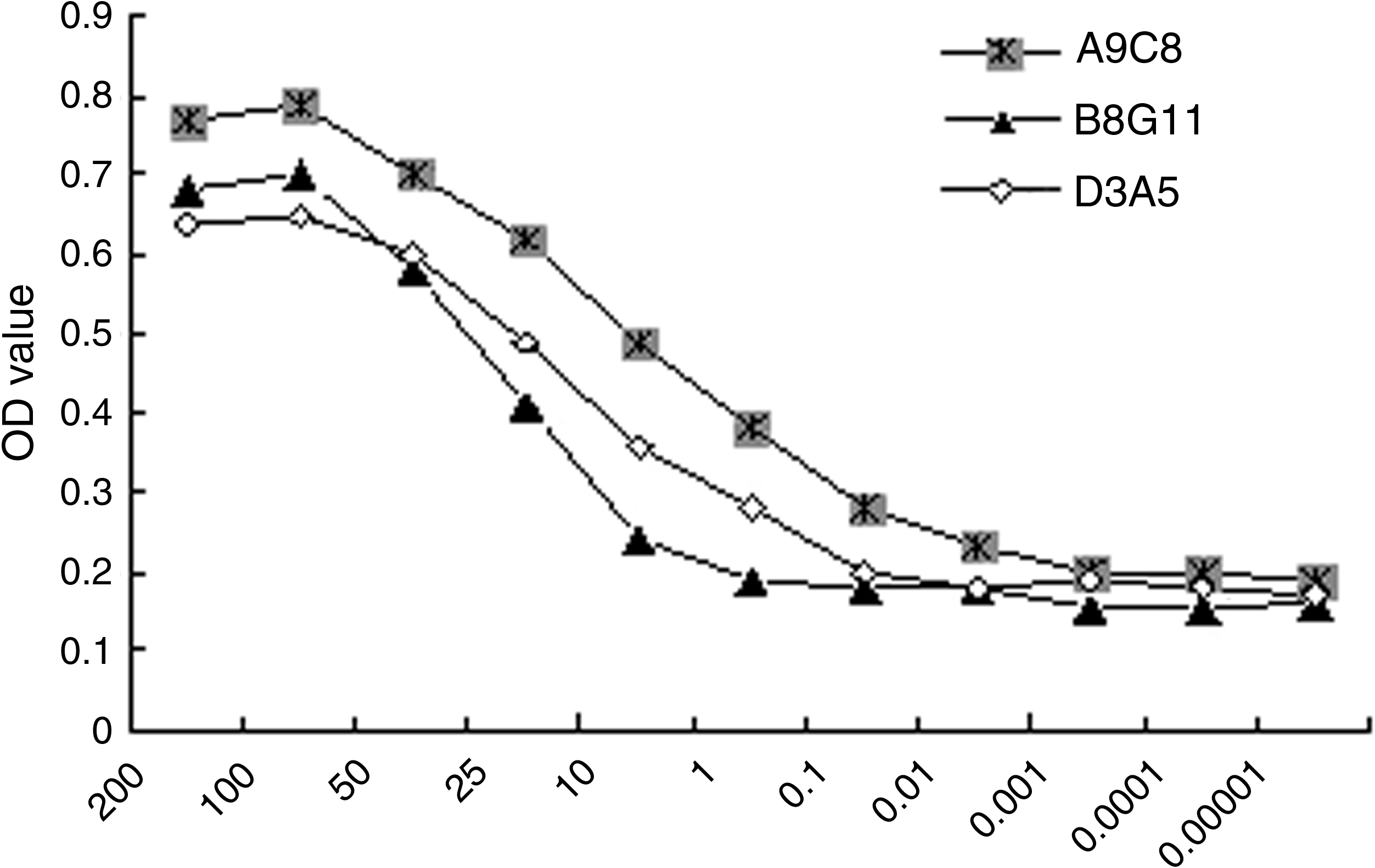
Relative affinity of anti-RpfB domain MAb. By using indirect ELISA assay, the OD values of reactions between serial diluted MAb and RpfB domain were plotted. The relative affinity of MAb was represented as the antibody concentration at an OD value corresponding to 50% of OD value at the plateau stage of antigen-antibody binding.
The three MAbs can specifically recognize the RpfB domain fusion protein. The titers of MAbs D3A5, B8G11, and A9C8 were significantly increased after purification from ascitic fluid by indirect ELISA assay.
Cross-reactivity assay of anti-RpfB domain MAb
As shown in Table 2, the ELISA result demonstrated that the anti-RpfB domain MAb can also react with M. luteus Rpf, Rpf domain, RpfB domain, and RpfA of MTB, as well as H37Ra strain. This indicates that the produced anti-RpfB domain MAb can recognize Rpf-like proteins and their Rpf domains.
Anti-Rpfb domain MAb can react with M. luteus Rpf, Rpf domain, RpfB domain, and RpfA of MTB H37Ra.
Inhibition of MTB growth by anti-RpfB domain MAb
As shown in the growth curve of both M. luteus and MTB H37Ra with treatment of RpfB domain of different concentration, RpfB domain promotes the resuscitation and growth of M. luteus significantly when its concentration is at 1000 pmol/L. At the concentration of 500 pmol/L, RpfB can significantly promote the resuscitation and growth of MTB H37Ra. This promotion effect is significantly inhibited when 1:1000 anti-RpfB domain MAb is added (Figs. 4, 5).
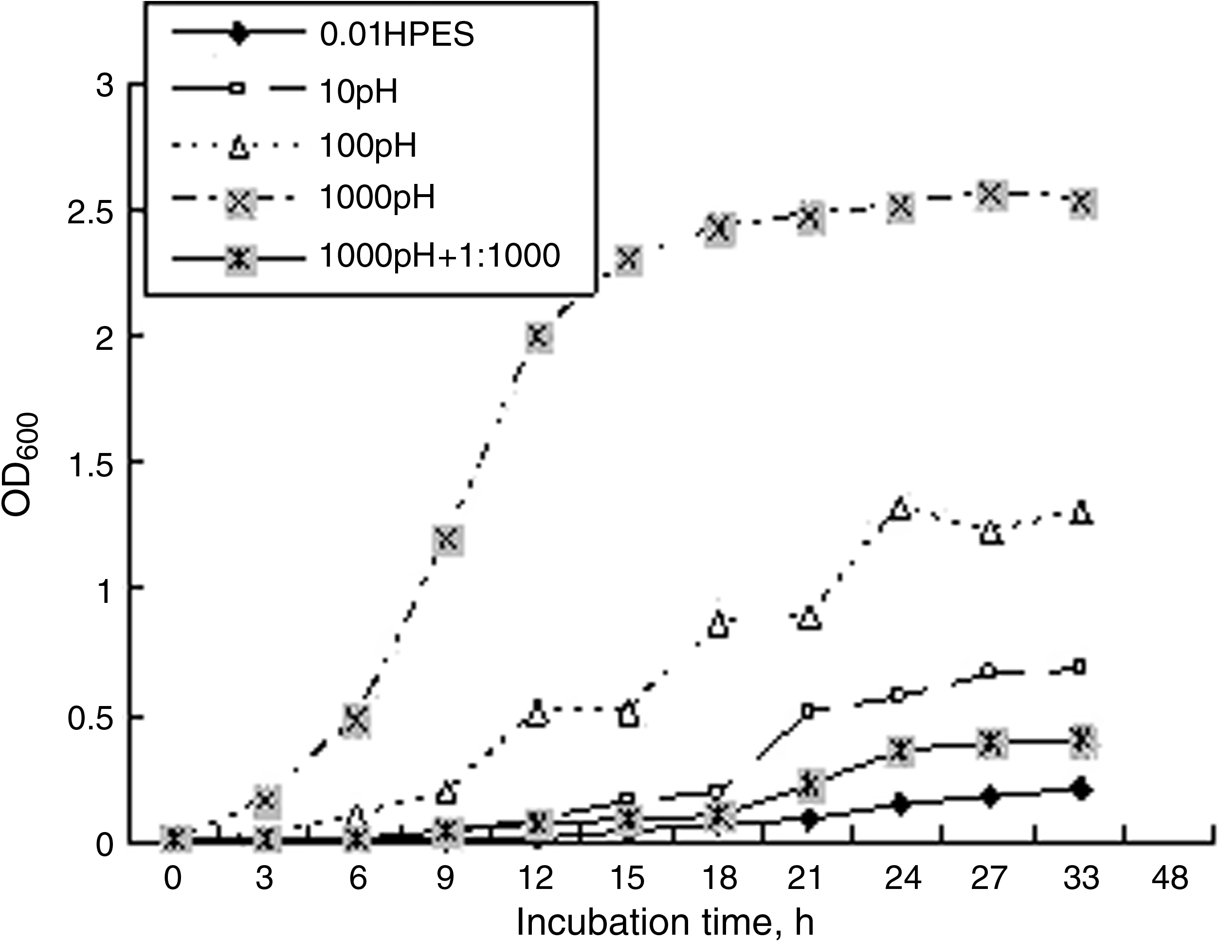
Growth curve of M. luteus in the presence of RpfB domain protein and anti-RpfB domain MAb. As shown in the growth curve of M. luteus with treatment of RpfB domain of different concentrations, RpfB domain promotes the resuscitation and growth of M. luteus significantly when its concentration is at 1000 pmol/L. This promotion effect is significantly inhibited when 1:1000 anti-RpfB domain MAb is added.

Growth curve of MTB H37Ra in the presence of RpfB domain protein and anti-RpfB domain MAb. In the growth curve of both MTB H37Ra with treatment of RpfB domain of different concentrations, RpfB domain promotes the resuscitation and growth of MTB H37Ra significantly when its concentration is at the concentration of 500 pmol/L. This promotion effect is significantly inhibited when 1:1000 anti-RpfB domain MAb is added.
Discussion
Although the infection rate of Mycobacterium tuberculosis is very high, the majority of infected individuals do not develop clinical symptoms. This is because the mycobacterium tuberculosis exists as dormant bacteria in the body. Rpf was first identified in M. luteus.(6) Outside of cells, it promotes the resuscitation of the dormant bacteria at the level of pictogram (10−12 g) by autocrine and paracrine. Since Rpf functions like growth factors in eukaryote, it is also called cytokines of bacteria. There are five Rpf-like genes in MTB genome, and the five Rpf-like proteins encoded show redundancy in functions.(10) Deletion of any one of them does not have a significant effect on MTB growth. Only when multiple (three) Rpf-like genes are being deleted, can significant changes happen, in which the mutants are unable to resuscitate spontaneously in vitro.(11) This indicates that single Rpf antibody cannot completely block Rpf proteins to inhibit MTB resuscitation and growth. Only when multiple anti-Rpf antibodies work together, can the antibodies effectively block Rpf proteins and inhibit MTB resuscitation and growth. However, the optimization, selection, and joint application of multiple proteins undoubtedly make the vaccine research and development more difficult.
Yeremeev and colleagues(7) have demonstrated that the Rpf proteins in M. luteus have strong immunogenicity. After being administered as subunit vaccines to C57BL/6 mice, Rpf proteins elicit IgG1 and IgG2a responses, T-cell proliferation, and the upregulation of gamma interferon, interleukin-10 (IL-10), and IL-12 but not IL-4 or IL-5. The vaccinated serum was also able to inhibit the growth and proliferation of virulent M. tuberculosis H37Rv.(7) Previous studies have shown that the Rpf domain in M. luteus have the same biological functions as the full Rpf proteins.(9) Therefore, the Rpf domain is likely an ideal protective antigen.
In this study, RpfB domain was purified under denaturing conditions by using metal chelating affinity chromatography and then renatured by slow dialysis. The purified protein was used as antigen to immunize mice, and three lines of monoclonal antibodies were produced. The obtained MAbs have high specificity and relatively high affinity. The hybridoma cells were intraperitoneally injected into mice and ascitic fluid was harvested. Higher purity MAbs were obtained after purification from ascitic fluid by caprylic acid-ammonium sulfate precipitation. The produced monoclonal antibody against RpfB domain can recognize many Rpf-like proteins and their domains. Since Rpf-like proteins are highly homologous and Rpf-like protein domains are highly conserved,(9) it is speculated that MAb against RpfB domain is likely able to recognize all the Rpf-like proteins in the MTB Rpf family. Furthermore, experiments are needed to confirm this speculation. Based on this, new methods to detect MTB-related antigens can be established to detect Rpf proteins at different MTB infection stages. It will not only overcome the defect that Rpf proteins are difficult to detect due to the temporal changes of expression, but also increase the sensitivity and specificity of detection by detecting multiple antigens in combination.
In this report, the role of RpfB domain in promoting resuscitation and growth of M. luteus and dormant MTB H37Ra was studied; at the same time, the inhibition of this resuscitation and growth by anti-RpfB domain MAb were also investigated. Our results showed that anti-RpfB domain MAb can significantly inhibit the resuscitation and growth of M. luteus and MTB H37Ra induced by RpfB domain. It was also shown that the specific antibody against Rpf domain might inhibit the reactivation of in vivo dormant or latent MTB to limit the recurrence of occult infection. The production of anti-RpfB domain MAbs provides a powerful experimental tool to further study the biological and immunological characteristics of RpfB domain and to evaluate whether RpfB domain should be considered as a candidate subunit component for tuberculosis vaccine.
Footnotes
Acknowledgments
This study was supported by the National Major S&T Projects of China (2008ZX10003-013) and the National Natural Science Foundation of China (30972767, 30801055), and funded by the Department of Microbiology, School of Basic Medicine and the Department of Clinical Laboratory of Xijing Hospital in the Fourth Military Medical University (Xi'an, China).