
Abstract
Human tumor necrosis factor related apoptosis inducing ligand (TRAIL) can selectively induce apoptosis in a variety of transformed cells and is currently being developed as a cancer therapeutic drug. Here we expressed the TRAIL protein including extracellular (114–281aa) without any tag protein named TRAIL-NT, and prepared anti-TRAIL polyconal antibodies (Poly-Ab). The human TRAIL extracellular gene was amplified from PBMC and cloned into pGEM-T-Easy vector for sequence analysis. The expression vector pET-28a/TRAIL was constructed using the DNA recombinant method, and the recombinant protein without any tag protein was expressed in Escherichia coli BL21(DE3). The TRAIL-NT protein was purified by cation ion-exchange column and identified by SDS-PAGE and Western blot analysis. The proliferation inhibition activity of TRAIL-NT was detected by the MTT method, Wright-Giemsa staining assay, and FACS. The polyclonal antibody of TRAIL-NT was obtained after the BALB/C mice were immunized with purificated TRAIL-NT protein. Results showed that the target protein expressed in E. coli BL21(DE3) has the same molecular weight as that expected and could be recognized by anti-TRIAL Poly-Ab. The TRAIL-NT protein could also inhibit proliferation and induced apoptosis of Jurkat cells but no cytotoxicity to human liver cells and PBMC was observed. This preliminary research laid a solid foundation for further research on its biological activity and application in anti-tumor therapy.
Introduction
In our research, we expressed and purified the TRAIL extracellular region (114aa to 281aa) without any tag protein in a prokaryotic expression system as well as prepared an anti-TRAIL polyclonal antibody. The bioactivity of the purified TRAIL protein, named TRAIL-NT, was identified and the titer of polyclonal antibody was tested.
Materials and Methods
Amplification of TRAIL gene by RT-PCR
The primers were designed according to the cDNA sequence of TRAIL in GenBank and synthesized by Invitrogen Biotechnology (Carlsbad, CA). Sense primer: 5′-CATG
Expression of TRAIL-NT protein
The TRAIL gene was digested from the sequencing vector and ligated to pET-28a prokaryotic expression vector with T4 ligase (NEB, Ipswich, MA). TRAIL-NT protein was expressed in E. coli BL21(DE3) induced by IPTG (0.1 mM, Sigma, St. Louis, MO) and purified by Cation Ion-Exchange column (Pharmica, Oak Ridge, NJ).
Western blot analysis
TRAIL-NT protein expressed in E. coli BL21(DE3) was resolved by SDS-PAGE in 15% polyacrylamide gels and transferred to a nitrocellulose membrane with a horizontal electrophoresis transfer system (Bio-Rad, Laboratories, Hercules, CA). The membrane was blocked with 5% non-fat milk for 1 h and then incubated with poly-anti-TRAIL antibody (eBioscience, San Diego, CA) at room temperature for 1 h. After washing with PBST two times, the membrane was incubated with HRP-conjugated secondary antibody. Western blot was developed using the enhanced chemiluminesense (ECL) reagents. TRAIL protein with His tag was used as a positive control.
Proliferation inhibition assay
Jurkat cells, Chang liver cells, and PBMCs (ATCC, Manassas, VA) were used to test the proliferation inhibition ability of TRAIL-NT protein. Briefly, 100 μL of these cells (8 × 10⁵/mL) were dispensed into 96-well cell culture plates. In one plate, TRAIL-NT was diluted (final concentrations: 0.02, 0.2, 2, 20, 200, 2000 ng/mL) and added to each well in triplicate. The plates were kept in an incubator at 37°C, 5% CO2 for 12 h. In the other plate, TRAIL-NT protein (100 ng/mL) was incubated with Jurkat cells at different times (2h, 4 h, 6 h, 8 h, 10 h, 12 h) and then 10 μL of 3-(4, 5-dimethylthiazol-2-yl)-2, 5-diphenyl tetrazolium bromide (MTT, 10 mg/mL, Sigma) was added. The plates were incubated for an additional 4 h at 37°C. Following this incubation, 100 μL solubilization solution was added and incubated at 37°C for 12 h. The plate was then read at 570 nm using a plate reader (Anthos, Eugendorf, Austria). Unrelated protein was used as a negative control. The following formula was used to estimate the proliferation inhibition ability of TRAIL-NT:
Apoptosis analysis
Jurkat cells were treated with TRAIL-NT (20 ng/mL) for 16 h and then washed with PBS once. PBS was used as reagent control. Every sample was divided into two equal portions. One part was treated with 75% ethanol at −20°C for 24 h and then incubated with 50 mg/mL RNaseA at 37°C for 30 min. FITC-conjugated Annexin V and PI were used to stain the cells for 30 min. Every sample was analyzed with flow cytometry (FACS Calibur, Becton-Dickinson, Franklin Lakes, NJ) using the software program CellQuest. Ten thousand events were counted. The other part of the sample was distributed on a slide with a cell centrifuge, fixed with methanol for 5 min, and stained with Wright-Giemsa staining buffer. The slides were observed under immersion objective and photographed.
Preparation and identification of anti-TRAIL polyclonal antibody
Six 5-week-old female BALB/c mice were subcutaneously immunized with 100 μg of purified recombinant TRAIL in complete Freund's adjuvant per animal. The immunization was repeated three times at 7-day intervals, then strengthened once using 100 μg of TRAIL in incomplete Freund's adjuvant. Three days after the final booster the mouse was bled retro-orbitally to obtain a total of 3 mL serum. Binding of anti-TRAIL polycolonal antibody to TRAIL was analyzed by ELISA. ELISA plates (Nunc, Roskilde, Denmark) were coated with 2 μg/mL of TRAIL in 0.1 M sodium carbonate buffer (pH 9.6) and incubated at 4°C overnight. The plate was then blocked with 5% milk powder in PBS for 1 h at 37°C. After washing the plates three times with PBST, serum dilutions from 10 to 107 were added and incubated for 1 h at 37°C. Pre-immunization blood was used as a control. The plate was washed three times with PBST and then 0.1 mL of 1:4000 diluted goat anti-mouse IgG antibody labeled with HRP was added and incubated for 1 h at 37°C. After three washes with PBST, the peroxidase reaction was developed with color development.
Results
Expression and purification of extracellular region of TRAIL
The extracellular gene (501 bp) of human TRAIL was cloned from volunteer PBMCs, ligated to pGEM-T-Easy, and verified to be right by sequence analysis. Then the gene was subcloned into prokaryotic expression vector pET-28a (pET-28a/TRAIL(114–281)) and expressed in E. coli BL21(DE3) induced with IPTG. The results of SDS-PAGE (15%) showed that target protein existed both in the supernatant and in the precipitation of lysed E. coli BL21(DE3) (transformed with pET-28a/TRAIL(114–281)). The target protein was purified with strong cation-change column. TRAIL-NT protein purified has a high purity (Fig. 1).
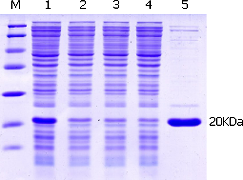
SDS-PAGE analysis of the expression of TRAIL in E. coli BL21(DE3). TRAIL-NT protein was purified by strong cation-change column. The eluate was identified by SDS-PAGE, then stained with Coomassie brilliant blue R-250 after electrophoresis. Lane M, protein marker; lane 1, pET-28a/TRAIL (before absorbed); lanes 2–4, pET-28a/TRAIL (after absorbed); lane 5, TRAIL-NT washed by imidazole.
Western blot analysis
To maintain its natural conformation, TRAIL was expressed without any exogenous tagged proteins. The results of Western blot showed that the expression products of E. coli BL21(DE3) (pET-28a/TRAIL(114–281)) can react with poly-anti-TRAIL antibody (TRAIL-His protein is the positive control). Therefore, we can safely say that the extracellular gene of human TRAIL without any tag protein was achieved successfully (Fig. 2).

Identification of soluble TRAIL protein by Western blot. TRAIL-NT and TRAIL-His was resolved by SDS-PAGE and transferred to nitrocellulose followed by incubation with anti-TRAIL polyclonal antibody, respectively. Second antibody was added and washed. The membrane was developed with ECL reagents. Lane 1, TRAIL-His; lane 2, marker protein; lane 3, TRAIL-NT.
Cytotoxicity assay
The cytotoxicity of TRAIL-NT was measured using MTT assays. The results showed that TRAIL-NT killed Jurkat cells in both time- and dose-dependent manners, but did not harm Chang liver cells and PBMCs. The protein can inhibit the proliferation of Jurkat cells potently in the range of 20 pg/mL to 2000 ng/mL and the maximum inhibition rate is 71%; IC50 is 12 ng/mL. TRAIL-NT has a little or no cytotoxicity to Chang liver cells and PBMCs, even in the largest concentration of 20 μg/mL (Figs. 3 and 4).

Identification of proliferation inhibition activity of TRAIL-NT by MTT. Jurkat cells, Chang liver cells, and PBMCs were dispensed into 96-well culture plates. TRAIL-NT protein with concentrations from 0.02 to 2000 μg/mL were added to each well. After 12 h reaction, MTT was added. Solubilized solution was added to resolve salt crystal formations. The plate was then read at 570 nm using a plate reader. All experiments were done in triplicate and repeated at least three times.
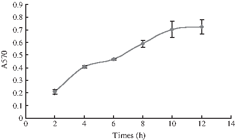
The relationship of the time-response of TRAIL-NT to Jurkat cells. Jurkat cells were dispensed into 96-well culture plates. TRAIL-NT protein with concentration of 100 μg/mL were added to each well. After 2, 4, 6, 8, 10, and 12 h reaction, MTT was added. Solubilized solution was added to resolve salt crystal formations. The plate was then read at 570 nm using a plate reader. All experiments were done in triplicate and repeated at least three times.
Apoptosis identification
Human lymphoma cell line Jurkat cells were incubated with TRAIL-NT for 16 h and then collected for further analysis. PBS was used as a reagent control. The results of FITC-conjugated Annexin V and PI staining showed that the automatic apoptosis rate of Jurkat cells was 4.19% (treated with PBS); the inducing apoptosis rate is 40.68% (treated with TRAIL-NT, 20 ng/mL) (Fig. 5). Wright-Giemsa staining results showed that morphology of Jurkat cells treated with TRAIL-NT was changed, characterized by cell shrinkage, chromatin condensation, and karyopyknosis. Jurkat cells treated with PBS did not show these phenomena (Fig. 6). The results above suggest that TRAIL-NT can induce apoptosis in target cells.
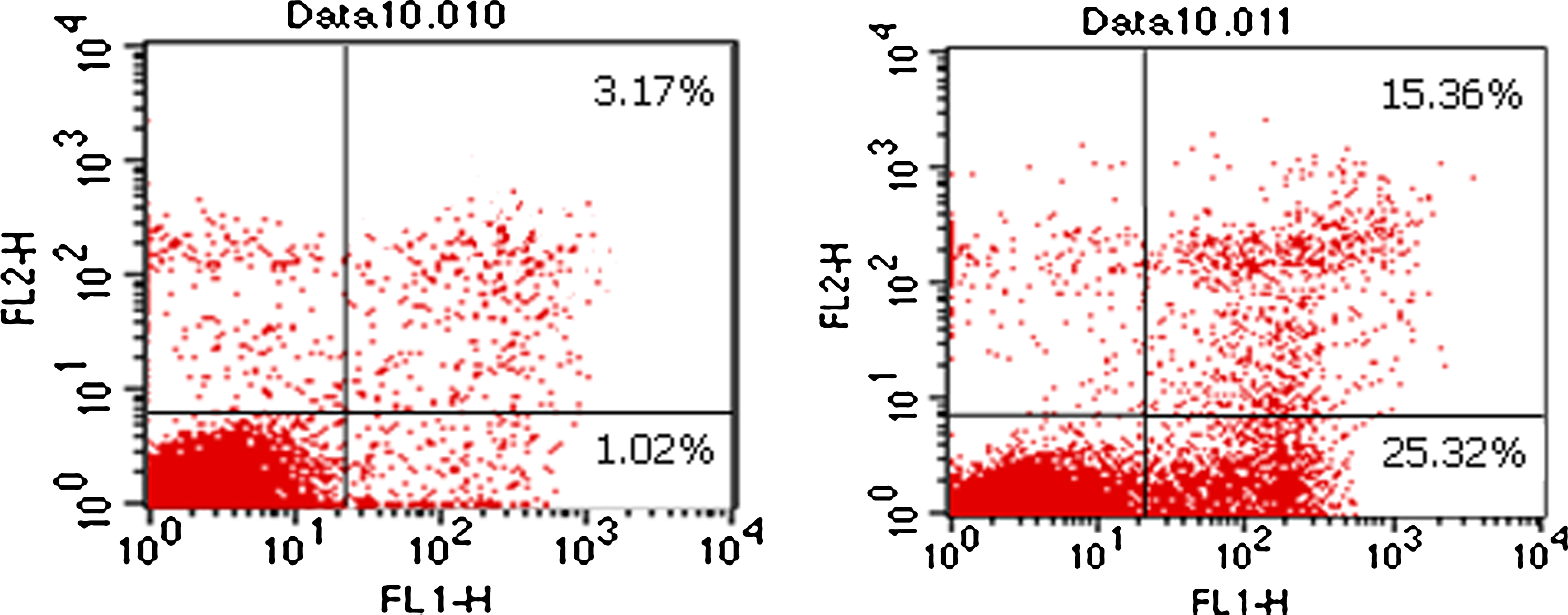
Flow cytometric analysis of apoptosis. Jurkat cells were incubated with 1640 (
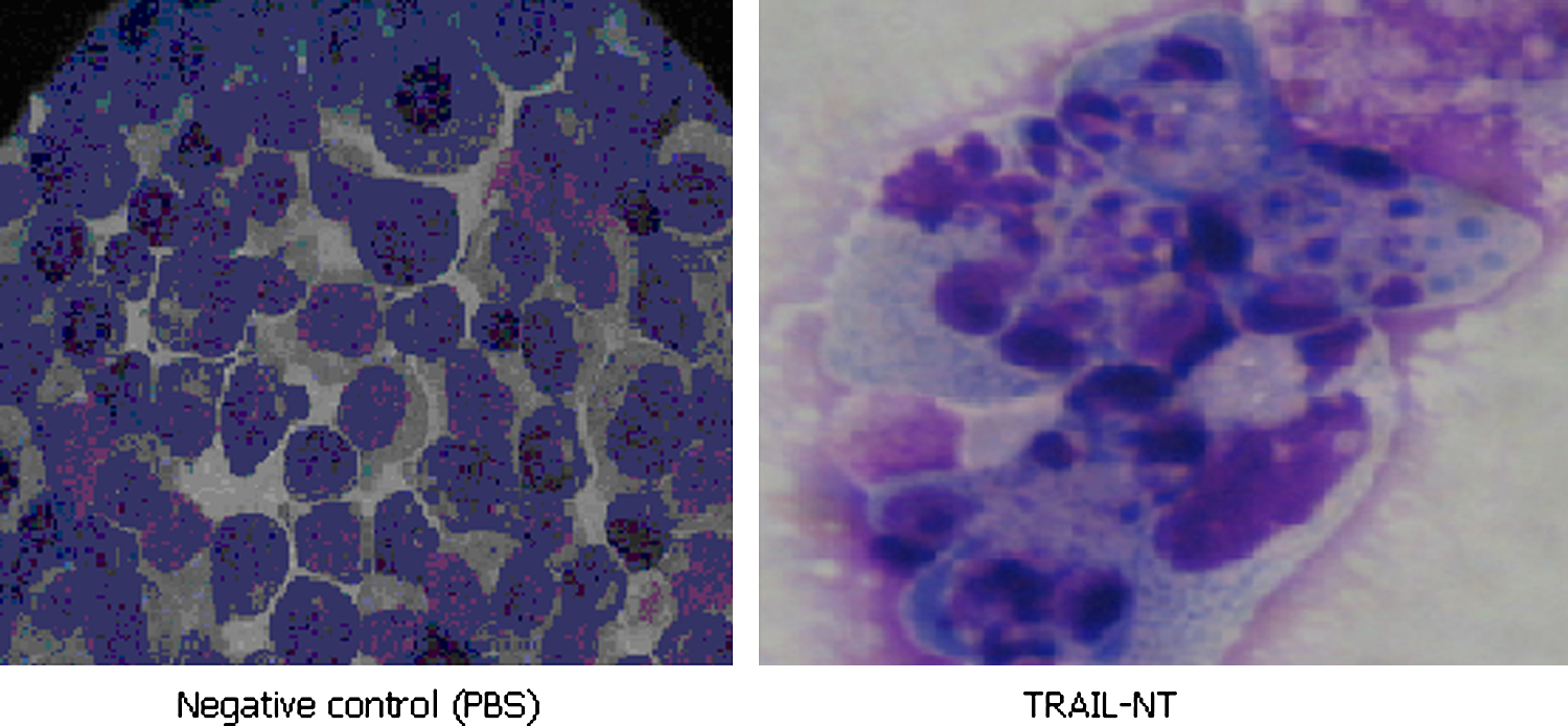
Apoptosis analysis of Jurkat cells (2000x) Jurkat cells were treated with TRAIL-NT (20 ng/mL) for 16 h. PBS was used as reagent control. The sample was distributed on a slide and then stained with Wright-Giemsa staining buffer. The slides were observed under immersion objective and photographed.
Identification of anti-TRAIL polyclonal antibody by ELISA
The result shows that the prepared anti-TRAIL polyclonal antibody not only binds strongly to DR5 protein but also that the titer reaches up to 10−5 (Fig. 7).
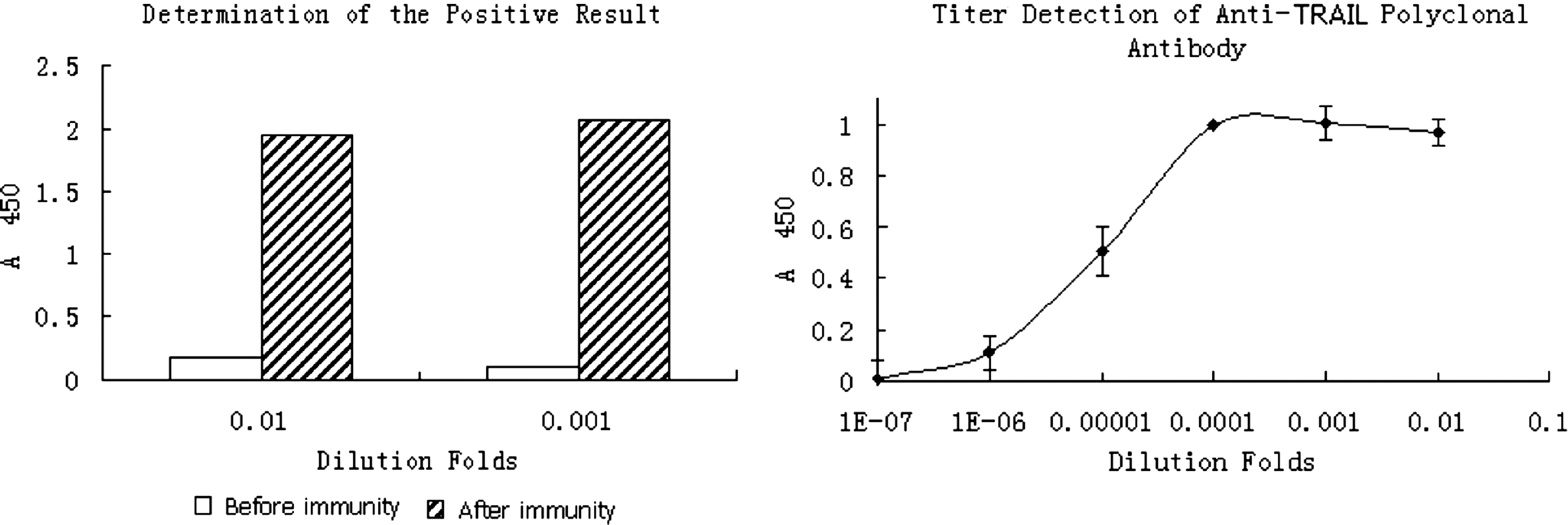
Identification of anti-TRAIL polyclonal antibody. The anti-TRAIL polyclonal antibody of activity was determined by ELISA method. TRAIL protein was coated to an ELISA plate at a proper concentration. The polyclonal antibody could bind to TRAIL, while the negative control (before immunity) showed no binding activity.
Discussion
Like other members of the TNF ligand family, TRAIL induces apoptosis in a variety of cell lines in vitro, including several tumor cell lines resistant to chemotherapeutic agents or ionizing radiation due to mutations in the p53 tumor suppressor gene.(4,5) However, while the utility of FasL and TNF-α is limited by their acute toxic effects on normal tissue, TRAIL preferentially induces apoptosis in tumor cell lines, but not in normal cells,(6) suggesting that it may prove to be a powerful cancer therapeutic. TRAIL can induce tumor cell apoptosis through several ways, including the specific binding of two death receptors that activate in mitochondrial and non-mitochondrial ways. In recent years, some results have shown that TRAIL can induce apoptosis through an independent caspase3 pathway.(7) TRAIL has a good synergistic effect in combination with some chemotherapy drugs and Chinese medicines.(8–12) But there are confirmed reports of some versions of recombinant TRAIL that have toxic side effects on normal cells for caused death receptor protein excessive aggregation. Further research revealed that the reason for this was exogenous tag proteins, which affected TRAIL protein native conformations.(13) To the contrary, some other forms of recombinant TRAIL protein showed no toxicity to normal cells,(14) so TRAIL as an anti-cancer drug is still expected.
In our research, we have developed a non-fusion TRAIL protein named TRAIL-NT. The protein maintained its natural formation because it is not labeled by any tag protein. TRAIL-NT was purified by a strong cation-change column from the supernatant of lysed bacteria and identified by both SDS-PAGE and Western blot analysis. TRAIL-NT has the same molecular weight as that expected and could be recognized by anti-TRIAL Poly-Ab. The protein could inhibit the proliferation and induce apoptosis of Jurkat cells, but had no effect on normal cells, including Chang liver cells and PBMCs. The inhibition rate with time- and dose-dependent reached 50% with a concentration of 0.2 μg/mL. In addition, we also prepared the anti-TRAIL polyclonal antibody. The polyclonal antibody has a high titer and good bioactivity identified by the ELISA method. This prospective research laid a solid foundation for further research on its biological activity and application in anti-tumor therapy.
Footnotes
Acknowledgments
This work was supported by the National Science Foundation of China (no. 30971508) and HNIFOS (no. 074200510014).
Author Disclosure Statement
The authors have no financial interests to disclose.