
Abstract
Fenoxaprop-ethyl is a selective aryloxyphenoxypropionate herbicide used widely to control annual and perennial grasses. A monoclonal antibody (MAb) against fenoxaprop-ethyl (FE), designated as 3E6B9C, was produced and had very low cross-reactivity with some of its structural analogs, such as clodinafop-propargyl, diclofop-methyl, lactofen, and quizalofop-p-ethyl. An indirect competitive enzyme-linked immunosorbent assay (icELISA) was developed. The concentration of R-(+)-fenoxaprop-ethyl (R-FE) producing 50% of inhibition (IC50) and the working range of icELISA were 3.1 ng/mL and 0.6-29 ng/mL, respectively. This assay is also sensitive to R-fenoxaprop, S-(-)-fenoxaprop-ethyl, and metamifop with IC50 of 3.4, 2.7, and 3.5 ng/mL, respectively. The recoveries of R-FE in soil samples with the icELISA were 86–102%.
Introduction
The herbicidal activity of FE is primarily from its optical isomer R-FE, which is extensively produced and applied in crop protection rather than FE at present. Some reports showed that R-FE possessed certain hepatotoxicity in mice, Daphnia magna, porcine sperm, and tadpole.(3–6) Therefore, it is desirable to establish a quick and easy method to monitor the residual R-FE in the environment and food samples, and develop its reasonable application patterns for plant protection.
Gas chromatography (GC),(7) high performance liquid chromatography (HPLC), HPLC mass spectrometry (HPLC-MS),(8) and spectrophotometry(9) have been employed to determine residual R-FE in reported studies. Although these chromatographic and spectrophotometric methods have a good sensitivity and accuracy, they are expensive and time-consuming, and they require extensive sample preparation. Enzyme-linked immunosorbent assay (ELISA) is rapid, sensitive, selective, and cost-effective, and has been extensively used in the detection of environmental contaminants.(10–13) Although a pAb-based ELISA for determination of R-FE was recently reported, its IC50 value was only 84 ng/mL the effect of ionic strength and methanol in buffer on the assay performance, and spike recoveries in tap water were studied.(14) In the present study, a MAb called 3E6B9C against FE was produced, and an indirect competitive ELISA (icELISA) was developed and optimized.
Materials and Methods
Reagents and apparatus
Clodinafop-propargyl, R-FE, S-(-)-fenoxaprop-ethyl (S-FE), diclofop-methyl, lactofen, metamifop, 6-chloro-2,3-dihydrobenzoxazol-2-one (CDHB), and quizalofop-p-ethyl were supplied by the Institute for Control of Agrichemicals, Ministry of Agriculture of China (ICAMA). Cell culture media (Dulbecco's modified Eagle's medium, DMEM) and fetal bovine serum (FBS) were obtained from Gibco BRL (Paisley, Scotland). Reagents purchased from Sigma (St. Louis, MO) were cell freezing medium dimethyl sulfoxide (serum free); hypoxanthine, aminopterin, and thymidine (HAT); hypoxanthine and thymidine (HT) medium supplements; l-glutamine, penicillin, streptomycin, and goat anti-mouse IgG conjugated with horseradish peroxidase (HRP); complete and incomplete Freund's adjuvant, N-hydroxysuccinimide (NHS), dicyclohexylcarbo-diimide (DCC), bovine serum albumin (BSA), ovalbumin (OVA), and o-phenylenediamine (OPD). Polyethlene glycol 2000 was purchased from Fluka (Buchs, Switzerland). Mouse antibody isotyping kit was obtained from Pierce (Rockford, IL). All other chemicals used were of analytical grade.
Cell culture plates and 96-well plates used were from Corning Costar (Corning, NY); an automated plate washer (Wellwash 4 MK2), a microplate reader (Multiskan MK3), and a direct heat CO2 incubator (311) were from Thermo (Vantaa, Finland). An electric heating constant-temperature incubator was from Tianjin Zhonghuan Experiment Electric Stove Co. (Tianjin, China).
The following solutions were used: (1) coating buffer (0.05 M carbonate buffer [pH 9.6]); (2) phosphate-buffered saline (PBS, 0.1 M phosphate buffer containing 0.9% NaCl [pH 7.5]); (3) PBS with 0.1% (v/v) Tween-20 (PBST); (4) PBST containing 0.5% (w/v) gelatin (PBSTG); (5) citrate-phosphate buffer (0.01 M citric acid and 0.03 M Na2HPO4 [pH 5.5]); (6) substrate solution (4 μL of 30% H2O2 added to 10 mL of citrate-phosphate buffer containing 2 mg/mL OPD); and (7) a stop solution (2 M H2SO4).
Myeloma cell line and medium
The HAT-sensitive Balb/c mouse myeloma cell line SP2/0-Ag14 obtained from the China Institute of Veterinary Drug Control (Beijing, China) was used in fusion experiments. DMEM containing 10–20% (v/v) FBS was supplemented with 0.2 M glutamine, 50,000 U/L penicillin, and 50 mg/L streptomycin. The standard medium was used for growing of myeloma and hybridoma cells.
Preparation of immunogen and coating antigen
Hapten R-FA (Table 1) was synthesized via alkaline hydrolysis of R-FE according to the procedure described earlier.(14,15) For conjugation, R-FA (20.0 mg), NHS (10.3 mg), and DCC (14.8 mg) were dissolved in 1 mL of DMF. The solution was stirred at room temperature for 6 h, and then the mixture was centrifuged at 8000 rpm for 5 min. The supernatant was added drop-wise to protein solution (131 mg of BSA or 88 mg of OVA in 10 mL of carbonate buffer, 50 mM [pH 9.6]), and was stirred again overnight at 4°C. Subsequently, the reaction mixture was dialyzed against five changes of PBS for 3 days, and stored at −20°C after lyophilization. The molar ratios of hapten-to-BSA and hapten-to-OVA were 11:1 and 9:1, respectively, which were determined through the UV-VIS method.
No inhibition was observed up to 10,000 ng/mL of analytes.
Production and characteristics of MAb against R-FE
Five Balb/c female mice (6–7 weeks old) were injected subcutaneously with 0.1 mg of R-FA-OVA conjugate dissolved in 0.1 mL PBS mixed with 0.1 mL Freund's complete adjuvant. Two subsequent injections were carried out at 2-week intervals using Freund's incomplete adjuvant. One week after the third injection, mice were eye-bled and sera were tested for anti-R-FE antibody titer and for R-FE recognition properties in icELISA. Two weeks after the third injection, the mouse with the highest titer and best specificity was boosted intraperitoneally with 0.1 mg R-FA-OVA conjugate in 0.1 mL PBS. Three days later, the spleen cells collected from the mouse were mixed with the SP2/0 cell line at a ratio of 10:1 and fused with PEG-2000. The plates were incubated at 37°C in a CO2 incubator (5% CO2 in air), and then the hybrid cells were grown selectively in the DMEM supplemented with HAT. Seven days after fusion, the supernatant was tested by icELISA, positive hybridomas were cloned by limiting dilution, and clones were further selected by icELISA.
The clone (designated as MAb 3E6B9C), having a high antibody titer and good sensitivity in the culture supernatant, was expanded in mice for production of ascites. The antibody was purified by ammonium sulfate precipitation. The immunoglobulin isotype was determined with a mouse antibody isotyping kit (Pierce, Rockford, IL), and the affinity of MAb 3E6B9C to R-FE were determined with icELISA.
Establishment of icELISA
In all procedures, microtiter plates were washed on an automated plate washer with 250 μL of PBST per well three times. A microtiter plate was first coated with 100 μL R-FA-BSA conjugate in coating buffer per well for 3 h at 37°C. After four washes with PBS, each well was blocked with 200 μL 3% non-fat dry milk in PBS for 30 min at 37°C. After four washes with PBST, 50 μL of aliquots of various concentrations of the standard in PBSTG were pipetted into each well followed by the addition of 50 μL of sera, and supernatant or purified MAb solution diluted in PBSTG. The plate was then incubated for 0.5 h at 37°C, the unbound antibody was removed by washing the plates four times with PBST, and then 100 μL of goat anti-mouse IgG–peroxidase conjugate in PBSTG was added to each well followed by incubation at 37°C for 0.5 h. After the plate was washed with PBST again, 100 μL of substrate solution per well was added. The reaction was stopped by adding 50 μL of 2 M H2SO4 per well. Absorbance was read at 492 nm with the microplate reader. The data were imported into and analyzed with OriginPro 7.5 (OriginLab) to fit the equation:
where×0=center; p=power; A1=initial Y value; A2=final Y value.
Specificity of antibody
The specificity of the MAbs was evaluated by cross-reactivity with aryloxyphenoxypropionate herbicides via icELISA. The relative cross-reactivity was calculated by the following formula:
Cross-reactivity (%)=(IC50 of R-FE/IC50 of other compound)×100
Spiking tests
Soil samples collected from Beijing were used in the spiking tests with pH of 6.9, moisture of 13.1%, and an organic matter content of 1.1%. They had been air-dried in the dark for 1 week and consisted of 41.9% sand, 52.6% silt, and 5.5% clay. R-FE stock solution (1 mg/mL) was serial-diluted to 0, 500, 1000, 2000, 4000 and 8000 ng/mL in methanol, and then 100 μL of varied concentrations of the standard solutions were gradually added to the 1 g of dried soil and mixed by vortex. Methanol (3 mL) was added into the spiked samples 3 hours later, and the solution was stirred on a vortex shaker again for 3 min followed by ultrasonic extraction for 10 min and centrifugation at 4000 rpm for 5 min. The organic layer was filtered through anhydrous sodium sulfate for dehydration. The same extraction step was repeated twice. All extracts were combined and evaporated to near dryness on a vacuum rotary evaporator at 40°C. The residue was dissolved in 1.0 mL of PBSTG, and diluted 60 times by PBSTG for detection with icELISA. A standard curve was obtained with 12 R-FE concentrations including 0, 0.19, 0.39, 0.78, 1.56, 3.13, 6.25, 12.5, 25, 50, 100, 200 ng/mL diluted in PBSTG. Three separate fortifications and extractions were conducted for the soil samples, and three replicate ELISAs with three well replicates were conducted too. The concentrations were computed from the linear part of the standard curve (actually from 20–80% inhibition).
Results and Discussion
Characteristics of monoclonal antibody against R-FE conjugate
Hapten R-FA was synthesized via alkaline hydrolysis of R-FE and was conjugated to carrier proteins by the active ester method.(17) A monoclonal antibody, designated as MAb 3E6B9C, was generated with R-FA-OVA conjugate as the immunogen. The dissociation constant (Kd) of the antibody was determined using the method of Beatty.(18) The Kd value was 3.1×10⁻¹⁰ M. The MAb 3E6B9C is an IgG1 isotype that has κ light chains, and showed more than 90% cross-reactivity with S-FE, R-FA, and metamifop, whereas there was <0.52% cross-reactivity with other metabolic products and commonly used aryloxyphenoxypropionate herbicides (Table 1).
Hapten R-FA has been reported by Moon and colleagues(15) as a coating antigen to screen with anti-metamifop anti-sera in an homologous and heterologous ELISA format for the analysis of metamifop. However, R-FA-BSA as a coating antigen did not offer a sensitive assay. A pAb-based ELISA for determination of R-FE was also reported by Wang and co-workers,(14) whose cross-reactivity with metamifop was 51.7%, but the cross-reactivity with S-FE was not mentioned. The MAb 3E6B9C in the present study showed a high cross-reactivity with S-FE and other structural analogs containing a [(6-chloro-2-benzoxazolyl)oxy] phenoxy portion. In general, there is some correlation between the position in the hapten molecule used for the conjugation to carrier protein and the recognition of the hapten by the prepared antibody, and antibody could distinguish chiral isomers in reported research.(16) However, we found that the MAb 3E6B9C could not distinguish the chiral isomers of FE in this study. The possible reason could be that chiral carbon on hapten is close to the site of conjugation and could be shielded by the carrier protein.
Optimization of icELISA
The optimal concentrations of coating antigen and MAb were screened by checkerboard titration. The coating antigen (1 mg/mL) and purified MAb 3E6B9C (0.13 mg/mL) were at a dilution ratio of 1:3000 and 1:700. The goat anti-mouse IgG–peroxidase conjugate (1 mg/mL) was at a dilution ratio of 1:1000.
Figure 1 shows the R-FE standard curve in icELISA, of which the×0, A1, and A2 were 2.26, 1, and 0.12, respectively, with a p value of 0.88 and R2 of 0.999. The IC50 value was defined as the concentration that gave a half maximal inhibition and the detection range was defined as the lowest and highest quantification limits of about 80 and 20% B/B0. The IC50 value and detection range were 3.1 ng/mL and 0.6–29 ng/mL, respectively (Fig. 1). Comparing IC50 value, the assay developed by us was approximately 30 times more sensitive than that reported by Wang and colleagues,(14) and about six times more sensitive than that reported by Moon and associates.(15)
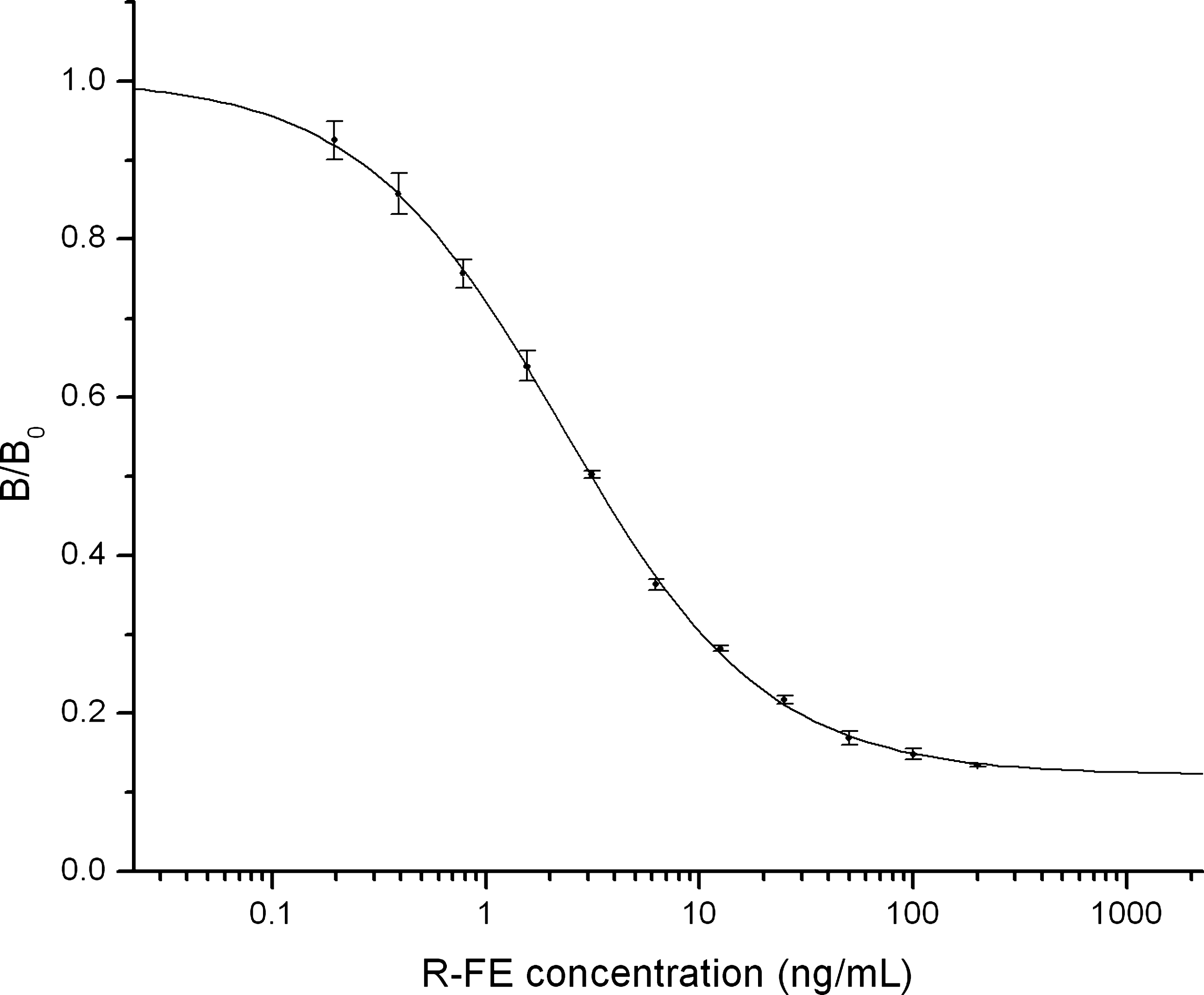
Standard inhibition curve of R-FE by icELISA.
Recoveries of R-FE in soil samples
Soil samples spiked at concentrations of 0, 50, 100, 200, 400, and 800 ng/g were determined with the icELISA (Table 2). The average recoveries of R-FE with the icELISAs were 86–102%. Furthermore, the developed assay was very sensitive, and the samples could be diluted 1000 times to minimize possible matrix interferences. So, the developed icELISA will be a reliable and sensitive method to detect rapidly R-FE in soil samples.
Data are means±standard deviations (SD) of three determinations.
Not detected.
Conclusion
To our knowledge, this is the first report on monoclonal antibody-based icELISA for FE. The MAb 3E6B9C had very weak cross-reactivity with other commonly used aryloxyphenoxypropionate herbicides except metamifop. The icELISA using MAb 3E6B9C had an IC50 value of 3.1 ng/mL, and the working range of 0.6–29 ng/mL based on 20–80% of inhibition of R-FE. This assay is also very sensitive to R-FA, S-FE, and metamifop (IC50, 3.4, 2.7, and 3.5 ng/mL, respectively). Spiking tests of R-FE in soil samples showed that the developed icELISA was a quick, reliable, and sensitive method.
Footnotes
Acknowledgments
This work was supported by a grant from the National Natural Science Foundation of China (no. 20972186), and the National Key Project of Scientific and the National Basic Research Program of China (no. 2010CB126105).
Author Disclosure Statement
The authors have no financial interests to disclose.