
Abstract
Glucose-regulated protein (GRP78), an ER chaperone that belongs to the heat-shock protein (HSP) family, exist in all cells and plays important roles in maintaining cellular homeostasis. GRP78 participates in protein folding, transportation, and degradation. Lack of high affinity antibodies especially monoclonal antibodies (MAbs) suitable for Western blot and immunohistochemical staining has lagged. To gain further insight into its possible functions, we generated a novel MAb specific for hGRP78 in Western blot and immunohistochemistry and localized hGRP78 in some human cancer cell lines and cancer tissues. Immunoreactivity of GRP78 was prominent in Hela, Colo205, and A549 detected by 3F9 in Western blot analysis. 3F9 antibody recognized endogenous GRP78 in human cervical cancer, colonic cancer, esophageal cancer, and lung cancer. Thus, successful production of GRP78 monoclonal antibodies provides a new powerful tool for investigation of GRP78 function.
Introduction
Considering the important function of GRP78, we generated a monoclonal antibody (MAb) specific for human GRP78, named 3F9, which could be used in Western blot and immunohistochemistry. Immunoreactivity of GRP78 was prominent in Hela, Colo205, and A549 detected by 3F9 in Western blot analysis. 3F9 antibody recognized endogenous GRP78 in human cervical cancer, colonic cancer, esophageal cancer, and lung cancer. Thus, our preliminary work provides a new powerful tool for investigation of expression profile of GRP78 protein and furthers the study on GRP78 functions.
Materials and Methods
Bacterial protein expression and purification
Full length of human GRP78 cDNA was subcloned into the pET-28a vector and expressed as a His-tagged fusion protein in BL21 Escherichia coli following induction with IPTG (1.2 mM) for 4 h at 35°C. Soluble protein recovered from bacterial cell lysates was purified using metal ion affinity chromatography with Ni-CAM HC resin (Sigma-Aldrich, St. Louis, MO). Recombinant protein was then dialyzed into phosphate-buffered saline (PBS) and concentrated using centricon-30 filtration units (Millipore, Bedford, MA), as described by the manufacturer. Purified GRP78 protein was quantified by BCA assay (Pierce Biotechnology, Rockford, IL) and used as antigen to immunize mice and for screening the monoclonal antibodies.
Production of hybridomas
Cell fusion was done to standard protocol in our laboratory. In brief, female BALB/c mice (6 weeks old) were immunized with 50 μg of recombinant protein in complete Freund's adjuvant and applied to the mice by subcutaneous (s.c.) injection. Then immunizations were carried out two more times with 50 μg of antigen mixed with incomplete Freund's adjuvant by s.c. injection at 3-week intervals. The titer of anti-GRP78 antibodies in blood sera was determined by ELISA. Finally, one of the immunized mice was boosted with additional 50 μg of antigen by intraperitoneal (i.p.) injection without adjuvant. Splenocytes from the boosted mouse and SP2/0 myeloma cells were fused in the presence of PEG (MW4000, Merck, Darmstadt, Germany) three days later. The positive hybrids were screened by ELISA and subcloned at least three times before being cultured in larger scales. Antibodies in the supernatants of hybridomas cultured in serum-free medium (Hyclone, Logan, UT) were purified using affinity purification by ProteinG-Sepharose CL-4B (GE Healthcare, Piscataway, NJ) chromatography. The IgG fractions were pulled together and dialyzed in PBS (pH 7.4).
ELISA assays
The 96-well plates were incubated with 0.5 μg/well of recombinant GRP78 or with His-tagged protein as a control in PBS (pH 7.4) overnight at 4°C. Then the plates were washed three times with PBS containing 0.1% Tween-20. Subsequently, 100 μL hybridoma media were added to the plates and were incubated for 1 h at 37°C followed by four washes. After washing, 100 μL of horseradish peroxidase (HRP)–conjugated goat anti-mouse IgG antibodies (1:5000 v/v, Promega, Madison, WI) were added to each well and plates were incubated at 37°C for 1 h. After three more washes, 100 μL of 2,2’-Azino-di(3-ethylbenzthiazoline-6-sulfonic acid) (ABTS) substrate were added to each well and plates were read at 405 nm on a micro-plate reader 15 min later.
Western blot analysis
Cell lysates were fractionated by SDS-PAGE, electrophoretically transferred to a PVDF membrane (Millipore, Bedford, MA), and probed with anti-GRP78 MAb to detect the expression of GRP78. Resulting immunocomplexes were detected using an ECL kit (GE Healthcare) and then exposed to Agfa x-ray film. Anti-β-actin MAb was used as internal standard for all samples.
Immunohistochemistry
Sections of various human cancer tissues were deparaffinized with dimethyl benzene and rehydrated through graded alcohols. Endogenous peroxidase activity of slides was blocked with 3% H2O2-methanol for 10 min, and normal goat serum (10%) was applied for blocking non-specific binding. Anti-GRP78 monoclonal antibody was dropped on the slices for 1 h at room temperature. The slices were then dipped into biotin-conjugated goat anti-mouse IgG (Promega, Madison, WI) for 1 h at room temperature followed by streptavidin-HRP complex incubation for 30 min. Antibody complexes were visualized by DAB chromogen. Sections were counterstained with Mayer's hematoxylin for 30 sec, dehydrated through graded ethanol, cleared in dimethyl benzene, and examined using light microscopy.
Results
Generation and characterization of MAbs
One positive hybridoma clone secreting MAbs against GRP78 was obtained and was designated 3F9. The immunoglobulin isotype of this MAb was IgG2a (κ). This hybridoma clone could stably secrete specific MAbs, which reacted with His-GRP78, but not other His-tagged protein in ELISA. The MAb was produced by culturing the hybridoma in large scales and purified using affinity purification by ProteinG-Sepharose CL-4B.
Reaction of 3F9 against endogenous hGRP78 protein determined by Western blot analysis
Next, 3F9 MAb was tested in Western blot analysis using cancer cell lysates of Hela (human cervical carcinoma), Colo205 (human colonic carcinoma), and A549 (human lung carcinoma). Cultured cells were harvested and prepared using lysis buffer. Cell lysates (50 μg) were separated by SDS-PAGE and transferred onto PVDF membrane. After exposure to P3F9 MAb, there was a band of 78 kDa on the film. Anti-β-actin MAb was used as internal standard for all samples (Fig. 1).
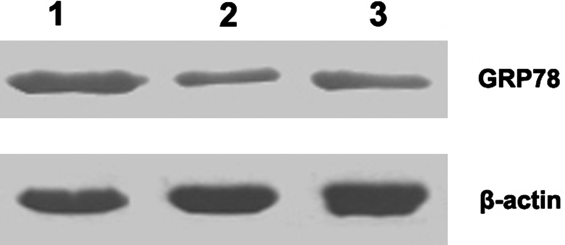
Western blot analysis of reactivity of MAb endogenous hGRP78 proteins. Western blot analysis of hGRP78 expression in total cell lysates of Hela (lane 1), Colo205 (lane 2), and A549 (lane 3). Cell lysates (50 μg) were separated by SDS-PAGE, transferred onto PVDF membrane, and exposed to P3F9 MAb. There was a band of 78 kDa on the film. 3F9 specifically blotted a 78 kDa band.
Application of MAb in immunohistochemistry
Paraffin blocks of human cervical cancer, colonic cancer, esophageal cancer, and lung cancer were sectioned for immunohistochemistry. Sections of human cancer tissues were deparaffinized with dimethyl benzene and rehydrated through graded alcohols before incubation with 3F9 antibody; the immunocomplexes were visualized by DAB. As shown in Figure 2, 3F9 antibody recognized endogenous GRP78 in human cancer tissue, which is visualized by strong DAB staining in the cytoplasm of cervical cancer (Fig. 2A), colonic cancer (Fig. 2B), esophageal cancer (Fig. 2C), and lung cancer (Fig. 2D).
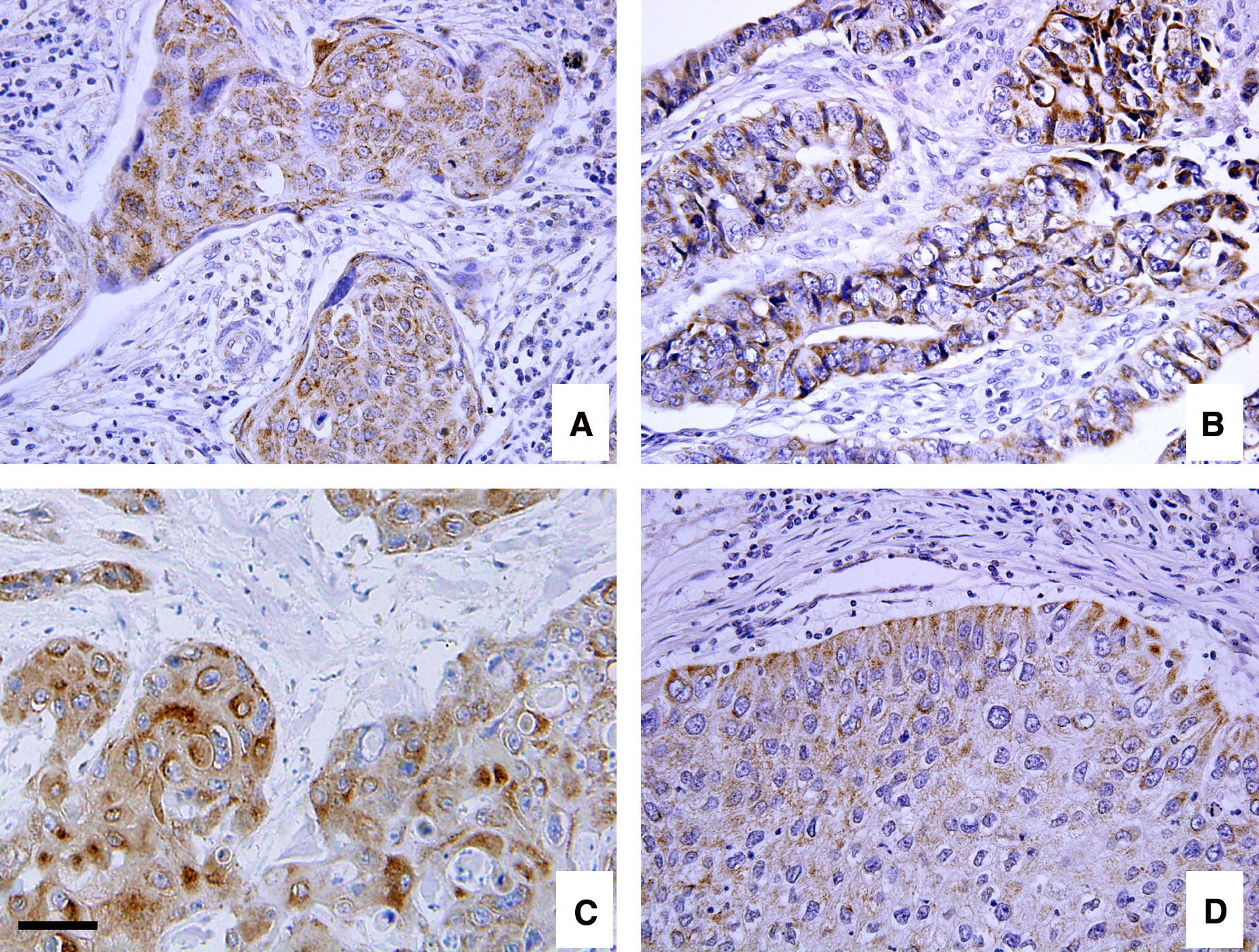
Reactivity of 3F9 antibody to endogenous hGRP78 in formalin-fixed paraffin-embedded sections. The paraffin block was sectioned for immunocytochemistry to test reactivity of 3F9. 3F9 antibody recognizes endogenous hGRP78 in human cancer tissues, which is visualized by strong DAB staining in the cytoplasm of cervical cancer (
Discussion
The unfolded protein response (UPR) is activated for the survival of tumor cells under ER stress. When expressed at high levels, ER chaperones serve as pro-survival components protecting the cells against death induced by ER stress.(3) Some ER chaperones are often upregulated in the UPR, thereby markedly increasing the capacity to handle misfolded proteins.(4) Studies have shown that GRP78, glucose-regulated protein 94 (GRP94), calreticulin (CRT), and Herp protect cancer cells against ER stress-induced apoptosis.(5–7)
GRP78, an ER chaperone that belongs to the heat-shock protein (HSP) family, exists in all cells and plays important roles in maintaining cellular homeostasis. The primary functions of GRP78 are related to its capacity to bind to hydrophobic patches on nascent polypeptides in the ER. GRP78 participates in protein folding, transportation, and degradation. GRP78 also can reside in cell surface and recognizes extracellular ligands, transducing proliferative signals, especially in certain tumors. Because GRP78 is best characterized with respect to its role in cancer progression, drug resistance, and possibly metastasis, it provides further insight into the event of carcinogenesis and cancer cell chemoresistance, indicating its prognostic predicting significance and validating potential therapeutics for clinical usage.(1,8,9)
GRP78 expression is highly upregulated in a variety of cancer cell lines and human cancer specimens, including breast cancer, lung cancer, liver cancer, and prostate cancer and protects cancer cells through multiple mechanisms. GRP78 can inhibit the activation of pro-apoptotic components, BIK and BAX, and suppress procaspase-7 and procaspase-12 cleavage.(9–11) But until now, there was no ideal MAb for GRP78 that could be used for Western blot analysis and immunohistochemistry. The establishment of 3F9 MAb presents a novel powerful tool for the future study of GRP78.
Footnotes
Acknowledgment
This work was supported by the National Science Foundation of China (No. 81000713).
Author Disclosure Statement
The authors have no financial interests to disclose.