
Abstract
An immunoassay system was established for the estimation of the quantity of an antitumor cyclohexapeptide, deoxybouvardin (RA-V) from Bouvardia ternifolia (Cav.) Schlecht, Rubia cordifolia L., and R. akane Nakai (Rubiaceae). First, RA-V was converted into a protein conjugate to make it an effective antigen. In the conjugate the molecular ratio between RA-V and the carrier protein was 5.9:1. The splenocytes from the mouse immunized with the conjugate were then fused with mouse myeloma cells to produce hybridoma, secreting monoclonal antibodies (MAbs) against RA-V. Two clones were isolated, one producing MAb IgG1 and the other MAb IgG2b, both having a κ light chain. The resultant MAbs were evaluated for their sensitivity and cross-reactivity.
Introduction
In our previous study,(1) we established an immunoassay system for the quantitative analysis of an antitumor cyclohexapeptide RA-VII (Fig. 1) from Rubia cordifolia L. and R. akane Nakai (Rubiaceae) by using RA-VII as the antigen. The MAbs obtained, however, also reacted with other compounds of this series of hexapeptide. In the present study, we established another immunoassay system using a cyclohexapeptide, deoxybouvardin (RA-V), as the original antigen (Fig. 1),(2) whose R 1 is H. The obtained MAbs showed greater specificity, reacting only with RA-V and RA-VII.
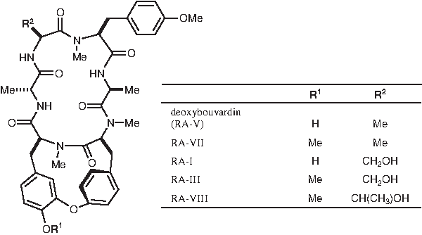
Structures of RA series of compounds.
Materials and Methods
Materials used
The hexapeptides used in the present study—RA-V, RA-VII, RA-I, RA-III, and RA-VIII—were prepared as reported previously.(3,4) Glutaric acid was obtained from Tokyo Kasei (Tokyo, Japan). Enriched RPMI 1640 medium, hypoxanthine-aminopterin-thymidine (HAT) supplement, hypoxanthine-thymidine (HT) supplement, penicillin-streptomycin-glutamine solution, 10 mM MEM non-essential amino acid solution, and 100 mM MEM sodium pyruvate solution were from Invitrogen (Carlsbad, CA). Bovine serum albumin (BSA), human serum albumin (HSA), and peroxidase-labeled anti-mouse IgG solution were from MP Biomedicals (Santa Ana, CA). Freund's complete and incomplete adjuvant solutions, polyethylene glycol (PEG) 1500, Nutridoma-SP, and Isostrip were from Roche Diagnostics (Basel, Switzerland); fetal bovine serum (FBS) was from Irvine Scientific Sales (Santa Ana, CA); and o-phenylenediamine was from Wako Pure Chemical Industries (Osaka, Japan). All other chemicals used were standard commercial products of analytical grade.
Flat-bottomed 96-well microtiter plates (ELISA plate code 3801-096) were obtained from Asahi Glass Co. (Tokyo, Japan); and PD-10 desalting column, HiTrap protein G HP affinity column, and HiLoad 16/60 Superdex 200 prep-grade gel filtration column were from GE Healthcare UK (Buckinghamshire, United Kingdom). BALB/c mice were obtained from CLEA Japan (Tokyo, Japan). Optical density was recorded at 492/600 nm on a microplate reader MPR-A4i (Tosoh Corp., Tokyo, Japan). The matrix-assisted laser desorption/ionization time of flight mass spectrometry (MALDI-TOF MS) was assayed on a Voyager Elite (Applied Biosystems, Framingham, MA), and the data were analyzed using GRAMS/386 software (Galactic Industries, Salem, NH).
Preparation of antigen, carrier protein conjugate of RA-V
The procedures for the preparation of carrier protein conjugate of RA-V and separation of MAbs were generally the same as described previously(1) (Fig. 2).
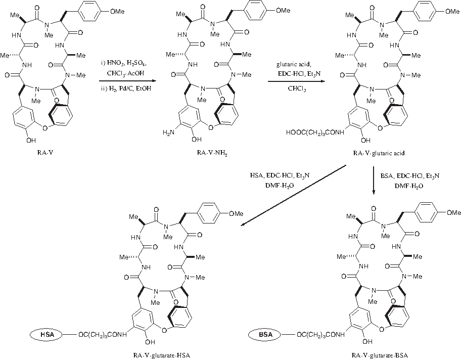
Preparation of immunogen or RA-V carrier protein conjugate by using BSA or HSA as a carrier protein. Glutaric acid plays a bridging role for the coupling of RA-V to the carrier protein.
After stirring at 0°C for 30 min, a solution of RA-V (9.7 mg, 0.013 mmol) in CHCl3:AcOH (1:1, v/v, 1.0 mL) was treated with nitric acid (50 μL) and sulfuric acid (50 μL) at room temperature for 2 min. After washing, drying, and removal of solvent followed by silica gel column chromatography as described previously, the reaction mixture gave RA-V–NO2 as a yellow solid (9.9 mg, 95%). Then, reduction of thus prepared RA-V–NO2 (9.4 mg, 0.012 mmol) in EtOH:1,4-dioxane:1 N HCl (1.0:0.5:0.1, v/v/v, 1.6 mL) with H2, in the presence of 10% Pd/C (10.0 mg) for 2.5 h, gave, after subsequent treatment, RA-V–NH2 as a colorless solid (7.8 mg, 84%).
Glutaric acid (9.5 mg, 0.071 mmol), triethylamine (Et3N) (10.0 μL, 0.071 mmol), and 1-(3-dimethylaminopropyl)-3-ethylcarbodiimide hydrochloride (EDC·HCl, 6.9 mg, 0.036 mmol) were added to a solution of RA-V–NH2 (6.5 mg, 0.0084 mmol) in CHCl3 (1.0 mL) at 0°C and the mixture was stirred at 0°C for 1 h and then at room temperature for 23 h. After subsequent treatment and octadecylsilyl (ODS) column chromatography followed by high performance liquid chromatography (HPLC), RA-V–glutaric acid was obtained as a colorless solid (3.4 mg, 46%).
H2O (0.3 mL), EDC·HCl (3.3 mg, 0.017 mmol),(5) and Et3N (2.4 μL, 0.017 mmol) were added to a solution of RA-V–glutaric acid (3.4 mg, 0.0039 mmol) in N,N-dimethylformamide (DMF, 0.1 mL) sequentially at 0°C, and the mixture was stirred at 0°C for 1 h. BSA (4.4 mg) was then added to the mixture. After stirring at 0°C for 3 h and then at room temperature for 5 days, the mixture was passed through a PD-10 desalting column, and subjected to lyophilization and purification processes to generate RA-V–glutarate–BSA conjugate (4.3 mg) to be used as an antigen in mice. Following the same procedure but using HSA (3.1 mg) instead of BSA, RA-V–glutarate–HSA conjugate (2.7 mg) was obtained to be used in ELISA.
Characterization of RA-V–glutarate–carrier protein conjugate
The amount of hapten in the prepared RA-V–glutarate–carrier protein conjugate was estimated by MALDI-TOF MS. Briefly, a mixture of ∼2 μg of the conjugate and 2 μL sinapinic acid solution (10 mg/mL) in MeCN:H2O (1:1) containing 0.1% trifluoroacetic acid was subjected to the delayed extraction MALDI-TOF MS operated in the linear mode. The accelerating voltage was 25 kV and the grid voltage was about 90% of the accelerating voltage. The data were analyzed using the GRAMS/386 software. The molecular weight of each component of the conjugate was calculated from the peak centroid of the [M+H]+ peak.
Direct ELISA procedures
Direct ELISA was performed in the usual manner as follows. A 100 μL portion of RA-V–glutarate–HSA conjugate in 50 mM carbonate buffer (pH 9.6, 4.0 μg/mL) was placed in each well of a 96-well flat-bottomed polystyrene immunoplate and left at 4°C overnight. The plate was washed three times with 0.05% Tween-20 containing phosphate-buffered saline (TPBS). The washings were discarded. Each well was then treated with 5% skimmed milk containing PBS (SPBS, 300 μL) at 4°C overnight to reduce nonspecific adsorption. Again, the supernatant was discarded and the plate was washed three times with TPBS. Each well was then treated with MAb solution in PBS (100 μL) at 37°C for 1 h. After the solution in the plate was discarded, each well was washed three times with TPBS and treated with a dilute peroxidase-labeled anti-mouse IgG solution (1:1000, 90 μL) as the secondary antibody at 37°C for 1 h. After washing the plate three times with TPBS, a portion of 100 mM citrate buffer (pH 5.0, 80 μL) containing 0.006% H2O2 and 0.1% o-phenylenediamine was added to each well, and the plate was incubated at 37°C for 15 min. The optical density was recorded at 492/600 nm on a microplate reader.
Immunization
For intraperitoneal injection, three antigen preparations were made. Thus, a solution of RA-V–glutarate–BSA conjugate in PBS (150 μg/0.3 mL) was mixed with: (A) an equal amount of Freund's complete adjuvant solution (150 μg/0.6 mL), (B) an equal amount of Freund's incomplete adjuvant solution (150 μg/0.6 mL), and (C) an equal amount of PBS (150 μg/0.6 mL).
Three male BALB/c mice were used. The antigen injection was given once every 2 weeks according to the following schedule. The amount to be injected at a time was 0.2 mL/mouse or 50 μg antigen/mouse. First, A was given five times with the first week injection given when the mice were 6 weeks old (injections at weeks 1, 3, 5, 7, and 9), then B was given five times starting at week 11 of antigen injection (injections at weeks 11, 13, 15, 17, and 19), and finally C was given once, 2 weeks after the final injection of B.
Preparation and cloning of hybridoma
Three days after the injection of C, the mouse giving the highest antibody titer was sacrificed. Its splenocyte cells were separated and fused with the cells of HAT-sensitive mouse myeloma cell line, P3/NS1/1-Ag4-1 (NS-1), by the PEG method.
Several weeks after culturing the hybridoma in five 96-well plates, the supernatants of 48 wells where the hybridoma showed good growth were assayed for their anti-RA-V MAb by direct ELISA. Of these, two gave high anti-RA-V MAb activities. The relevant hybridoma cells were then cloned three times by the limited dilution method as follows. The hybridoma suspensions (∼0.2 mL) from the above two chosen wells were each diluted with RPMI 1640 medium (×102, ×103, and ×104), and a portion of the suspension (100 μL) was added to each medium. After 2 weeks, the cells from two of the wells, each containing one single colony, were separately subcultured in a 24-well plate. Each lot of the hybridomas was then cultured in a larger scale serum-free medium, RPMI 1640 medium supplemented with 1% Nutridoma-SP additives containing penicillin–streptomycin–glutamine solution, 10 mM MEM non-essential amino acid solution, and 100 mM MEM sodium pyruvate. Two clones were thus established and named 2H10 and 4E6.
Purification and characterization of MAbs
The Isostrip, mouse MAb isotyping kit, was used for the identification of the subclass of the MAbs produced by the two hybridoma strains as previously described. The antibody produced by one of the two established clones, 2H10 (MAb 2H10) was found to be IgG1, and that by 4E6 (MAb 4E6) was IgG2b.
MAbs 2H10 and 4E6 from the supernatant of the cultures (each ∼600 mL) were each subjected to HiTrap protein G HP affinity column chromatography as described previously to give a crude IgG fraction (∼3.0 mg), which, after further purification procedure, was lyophilized to generate MAbs to be used in the present assay.
The reactivity of the MAbs with the other RA series compounds was assayed by the competitive ELISA. Competitive ELISA was performed in the same manner as direct ELISA, except that, after blocking and washing, a portion of test sample solutions of various concentrations of RA-V and related compounds RA-VII, RA-I, RA-III, and RA-VIII in 20% methanol–H2O (50 μL) were placed in the wells that had been treated with RA-V–glutarate–HSA conjugate. An equal volume of MAb solution (50 μL) was added to each well, and the plate was incubated at 37°C for 1 h. The antigen concentrations examined in this assay were in the range from 10 to 1000 μg/mL, due to their solubility.
Cross-reactivities (CR%) of the related RA series compounds were calculated according to Weiler's equation(6):
where A is the absorbance in the presence of a relevant compound and A0 the absorbance in the absence of a relevant compound.
Results
MALDI-TOF MS revealed that m/z of the presently prepared RA-V–glutarate–BSA conjugate was 72,302, and that, accordingly, the molecular ratio between RA-V–glutaric acid (MW 885) and the carrier protein BSA (MW 67,094) was about 5.9:1. Analogously, the assay showed that the antigen-carrier protein ratio in the RA-V–glutarate–HSA conjugate was 6.2:1.
The two hybridoma clones, growing quite fast and producing high antibody titers against RA-V, were established by the cloning procedures and named 2H10 and 4E6, which produced MAb 2H10 and MAb 4E6, respectively. MAbs 2H10 and 4E6 were identified as IgG1 and IgG2b, respectively, and were shown to contain a κ light chain by the test using a mouse the MAb isotyping kit Isostrip (Table 1).
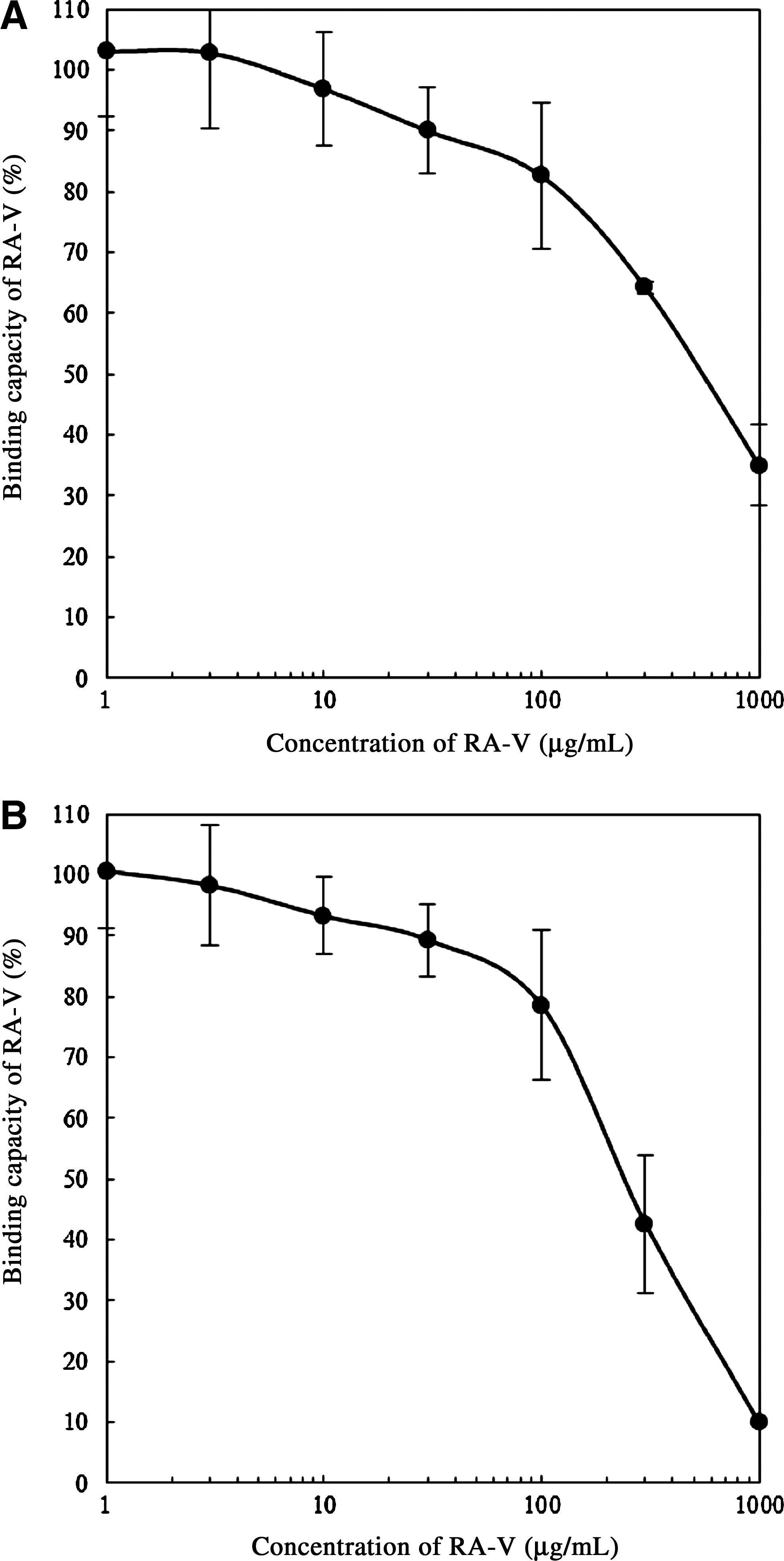
Using competitive ELISA, the calibration curves were prepared to relate the amount of RA-V in samples and the percent of the amount of RA-V bound by MAbs 2H10 and by 4E6, the results of which are given in Figure 3. The amount of RA-V giving 50% of binding, or the IC50 value for MAb 2H10, was 550 μg/mL, and that for MAb 4E6 was 230 μg/mL.
Reactivities of RA-V with (
The cross reactivity of MAbs 2H10 and 4E6 with the other RA series compounds of the present assay, RA-VII, RA-I, RA-III, and RA-VIII, was evaluated by analogous competitive ELISA. The inhibitory effect on RA-V was taken as 100%, and the cross reactivity of the MAbs with the other compounds was expressed as the concentration of assay compounds required to produce 50% inhibition of antibody binding compared with RA-V according to Weiler's equation.(6) The results are given in Table 2.
Cross-reactivities of RAs were determined using Weiler's equation.(6)
Control substance.
Discussion
As an easy and efficient quantitative assay of some biologically active substance in plants, whose content is often very small, a selective immunoassay has been proposed and is in use today. In this method, the small molecular weight target substance is converted into its suitable protein conjugate, which is used as an effective antigen for the production of specific MAb against the target substance. By using the obtained MAbs, selective immunoassay is performed for the quantitative estimation of the target substance.
In the present study, for the immunoassay of RA-V, RA-V was converted into RA-V–glutarate–carrier protein conjugate; by using the conjugate as an antigen, two clones of hybridoma, 2H10 and 4E6, producing specific MAbs 2H10 IgG1 and 4E6 IgG2b, respectively, were obtained. Detailed evaluation of the immunoassay using these MAbs revealed that the MAbs possess specificities and sensitivities as shown in Table 2, the sensitivity or IC50 for RA-V by MAb 2H10 being 550 μg/mL and that by MAb 4E6 being 230 μg/mL. The antigen specificity assay of the MAbs clearly showed that, of the analogs of RA-V examined (i.e., RA-VII, RA-I, RA-III, and RA-VIII), both MAbs 2H10 and 4E6 had reactivity only with RA-V and RA-VII. The results suggest that those MAbs specifically recognize the homolog compounds having Me in R 2 and react only with them, apparently regardless of whether R 1 is Me or H. Further studies, however, are needed to give a definitive conclusion.
Compared with the antibodies obtained in our previous study(1) in which RA-VII was used as the antigen, whose R 1 was Me, the present MAbs prepared by using an antigen homolog whose R 1 is H apparently had higher specificity. The present system, showing higher specificity and higher sensitivity to RA-VII, is definitely more useful than the previous system for the quantitative assay of the antitumor cyclohexapeptide RA-VII in the root of R. cordifolia and R. akane. Cyclohexapeptide RA-VII is a promising antitumor agent, which has gone through phase I clinical assay.(7,8)
Footnotes
Author Disclosure Statement
The authors have no financial interests to disclose.