
Abstract
Renalase is generated mainly by the kidneys, and renalase's expression in chronic kidney disease patients is reduced due to renal dysfunction. In this study, human renalase recombinant protein with prokaryotic expression was used for immunization of male New Zealand rabbits, and polyclonal antibodies against human renalase were obtained. To prepare and verify renalase polyclonal antibody, renalase recombinant protein was used as antigen and male New Zealand rabbits were immunized to obtain anti-serum for identification. On the basis of renalase antibody, the expression of renalase in renal tubular epithelial cells and renal tissue was detected. Two anti-renalase polyclonal antibodies were obtained. Renalase was constitutively expressed in human renal tubular epithelial cells, with the maximum expression in proximal convoluted tubules in renal tissue, and a small amount of expression in the glomeruli. Anti-renalase polyclonal antibodies were successfully prepared, which lays a foundation for further studies.
Introduction
Materials and Methods
Experimental animals
Two New Zealand big-eared rabbits, weighing about 2.5 kg, were purchased from the Shanghai Laboratory Animal Center. The animals were raised in clean-level animal rooms and cared for and fed according to relevant standards for the use of this experiment.
Regents
The following were the main reagents used in this study: recombinant renalase protein (prepared by this laboratory); Freund's complete adjuvant and Freund's incomplete adjuvant (Sigma, Shanghai, China); FITC-conjugated goat anti-rabbit IgG and HRP-conjugated goat anti-rabbit (Shanghai Immune Biotech, Shanghai, China); IgG, ELISA substrate (3',3',5,5'-tetramethylbenzidine [TMB], Shanghai Immune Biotech, Shanghai, China); human renal tubular epithelial cell HK2 cells (ATCC, Manassas, VA); and SABC kit (Boster, Wuhan, China). Renal tissue was from traumatically removed kidney.
Animal immunization
The study used purified renalase protein as antigen and two adult male New Zealand rabbits. A small amount of blood and serum was collected for use before basic immunization and immunization was conducted upon the completion of serum collection. Antigen protein (1000 μg/rabbit) was used for each immunization, and each rabbit was subjected to immunization once a week for 3 weeks. For the first immunization, Freund's complete adjuvant was made into emulsion with the antigen and used for back subcutaneous multi-point injection. The subsequent enhancing immunization used Freund's incomplete adjuvant. On the seventh day after the third immunization, the rabbit underwent carotid artery bleeding for collection of blood. Serum was separated by conventional methods: coagulated blood was first placed in an incubator at 37°C for 2 h, then at 4°C overnight. On the next day, it was centrifuged at 10,000 rev/min for 10 min, and then the supernatant was collected for water bath at 56°C for 30 min, and then stored at −70°C after repackaging. The collected anti-serum was subject to ELISA titer detection.
Indirect ELISA method to detect anti-renalase antibody titer
Coating buffer (50 μL/hole) was used to dilute renalase co-protein antigen, and the antigen concentration was 20 μg/mL, placed at 4°C for 16∼18 h. After coating was complete, the anti-renalase antibody titer was measured by ELISA.
Renalase's expression in renal tubular epithelial cells and renal tissues
The prepared anti-renalase polycolonal antibody served as the primary antibody, and the expression of renalase in renal tubular epithelial cells was detected using an indirect immunofluorescence method, with PBS as negative control. With the prepared anti-renalase's polyclonal antibody as the primary antibody, immunohistochemistry was carried out using an SABC kit to detect renalase expression in renal tissue. The steps were performed in accordance with the manufacturer's instructions, with PBS as negative control. Renal tissues subjected for testing were from a post-traumatic kidney.
Results
ELISA test on rabbit anti-serum titer after immunization
Twenty mL of antiserum was obtained from the two rabbits, respectively; the anti-serum titer test results are shown in Table 1.
Immunization was performed three times prior to ELISA.
Renalase's expression in renal tubular epithelial cells
Immunofluorescence detection showed constitutive expression of renalase in HK2 cells, as shown in Figure 1.

Renalase's expression in renal tubular epithelial cells (1000×). (
Renalase's expression in renal tissue
Immunohistochemistry detection showed significant expression of renalase in renal tubules and a small amount of expression in the glomeruli (Fig. 2).
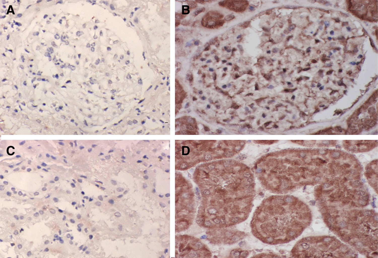
Renalase's expression in renal tissue (400×). (
Discussion
Discovered in 2005, renalase is mainly generated by the kidneys. Renalase is highly expressed in healthy human plasma, while such protein is nearly undetectable in patients receiving hemodialysis and patients with ESRD.(10) Renal tubular epithelial cells are the main cells expressing renalase, which can be secreted into the blood to regulate catecholamine metabolism, sympathetic nervous activity, and cardiovascular function. The human renalase gene is located on No. 10 chromosome q23.33, with nine exons, spanning over nearly 311,000 bp. Renalase protein is composed of 342 amino acids, with a molecular weight of about 37.8 kDa. Protein structure analysis shows that renalase N-terminal has one signal peptide, one flavin adenosine dinucleotide (FAD) binding site (AAs4-35), and one monoamine oxidase function domain (AAs75-339). Renalase knockout mice showed increased plasma catecholamine levels, hypertension, ventricular hypertrophy, and reduced ischemic tolerance, and the risk of myocardial damage was increased 3-fold. As renalase has myocardial protective effects, recombinant renalase replacement therapy can change these abnormalities.(11)
Studies have shown that renalase can degrade catecholamines, including dopamine, epinephrine, and norepinephrine,(12) to decrease myocardial contractility and heart rate, thereby reducing blood pressure. Renalase's expression in CKD patients is reduced due to renal dysfunction, and circulating catecholamines cannot be fully degraded, which may be one of the important mechanisms for hypertension and cardiovascular complications in patients with CKD.(13,14) In addition, studies have found that renalase is related to hypertension in ESRD patients and renal function in patients receiving a transplant.(15–17) Similar to erythropoietin, renalase replacement therapy may be a promising clinical treatment.(18,19) Study on the pathological significance of renalase and its related pathogenesis has become a hot topic and generation of a high-sensitivity antibody would be a good tool for such studies.
In the preparation of a polyclonal antibody, obtaining high-quality anti-serum is dependent on the quality, quantity, and purity of the antigen, which is also related to the specificity and sensitivity of the detection method. If possible, protein antigens of homologous biochemical properties would be the best candidates. At present, rabbit is the most commonly used experimental animal for polyclonal antibody, because genetically it is quite different from human and murine proteins. Each blood collection from a rabbit can provide more than 20 mL of serum without serious side effects. Horses are more often used for the preparation of a large amount of anti-toxin serum. In circumstances with a scarce amount of antigen and very little need for antibody, pure-strain mice can also be chosen.(20,21) This study adopted rabbits, used human renalase recombinant protein with prokaryotic expression for immunization, and eventually obtained polyclonal antibodies against human renalase.
With the renalase polyclonal antibody prepared in this study as the primary antibody, constitutive expression of renalase in renal tubular epithelial cells can be found through cytochemistry experiments. In studies using the immunohistochemical method and the prepared renalase polyclonal antibody, an abundant expression of renalase can be found in human renal tubules and a small amount of expression can be found in the glomeruli.
Compared to the monoclonal antibody, the polyclonal antibody has certain shortcomings, but due to its easy preparation, polyclonal antibody production is still a widely used technology. This study obtained an anti-renalase polyclonal antibody and laid the foundation for further experiments.
Footnotes
Acknowledgments
This research was supported by the Project of National Nature Science Foundation of China (81100528), Program for Excellent Young Talents of Shanghai Sixth People's Hospital (1402), Shanghai Sixth People's Hospital Consortium Subject (1423), and Shanghai Committee of Science and Technology Research Project (114119a6100).
Author Disclosure Statement
The authors have no financial interests to disclose.