
Abstract
Prostate-specific membrane antigen (PSMA), a type II integral membrane glycoprotein, is highly overexpressed in all forms of prostate cancer tissues. It has also been demonstrated in a wide range of neovasculature of non-prostatic solid tumors, including bladder, pancreas, lung, kidney, colorectal, and gastric cancers. Given the unique expression of PSMA, it is considered an alluring target for antibody-based imaging and therapy of cancer. In the present study, the production and characterization of camel heavy chain antibodies (HCAbs) specific for the external domain of the PSMA are reported. Due to the absence of the CH1 domain, HCAbs are smaller than their counterparts in conventional antibodies. In this study, camel antibodies were generated through immunization of Camelus dromedarius with a synthetic 28 amino acid peptide corresponding to the external surface domain of antigen and PSMA-expressing cell lines. Different binding properties to protein A and protein G affinity columns were deployed to separate three subclasses of camel IgG. The affinity purified HCAbs bound selectively to the synthetic peptide in enzyme linked immunosorbent assay (ELISA) and reacted specifically with PSMA-expressing cell line through imunocytochemistry study. Currently, we are attempting to develop recombinant variable domain of these heavy chain antibodies (VHH or nanobody) for tumor imaging and cancer therapy.
Introduction
Since there is little effective treatment for metastatic prostate disease, it is critical to recognize genes and/or gene products that represent diagnostic and prognostic markers, as well as targets for treatment. Prostate-specific antigen (PSA) is such a beneficial cancer marker in the clinical diagnosis and staging of prostate cancer. Since the PSA cannot distinguish benign prostatic hyperplasia (BPH) from prostatitis or prostate cancer in the range of 4 to 10 ng/mL, a cytologic and/or histologic assessment to confirm the accurate diagnosis is essential.(1)
Prostate-specific membrane antigen (PSMA) is a type II transmembrane glycoprotein with a molecular weight of about 110 kDa (Fig. 1). This protein represents neurocarboxypeptidase and folate hydrolase activity and is reported to be involved in the neuroendocrine regulation of prostate growth and differentiation. It is predominantly expressed by prostatic epithelial cells and its expression is increased in prostate cancer, particularly in poorly differentiated, metastatic, and hormone refractory carcinomas.(2–5)

Schematic of PSMA demonstrating extracellular, a hydrophobic transmembrane, and a short NH2-terminal cytoplasmic domain. Nine potential N-glycosylation sites have been seen in the extracellular domain of the PSMA(Y).
Moreover, PSMA is also expressed in neovasculature of extraprostatic solid tumors, including bladder, pancreas, lung, kidney, colorectal, and gastric cancers.
It is also identified in endothelial cells of capillary vessels in peritumoral and endotumoral areas of certain malignancies, such as renal cell carcinomas and colon carcinomas, but not in blood vessels of normal tissues. Further, PSMA is reported to be related to tumor angiogenesis.(6–8) Several antibodies against the extracellular fragment of PSMA have been described.(9–18) More recently, human and humanized antibodies with the capability of binding to PSMA have been described.(19–21)
Such antibodies have been deployed for imaging of prostate cancer cells.(22,23) In addition, they are considered to be one of the therapeutic approaches to treat prostate cancer, typically as a conjugate of a chemotherapeutic agent or radioactive isotope.(14,16,24,25)
Development of effective anti-PSMA antibodies to treat and/or prevent diseases involving PSMA expression, particularly those with cytotoxic effects and without the need to be conjugated to a chemotherapeutic agent or radioactive isotope, can be considered a desired advancement of such therapeutic antibodies.
Typically, monoclonal antibodies (MAbs) are produced in mice (murine Abs), although they have functional constrictions such as high immunogenicity and low penetration through solid tumor. Such restrictions have largely been overcome by the benefits of using the camel antibodies. In 1993, natural heavy chain antibodies (HCAbs) bereft of a light chain and CH1 domain were discovered in camelids.(26–29) The HCAbs (i.e., IgG2 and IgG3, 90 kDa), void of CH1 domain, are smaller than their counterparts in conventional mammalian antibodies (150–160 kDa).
The antigen-binding site of variable domain of camelid HCAbs has been mentioned as VHH, or nanobody.(30–32) It consists of the smallest known natural intact antigen-binding domain. These small-sized fragments (VHH) are well-expressed and have many particular features such as heat resistance, small size, very high solubility in aqueous, and are non-immunogenic in humans.(32,33) In this survey, the production and characterization of novel anti-PSMA specific polyclonal antibodies in dromedary camels are reported.
Materials and Methods
Animal farming
Two adult male Camelus dromedarius, purchased locally, were housed in the Guilan University of Medical Sciences animal facility, where they were given free access to water and food resources. During all the stages of these experiments, guidelines prepared by the Guilan University of Medical Sciences were precisely followed for the care of the animals.
Cell culture
At this stage, prostate cancer cell line overexpressing human PSMA (LNCaP) and the prostate cancer cell lines, which do not express the human PSMA (DU145 and PC-3), were cultured routinely in high glucose DMEM (Gibco, Grand Island, NY) while 10% fetal bovine serum (Gibco) and penicillin and streptomycin antibiotics were added as a supplementary. All cell lines were gained from the National Cell Bank of Iran at Pasteur Institute.
Immunization of Camelus dromedarius with LNCaP cell line and immunogen
With the use of recombinant PSMA (R&D Systems, Minneapolis, MN), LNCaP cells, and a 28-amino acid peptide (NFTEIASKFSERLQDFDKSNPIVLRMMN-COOH), which were in accordance with the external surface domain of PSMA, the dromedaries were immunized subcutaneously and intramuscularly. The peptide was synthesized and conjugated to bovine serum albumin (BSA) by Biomatik (Cambridge, Canada).
Each animal received eight doses of LNCaP cells (∼108 cells in 1 mL PBS per injection) and 1 mg of peptide conjugated to BSA emulsified in 2 mL complete Freund's adjuvant for the first and in 2 mL incomplete Freund's adjuvant for the following injections, at weekly intervals. Moreover, animals received 10 μg of recombinant PSMA at weeks 5 and 6 of immunization.(28)
Fractionation of IgG subclasses from serum of preimmunized and immunized camels
Before immunization (day 0) and also after passing 6, 7, and 8 weeks of immunization, pre-immune and immune sera were collected. Afterward, the high-titer antibodies, which were collected 6 weeks after the primary immunization, were chosen; then, applying differential absorption on protein G and A (Hitrap Pharmacia) that was previously reported by Omidfar and colleagues,(28,34) IgG subclasses were purified. Separation of the different immuoglobin (IgG) subclasses obtained from camel immune serum was carried out with modification. After collecting the serum, the total immunoglobulin was precipitated with ammonium sulfate and then dissolved in a minimum amount of phosphate buffer. The precipitate was then dialyzed all night in an extensive manner against the buffer. The obtained clear solution was first loaded onto a protein G column, then IgG3 was eluted with acetate buffer (pH 3.5) and IgG1 was eluted with glycine HCl (pH 2.7). In order to recover the IgG2 subclass, the flow-through was loaded onto a protein A column, which was eluted with acetate buffer (pH 3.5). The eluent was stored at −20°C while it was dialyzed immediately against bicarbonate buffer (pH 9.5) and treated with sodium azide. Through the Bradford test, the protein concentration of column fractions was determined, and by means of SDS-PAGE analysis, the purity of the isolated serum IgG was confirmed.(28)
ELISA assay
Enzyme linked immunosorbent assay (ELISA) was employed to determine the specificity and reactivity of the various IgG subclasses toward the peptide. In order to coat wells of micro-titer plates, they were subjected to 1 μg/100 μL concentration of PSMA peptide-BSA (for specific binding), BSA (for non-specific binding), and an extraneous peptide (a 14 amino acid peptide, LEEKKGNYVVTDHC, for non-specific interaction of antibody with peptides). The plate was incubated overnight at 37°C, and the remaining areas on every well were blocked with BSA and treated with anti-peptide camel antibodies for 1 h at 37°C.
After being washed with PBS several times,(28,34) wells were treated with 100 μL of horseradish peroxidase (HRP) conjugated goat anti-camel antibodies for 1 h at 37°C. Subsequently, after five washes with PBS, 100 μL of tetramethylbenzidine (TMB) and H2O2 solutions as substrate and 50 μL of HCl (0.5 N) as a stop solution were added to determine and stop the enzyme activity, respectively. Finally, the absorbance was evaluated in an ELISA plate reader at 450 nm.
Immunocytochemistry assay
Prostate cancer cell line overexpressing human PSMA (LNCaP) and the human PSMA-negative prostate cancer cell lines (DU145 and PC-3) were applied as the targets. Succinctly about 2×10⁴ cells were seeded into each well of a 96-well culture plate, and the cells were incubated in culture medium at 37°C. Once they became roughly confluent, the medium was discarded in order to incubate the cells with 100 μL of pre-immune whole serum (1:1500), the total serum from immunized camels (1:1500), and purified IgG subclass antibodies (1 μg/mL) for 1 h on ice. Subsequently, the cells that were washed twice with PBS-BSA were incubated with 100 μL of 1:2000 diluted HRP-conjugated goat anti-camel antibodies for 1 h at 4°C. Then, after washing the wells five times according to the previous described method, the binding activity of each preparation was measured in the ELISA plate reader at 450 nm.(28,34)
Affinity determination
Determination of the mean affinity of HCAbs, which was produced and purified in the present study toward antigen, was performed by coating the wells of the microtiter plate with PSMA peptide–BSA conjugate. Then, the whole surface of the wells was blocked to avoid further non-specific binding and, using various dilutions of HCAbs, the wells were treated for 1 h at 37°C. After being washed several times and subjected to HRP–anti-camel goat antibodies for 1 h at 37°C. The optical density and affinity were considered according to the protocol described by our group(31) and others.(35)
Results
Production and separation of camel IgG subclasses
Immunoglobulin, which was produced from two camels, was precipitated with ammonium sulfate and then separated into different IgG subclasses that included conventional (IgG1) and heavy chain (IgG2 and IgG3) antibodies. The conventional IgG (G1) antibodies were composed of heavy chains and light chains with a molecular weight of 160–170 kDa (Fig. 2). The molecular weights related to two other HCAbs (IgG2 and IgG3) are 90 kDa (Fig. 2).
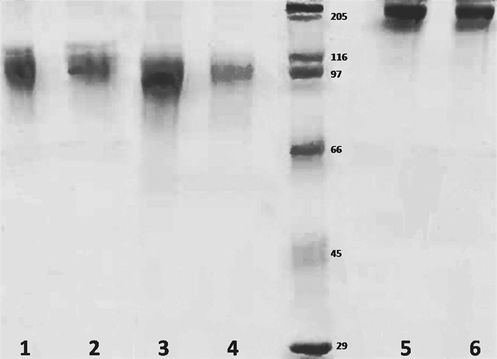
SDS-PAGE electrophoresis of IgG3 (lanes 1 and 2), IgG2 (lanes 3 and 4), and IgG1 (lanes 5 and 6). Inner numbers designate the molecular weight markers in kDa.
Reactivity of separated camel antibodies toward synthetic peptide
Purification of anti-PSMA subclass antibody from the serum of immunized camel was performed employing affinity column. By means of ELISA against peptide, the activity of IgG subclass antibody eluted in the camels was assessed. The reactivity of purified IgG1, IgG2, and IgG3 antibodies with the 28 amino acid PSMA conjugated to BSA, BSA alone, and a 14 amino acid extraneous peptide can be seen in Figure 3. A strong and specific reactivity towards BSA-PSMA peptide was observed in IgG subclasses.

Binding of anti-peptide antibody of C. dromedarius with BSA-peptide, BSA, and extraneous peptide. All assays were performed in duplicate.
Peptide affinity for HCAbs was calculated. The measurements were as follows: 1×10⁸ M−1 for IgG2 and 6×10⁷ M−1 for IgG3.
Reactivity of camel antibodies against PSMA positive and negative cell lines
Reactivity of whole antiserum and protein G and protein A purified subclasses was tested against PSMA-overexpressing and PSMA-negative human prostate cancer cell line, LNCaP, and DU145 and PC-3, respectively. Based on the results provided in Table 1, it can be observed that the whole antiserum and IgG subclasses only reacted with LNCaP, but not with DU145 and PC-3.
All data are illustrated as OD at 450 nm.
Discussion
In the present study, the first survey of the production and characterization of anti-PSMA–specific antibodies in camels has been presented. The heavy chain antibodies (HCAbs) that made up variable regions of heavy chain, CH2, and CH3 domains are shown in camelids (camels, llamas, and alpacas). Compared with other counterparts in conventional mammalian antibodies, HCAbs are smaller due to the absence of the CH1 domain.(26) Since the antigen-binding domains of these antibodies are located on the heavy chain of the protein molecule, production of recombinant smaller fragment of such antibodies was facilitated here. It could be that the provided antibody is a more appropriate substitution for the present conventional antibodies regarding the feasibility, effectiveness, and beneficial aspects of camel antibody production. Because of recent advances in the existing knowledge about molecular and tumor immunology, new immunotherapies have been achieved to treat human cancers including humoral and/or cellular approaches. Antibody therapy has been studied for various types of human cancers. In fact numerous studies have recognized cell surface antigens, which are tumor or lineage specific (e.g., CD20), receptors mutated in cancer cells (e.g., mutated EGFR), overexpressed receptors, and/or antigens on cancer cells compared with normal cells (e.g., EGFR, HER-2, and MUC1).(2,5,36,37)
PSMA (folate hydrolase 1, glutamate carboxypeptidase II) is a 750 amino acid type II cell surface membrane protein that was identified in prostate cancer epithelium for the first time. Subsequently, it was discovered that PSMA is in fact expressed by the neovascular endothelium of almost all solid tumor types but without being expressed by the tumor cells or normal vascular endothelium. PSMA is upregulated 10-fold in prostate cancer, especially in hormone refractory diseases; therefore, it can be considered an attractive target for immunotherapy and imaging of prostate cancer. It can also be regarded as an ideal sentinel molecule for use in targeting prostatic cancer cells. Moreover, PSMA has been exploited as a diagnostic target to detect prostatic cancer.(2,4,5)
In the present study, two camels were immunized with a synthetic peptide corresponding to the PSMA antigen and PSMA-expressing cell line. It was mainly aimed to develop high-titer HCAbs, which is particular for PSMA and the final objective was to develop the recombinant camelid single domain antibody. The production of the heavy chain camel antibodies (i.e., IgG2 and IgG3) specific for the human PSMA has been shown in this study (Fig. 2).
According to the performed analysis, the serum of camelid species contained approximately 68% HCAbs. As a result of some techniques, it was made evident that the camelid HCAbs are specific for the PSMA.
At the first stage, conventional and heavy chain (HC) affinity purified antibody from the serum of immunized camels was bound specifically to the PSMA peptide conjugate to BSA but did not bind to BSA alone or an impertinent peptide (Fig. 3). The whole antiserum, conventional, and HC affinity purified antibodies with human prostate cancer cell line that express high level of PSMA (LNCaP) showed significant positive reactions.
In contrast these compounds do not interact with the PSMA-negative prostate cancer cell lines (DU145 and PC-3) in an apparent way. However, it might be that antibodies of affinities in the range of 108 to 1010 M−1 are obtained according to the affinities of anti-PSMA antibodies being observed for more than 90% of the previous reports.
In the present research, the heavy chain anti-PSMA antibodies with comparably good affinity were produced in the range of 1×10⁸ M−1 and 6×10⁷ M−1 for IgG2 and IgG3, respectively.
Some previously performed reports about the use of HCAbs against tumor-associated cell-surface markers implied that camel HCAbs are more sensitive compared with the normal antibody to detect the antigen.(34) Perhaps, this is due to the presence of the longer complementarily determining regions (CDR) domain of the HCAbs, which allows better contact with the antigens and a higher capability to identify them.(32,38) Therefore, respect to the more feasible, effectual, and lucrative aspects in the production of this antibody, the current conventional double-chained antibodies may be replaced with camel antibodies if such an experimentation can be applied in a wide range.
Footnotes
Acknowledgment
This research was supported by the Endocrinology Metabolism Research Center of Tehran University of Medical Sciences, and the Pasteur Institute, Tehran, and I.R. Iran. The authors wish to express their gratitude to all who provided support during the course of this research.
Author Disclosure Statement
The authors have no financial interests to disclose.