




Abstract
During the course of codifying low pathogenicity avian influenza, viruses were tested for their capacity to induce type-I interferon (IFN) and to measure their content of IFN induction-suppressing particles (ISP). One isolate caused a >10-fold reduction in the yield of IFN from chicken embryonic cells co-infected with a virus that normally induces high yields of IFN. The apparent content of ISP was calculated to be ≈100-fold higher than the number of physical particles of virus measured as hemagglutinating particles. This unrealistic interpretation prompted us to test for a soluble IFN induction-suppressing activity in the allantoic fluid freed of the virus by centrifugation. Indeed, the IFN induction-suppressing activity remained in the virus-free supernatant. The original virus stock subsequently was found to be contaminated with a Gram-negative bacterium, leading us to test lipopolysaccharide (LPS) as the putative IFN induction suppressor. Pure LPS mimicked in a similar dose-dependent manner the IFN induction-suppressing activity of the original allantoic fluid-derived virus, and the allantoic fluid freed of all virus and bacteria. The inhibition of viral-mediated type-I IFN induction by LPS was observed for viruses from 3 different families. These observations suggest that exposure of a host to endotoxin may compromise the IFN induction response of the innate immune system and thus exacerbate virus infection.
Introduction
T
In an ongoing study, several AIV strains were screened for IFN-inducing potential by generating IFN induction dose (multiplicity of virus)–response (IFN yield) curves (Marcus 1986). In brief, confluent monolayers of primary CEC were prepared from 9-day-old embryonated eggs and developmentally aged by incubating in NCI medium plus 6% calf serum for 9 days without a change of medium (Sekellick and Marcus 1986). Maximal levels of IFN were induced in these aged CEC monolayers following attachment of virus for 1 h and a 24 h period of incubation at 40.5°C. The medium is then collected and infectious virus is removed via the addition of fetal bovine serum in the presence of perchloric acid. The resulting samples therefore contain only acid-stable, type-I IFN and are assayed as previously described (Sekellick and Marcus 1986). Figure 1 shows the maximum yield of acid-stable, type-I IFN induced by 6 different isolates of chicken egg allantoic fluid-derived virus arranged in the order of decreasing capacity to induce IFN. The virtual absence of IFN-inducing capacity of the A/Red Knot/744/03 (H9N2) isolate flagged it as a virus population that potentially contained ISP (Marcus and Sekellick 1985; Marcus and others 2005).
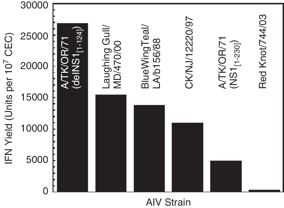
The maximum yield of interferon (IFN) induced by each of 6 influenza virus strains in developmentally aged chicken embryonic cells (CEC). IFN induction dose—response curves were generated for each strain and analyzed for the maximal yield of IFN (Marcus 1986; Sekellick and Marcus 1986). Because the Red Knot/744/03 isolate produced little, or no, IFN, it was subsequently tested for its capacity to suppress IFN (Marcus and Sekellick 1985; see Fig. 2).
ISP were assayed in aged monolayers of CEC. Cells were co-infected at m IFP = 5 with a good inducer of IFN (A/TK/OR/71-delNS1[1–124][H7N3]) (Marcus and others 2005) along with increasing multiplicities of the potential ISP (Red Knot/744/03), and incubated for 24 h at 40.5°C. This generated the IFN induction-suppression curve shown in Figure 2. About 90% of the yield of IFN initially was lost at an exponential rate. About 10% of the IFN yield appeared refractory to suppression at higher amounts of Red Knot virus. Based on a Poisson distribution of virus particles amongst the CEC of the monolayer, the amount of ISP added to cells that results in 0.37 of the maximal yield of IFN is assumed to contain an average of 1 ISP/cell (Marcus and Sekellick 1985). This stock of Red Knot/744/03 was found to suppress IFN induction at such a high dilution of virus as to suggest that there were about 100 times more ISP present than could be accounted for by the physical particles of AIV measured as hemagglutinating activity (Marcus and others 2009). This improbable interpretation of the data prompted us to search for a soluble moiety that possessed ISP activity.

The interferon (IFN) induction-suppressing activity of Red Knot/744/03. Control monolayers of aged CEC (Sekellick and Marcus 1986) were infected with an excellent inducer of IFN, TK/OR/71-delNS1(1–124) (see Fig. 1), at m IFP = 5 so as to produce a maximal yield of IFN from each cell (dashed line) (Sekellick and Marcus 1986). The test mono-layers of cells were simultaneously infected with increasing amounts (multiplicities) of Red Knot to determine the capacity of the Red Knot virus to suppress the yield of IFN as shown, presumably functioning as IFN induction-suppressing particles (“ISP”).
Consequently, the original allantoic fluid-derived Red Knot stock was freed of virus and any other particulate matter by centrifuging it at 40,000g for 4 h. In addition, the particle-free supernatant was filtered twice through 0.22-μm Millipore filters and tested for ISP activity. This supernatant was found to have lost little or no ISP activity (Fig. 3). This confirmed that the observed ISP activity of Red Knot/744/03 resided in a soluble component in the allantoic fluid of the virus stock.
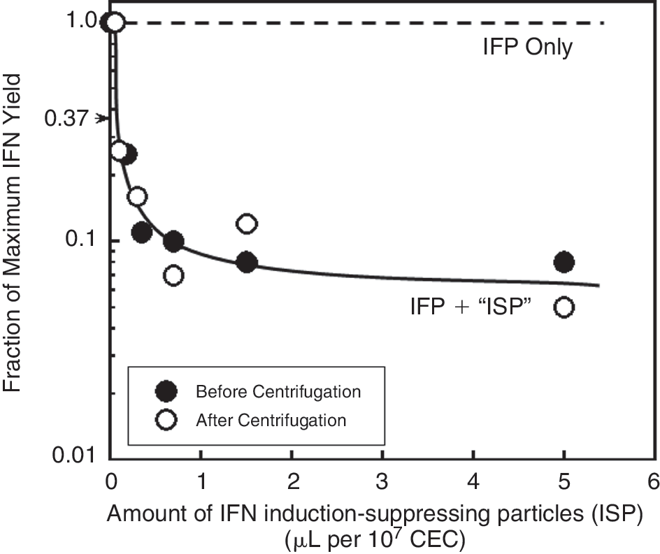
The interferon (IFN) induction-suppressing activity of the Red Knot virus stock before and after centrifugation at 40,000g for 4 h. An aliquot of the original allantoic fluid from Red Knot-infected eggs was tested before and after centrifugation. The data indicate that the degree of IFN induction-suppression by the supernatant material was virtually unchanged following centrifugation. A single curve accommodates, within experimental error, the variations of both data sets.
Fortuitously, a plaque assay of the original Red Knot stock, incubated for 1 day longer than the usual 3 days, unexpectedly revealed a low level of contamination with a rod-shaped Gram-negative bacterium (≈105 colony-forming units/mL). This prompted a test of lipopolysaccharide (LPS) as the putative IFN induction suppressor, despite its reputation as an inducer of IFN and other inflammatory cytokines (Guha-Thakurta and Majde 1997). A sample of pure LPS derived from Escherichia coli 026:B6 was obtained from Sigma-Aldrich (L#2654) and suspended in pyrogen-free saline buffer at a concentration of 1 mg/mL. Figure 4 demonstrates that pure LPS mimicked in a similar dose-dependent manner, the observed IFN induction-suppressing activity of the virus- and bacteria-free allantoic fluid (cf. Fig. 3). There was an initial exponential rate of loss of about 90% of the quantum yield of acid-stable type-I IFN, up to a dose of about 50 endotoxin units (EU)/mL. Higher doses of LPS revealed that about 10% of the IFN yield was refractory to the action of LPS. Thus, when a good inducer of IFN (A/TK/OR/71 delNS1) was attached to 107 CEC for 1 h in the presence of ≥50 endotoxin units (1 EU = 1.67 ng)/mL of LPS, there was a 90% reduction in IFN yield when measured 24 h later. A 50% reduction in IFN yield was obtained with 2.5 EU (4.2 ng)/mL LPS. Similar results were obtained if the IFN-inducing virus was attached to the cell prior to the addition of the LPS, indicating that LPS was not blocking uptake or entry of virus into the cell. Serum was present in the medium at all times, thereby eliminating the LPS-binding protein as a rate-limiting step in the action of LPS on CD14/TLR-4 receptors (Triantafilou and Triantafilou 2002).

Assay for interferon (IFN) induction-suppression by lipopolysaccharide (LPS). Monolayers of aged chicken embryonic cells (CEC) were exposed to TK/OR-delNS1(1–124) to induce maximal levels of IFN in all control cells. Other monolayers of cells infected with this virus were exposed to varying amounts of LPS during the 1 h virus attachment period. The monolayers then were washed, fresh medium added, incubated at 40.5°C for 24 h, and the supernatants assayed for acid-stable IFN (Sekellick and Marcus 1986). The amount of LPS is noted in both endotoxin units (EU) and the amount (ng) per 107 cells.
The presence of variable amounts of LPS in calf serum used in the days of cell culture before animal sera were tested for endotoxin offers a plausible explanation for the wide variation in the yields of IFN from chicken embryo cell cultures observed from batch-to-batch of sera (see table in Sekellick and Marcus 1986), and why the highest yields of IFN were obtained from cells without serum in the medium (Majde 1993).
Monolayers of CEC incubated for 2-days were exposed to a single high dose of LPS (300 EU/mL) that remained in the medium for the duration of the 9-day aging process. These cells were found to induce IFN at yields comparable to non-treated cells. Thus, the continuous presence of LPS did not perturb the maturation of the interferon system during developmental aging of embryonated chicken cells. Also, the cell-signaling events that result in the suppression of viral-mediated IFN induction occur rapidly following exposure to LPS. Thus, when administered prior to viral infection and then washed out, a dose of LPS equal to 50 EU [83.3 ng]/mL/107 cells demonstrated maximal suppression of IFN yield within a 15 min pre-incubation period (data not shown).
Following double filtration through 0.22-μm Millipore units to remove any bacteria, a new stock of A/Red Knot/744/03 was generated by passage in 9-day-old embryonated chicken eggs. Figure 5A shows that the bacterial-free virus induced over 2,000 IFN U/107 CEC. The IFN induction-suppressing activity could be restored by mixing the contaminant-free Red Knot/744/03 preparation with the supernatant from the centrifuged, bacterial-contaminated stock of Red Knot or with pure LPS. The IFN induction-suppression activity of LPS was not unique to IFN induction by the Red Knot virus. Figure 5B shows that IFN induction by A/TK/OR/71-delNS1(1–124), vesicular stomatitis virus (VSV), and Newcastle disease virus (NDV) was also suppressed by LPS in a similar fashion, indicating a global action of LPS on the cells. On average about 5% of the yield of IFN appeared to be refractory to the action of LPS (Fig. 5B). We postulate that 5%–10% of the cells in a preparation of primary chicken embryo cells may be intrinsically refractory to the action of LPS as a suppressor of IFN induction, possibly because they lack appropriate receptors (Triantafilou and Triantafilou 2002).
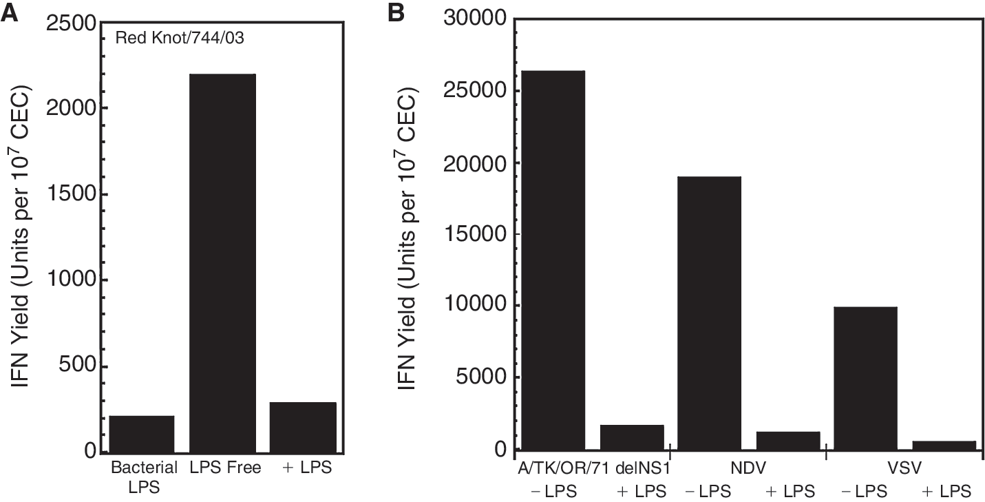
Lipopolysaccharide (LPS)-mediated suppression of interferon (IFN). From left to right, the histobars show the IFN-inducing capacity of (
The suppression of viral-mediated IFN induction by LPS has been reported previously using RAW cells, a line of mouse macrophages, and NDV as an IFN inducer, to demonstrate lowered levels of type-I IFN mRNA transcription (Juang and colleagues 1999). This study showed that LPS inhibited the phosphorylation and nuclear transport of 2 important virus-activated IFN-regulatory factors, IRF-3 and IRF-7. Our results indicate that the lower levels of type-I IFN mRNA reported after LPS treatment (Juang and others 1999) also are reflected in lower yields of IFN.
A second study investigated the cytokine expression profiles in mice following exposure to NDV (Guha-Thakurta and Majde 1997). A heat-inactivated NDV stock was unexpectedly found to induce elevated levels of IL-10, M-CSF, and type-II IFN, suggesting a possible LPS contamination that was later confirmed via a Limulus lysate assay. This study makes clear that given the relatively high thermal stability of LPS (Majde 1993), even heat-inactivated virus stocks are still at risk for containing functional LPS.
The capacity of LPS to modulate and suppress the normal innate immune response to viral exposure has been demonstrated in embryonic chicken cells that, in developmental terms for the IFN system, mimic acquisition of IFN-inducing capacity in the incubating egg (Sekellick and others 1990). Collectively, these data suggest the vulnerability in a clinical setting of the IFN induction element of the innate immune system to a co-infecting bacterium that produces endotoxin. Thus, LPS-producing bacteria may enhance the establishment and severity of viral infections (Shope 1931). Indeed, some deaths in the 1918–1919 influenza pandemic were attributed to co-infection with Haemophilus influenzae (Morens and others 2008), and more recently, the co-purification of LPS with plasmid DNA prepared from E. coli further points out the caution that should be exercised in using reagents derived from endotoxin-producing bacteria (Majde 1993; Wicks and others 1995). Our data and the reports cited here seem especially relevant in a clinical setting given the elevation to pandemic status of the novel H1N1 outbreak. Since type-I IFN also has been shown to play a role in innate immunity during bacterial and protozoan infections (Walberg and others 2008), abrogation of IFN induction by LPS may have even broader implications in clinical situations.
Footnotes
Acknowledgments
This study was supported in part by USDA grants 58-1940-0-007 through the Center for Excellence in Vaccine Research at the University of Connecticut, and USDA SCA award #58-6612-7-157. This study benefited from the services of the Animal Cell Culture Facility of the Biotechnology-Bioservices Center of the University of Connecticut.