
Abstract
In view of well-established immunomodulatory properties of Lactobacillus, present investigation was carried out to evaluate antioxidant and anti-inflammatory potential of Lactobacillus casei and Lactobacillus acidophilus, against inflammatory pathway and oxidative stress developed in an experimental model of arthritis. Collagen-induced arthritis (CIA) model was used. Oral administration of L. casei, L. acidophilus, standard antiarthritic drug indomethacin, and vehicle were started after induced arthritis and continued up to day 28. Interleukin (IL)-6, tumor necrosis factor (TNF)-α, IL-1β, IL-17, IL-4, and IL-10 levels were estimated in serum. In parallel, oxidative stress parameters were also measured from synovial effsuate. All rats were graded for arthritis score at the end of each week. L. casei, L. acidophilus, and indomethacin treatment significantly downregulated proinflammatory and upregulated anti-inflammatory cytokines at P<0.0001. They have significantly decreased oxidative stress in synovial effsuate (P<0.0001) and also arthritis score (P<0.05). Protection provided by L. casei and L. acidophilus was more pronounced than that of indomethacin. These lines of evidence suggest that L. casei and L. acidophilus exert potent protective effect against CIA. It further establishes effective anti-inflammatory and antioxidant properties of Lactobacillus. However, additional clinical investigations are needed to prove the efficacy of Lactobacillus in treatment/management of rheumatoid arthritis.
Introduction
The role of cytokines in arthritis has been a well-established fact (Fedelmann and others 1996; Yeon and others 2006; Agrawal and Malviya 2008). Due to hyperplasia, increased vascularity and inflammatory cell infiltration in joint area leads to release of inflammatory mediators from macrophages, CD4+ T cell, synovial cells, and fibroblasts. Cytokines such as interleukin (IL)-1, IL-1β, TNF-α, IL-6, IL-12, IL-17, and IL-15 have been reported to be high in both experimental models of arthritis and in patients suffering from arthritis (Szekanecz and others 2000; Sarkar and others 2009), while IL-4, IL-10, IL-11, and IL-13 are included in anti-inflammatory cytokines. Proinflammatory cytokines attract and activate other inflammatory cells, propagating a vicious inflammatory cycle that leads to bone damage (Feldmann and others 1996). These cytokines also increase the production of matrix metalloproteinases (MMPs), enzymes that can degrade all components of extracellular matrix, leading to destruction of cartilage (Muller-Landner and others 2005). Some cytokines such as interferon gamma (IFN-γ) and tissue growth factor beta (TGF-β) also play dual nature (Agrawal and Malviya 2008). Imbalances between proinflammatory and anti-inflammatory cytokines play an important role in initiation and pathogenesis of arthritis.
Along with these cytokines, reactive oxygen species (ROS) (nitric oxide, superoxide anion, hydrogen peroxide, and hydroxyl radical) are also responsible for joint damage (Sabina and Rasool 2007). Free radicals are highly reactive chemical species with unpaired electrons. These radicals formed within cells can oxidize biomolecules, leading to cell death and tissue injury. Although free radicals perform some useful functions, they are toxic in excess. Free radicals and ROS in pathogenesis of RA have been identified in increasing incidence (Hadjigogos 2003; Fleischmann and others 2004). ROS and reactive nitrogen species are potent mediators of inflammation that damage cartilages and bones (Lotz 1999; Tiku and others 1999).
Live microorganisms and their products are consumed as probiotics, which upon ingestion gives numerous health benefits (Amdekar and others 2010). In recent time, Lactobacillus casei, Lactobacillus acidophilus, and Lactobacillus delbreukii have been in use as model probiotics. These are used as dietary supplements for their immunomodulatory nature (Singh and others 2009). In vitro antioxidant and radical-scavenging properties of L. casei (Kapila and others 2008) are well known.
Previous study has shown that L. casei and L. acidophilus possess strong anti-inflammatory activity (Amdekar and others 2012). This study was designed to determine anti-inflammatory and effective antioxidant properties of L. casei and L. acidophilus in an experimental model of arthritis. Antiarthritic activity was assessed by observing arthritis score, measuring oxidative stress parameters in synovial effsuate along with pro- and anti-inflammatory cytokines status in serum. Nonsteroidal anti-inflammatory drug indomethacin was used as a reference for comparison.
Materials and Methods
Bacterial culture
L. casei (ATCC 334) and L. acidophilus (ATCC 314) were purchased from Hi Media. Lyophilized cultures were streaked over De Man Rogosa (MRS) Agar at 37°C in anaerobic condition.
Chemicals and drugs
Incomplete Freund's complete adjuvant was purchased from Genei. Bovine tracheal cartilage type II collagen was purchased from Roche Diagnostics GmbH. Cytokines assay kits (single assay kits) were purchased from Ray Biotech and DNA Bio. Indomethacin was purchased from Recon. All other chemicals used in this study were of analytical grade and purchased locally.
Experimental animals
Thirty male Wistar rats (300 g each) were procured from Indian Institute of Toxicological Research and maintained in an animal house of Institute of Biomedical Sciences, Bundelkhand University. Animals were housed in groups of 6 in stainless steel cages (34X47X17 cm3) with soft wood shaving as bedding and fed with commercial pellet diet (Amrut Feed). Water is given ad libitum and maintained under laboratory conditions (temperature 24°C–28°C, relative humidity 60%–70%, and 12 h light–dark cycle). The Ethics Committee of the Institute approved the use of these animals for experimental purposes. All procedures and techniques used in this study were in accordance with the ethics committee guidelines.
Induction of collagen-induced arthritis
The effects of Lactobacillus and standard drug were investigated on experimental animals while using a collagen-induced arthritis (CIA) model. A method proposed by Remmers and others (2004) was used with some modifications. Experimental protocol was of 28 days. Clinical symptoms of arthritis were evaluated (Larsson and others 2004) on 7th, 14th, 21st, and 28th day for each knee joint of every experimental animal. Arthritis scores were calculated on a 5-point scale: 0=no swelling or erythema; 1=slight swelling and/or erythema; 2=low-to-moderate edema; 3=pronounced edema with limited joint usage; and 4=excess edema with joint rigidity. Total score for each animal was then calculated and used as an articular index with a maximum value of 16.
Experimental design
Animals were divided into 5 groups (n=6) as follows: group I—normal rats; group II—arthritic rats (administered with 500 μL vehicle, ie, distilled water); group III— arthritic rats (administered with 2×10⁸ CFU/mL of L. casei suspended in 500 μL in distilled water); group IV—arthritic rats (administered with 2×10⁸ CFU/mL of L. acidophilus suspended in 500 μL in distilled water); group V—arthritic rats (administered with standard drug indomethacin at 10 mg/kg body weight). Doses of Lactobacillus were prepared by the serial dilution method. Simply, 0.1 mL from 10−7 dilution was used for spreading over the MRS agar. Suspension obtained by mixing 2 colonies from the plate of 10−7 dilution in 0.5 mL will be equivalent to 2×10⁸ CFU/mL. These treatments were started after induction of arthritis (ie, on day 8th) and continued up to 28th day.
Rats were then euthanized with diethyl ether. Blood samples were collected to isolate serum, which is to be used for cytokine assay. Synovial effsuate was collected from all rats by following the method of Sandya and others (2009).
Cytokine assay
IL-6, TNF- α, IL-1β, IL-17 (proinflammatory cytokines), IL-4, and IL-10 (anti-inflammatory cytokines) in picogram per milliliter (pg/mL) were estimated in the serum sample with the help of an ELISA reader (Lisa Plus). Assays were performed according to the manufacturer's recommendations.
Antioxidant enzyme assays
Oxidative stress markers were determined at the site of inflammation. Reduced glutathione (GSH) (Ellman and others 1959), catalase (Sinha and others 1972), superoxide dismutase (SOD) (Winterbourn and others 1979), lipid peroxidation/malonaldehyde (Ohkawa and others 1979), and glutathione peroxidase (GPx) (Flohe and Gunzler 1984) concentrations were estimated in the synovial effsuate. Protein was estimated (mg/mL) in serum samples using a method proposed by Lowry and others (1951), with bovine serum albumin as standard.
Statistical analysis
Results are expressed as a mean±SEM of 6 rats per group. Results obtained from different groups were analyzed by one-way ANOVA, followed by Bonferroni's multiple comparison test. Data were considered statistically significant if P<0.0001.
Results
L. casei and L. acidophilus have shown convincing protective properties against chronic inflammatory symptoms associated with CIA in male rats.
Arthritis score
The result of oral administration of L. casei and L. acidophilus is shown in Fig. 1. Symptoms of arthritis started appearing on 8th day in all groups, except in group I animals. Symptoms were severe from 11th day. Swelling and redness were prominent observations. Signs of inflammatory arthritis were also induced in Lactobacillus-administered groups, but these were less as compared to group II animals, whereas group V animals showed moderate symptoms of redness and swelling. Arthritis score was 0 for group I animals on all days. There was an abrupt increase in the arthritis score of group II animals from 7.22±0.09 (day 7th) to 11.50±0.35 (day 21st) and 13.87±0.06 (28th day). It was found to be statistically significant at P<0.0001 when compared to group I animals. Arthritis scores for group III and group IV animals were increased upto 7th day, but both groups have shown decreased in the arthritis score of 7.6±0.08 (group III) and 6.28±0.10 (group IV) on 21st day. At 28th day, arthritis scores were decreased to 7.47±0.02 and 6.2±0.04 for group III and group IV animals, respectively. These were statistically significant at P<0.05. Group V animals have shown an increase in score of 6.16±0.04 on 14th day, which was slightly increased to 6.32±0.06 on 21st day. At last, on 28th day, the arthritis score was decreased to 6.14±0.08, and it was found to be statistically significant at P<0.0001 with group I animals.
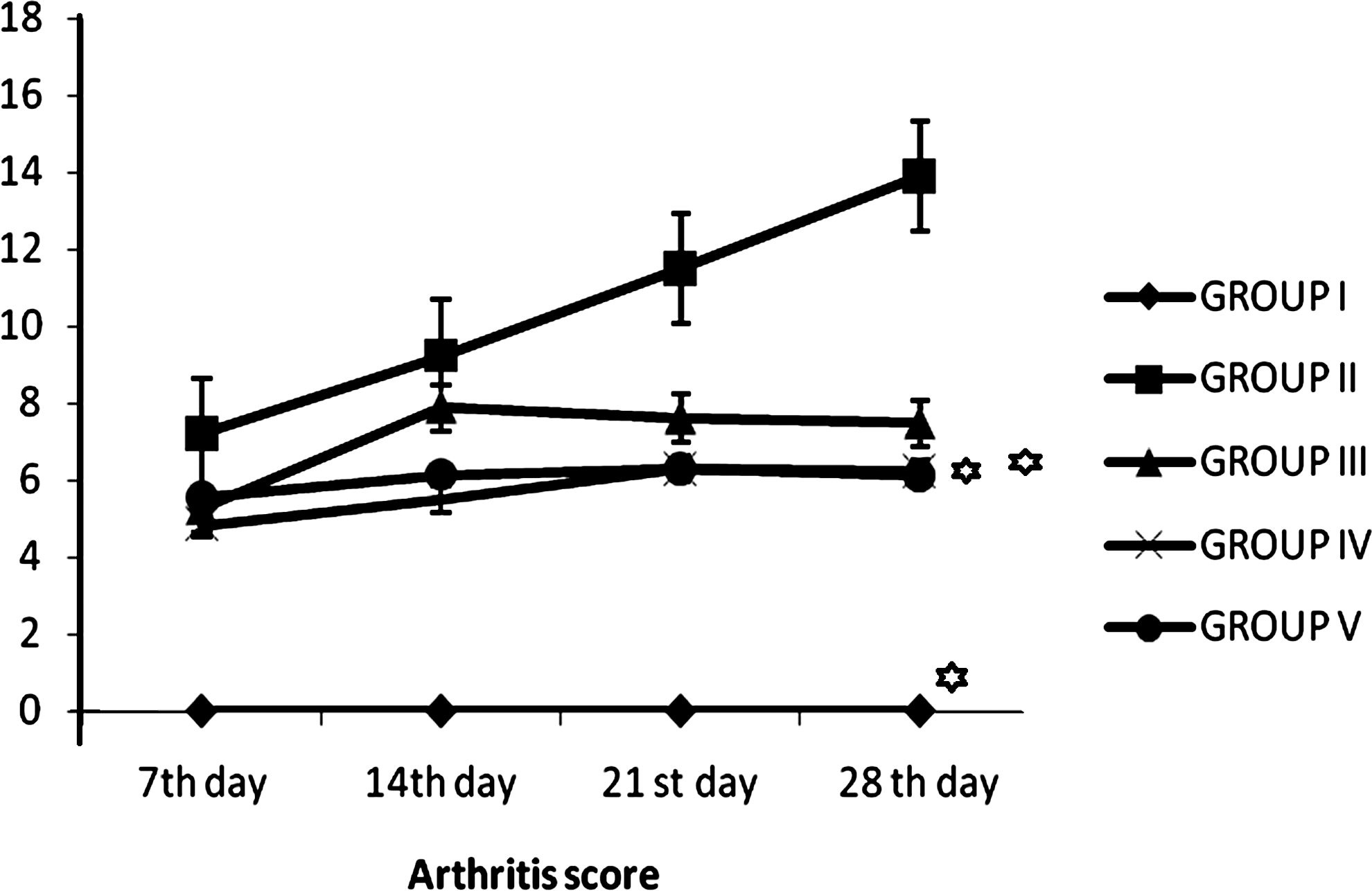
The arthritis score determined at the end of each week for collagen-induced arthritic Wistar rats. Results are expressed as mean±SEM of 6 rats per group. *P<0.05, group II versus all other groups. Group I: normal rats; group II: untreated arthritic rats; group III: arthritic rats administered with Lactobacillus casei; group IV: arthritic rats administered with Lactobacillus acidophilus; group V: arthritic rats administered with standard drug indomethacin.
Cytokines assay
The serum level of anti-inflammatory (IL-10 and IL-4) and proinflammatory (IL-6, IL-1β, TNF-α, and IL-17) cytokines were calculated by ELISA.
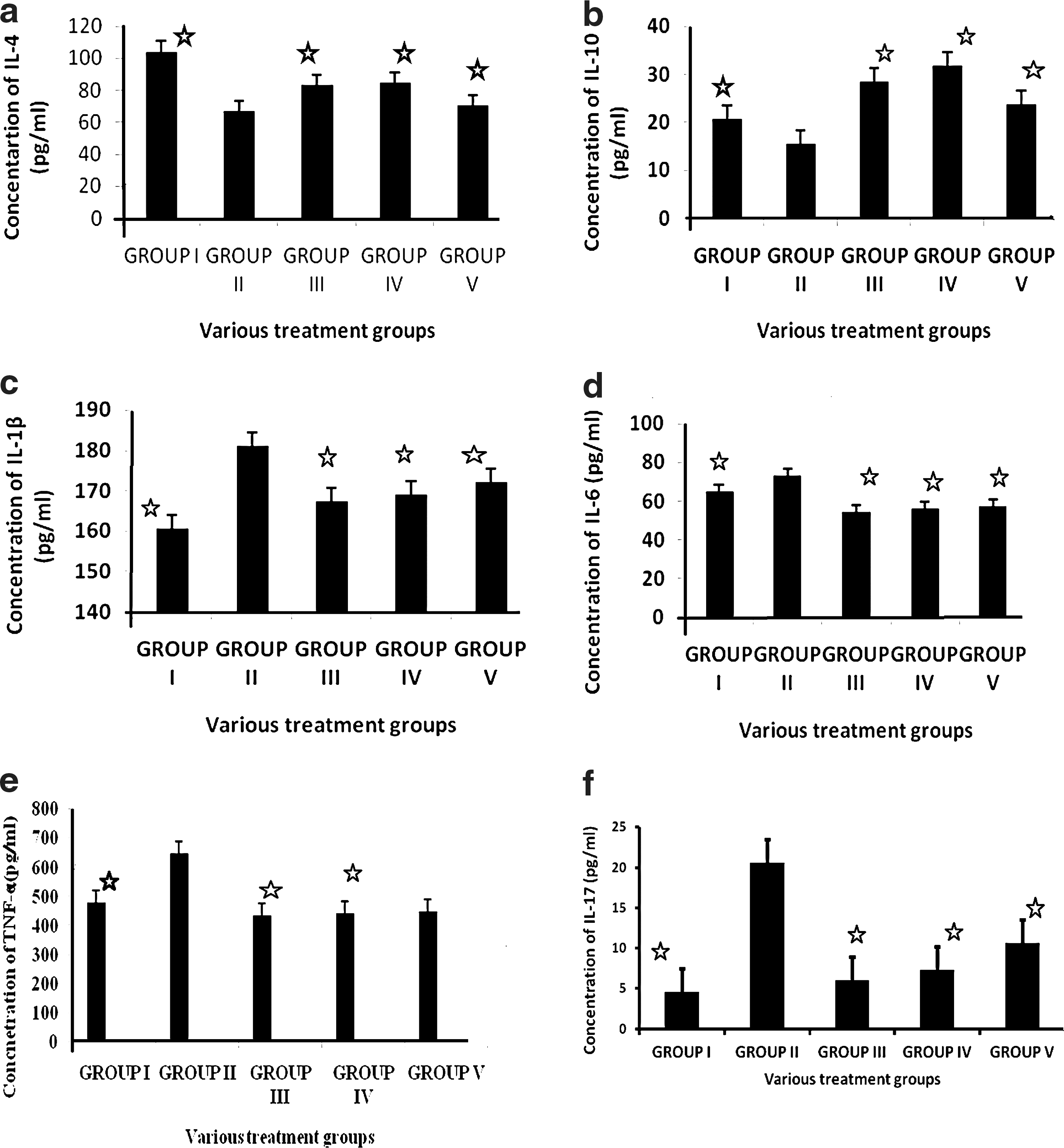
Lactobacillus treatment significantly increased IL-4 and IL-10 in group III and IV animals. IL-4 concentration for group III and IV animals were found to be 83.34±0.20 pg/mL and 84.82±0.21 pg/mL, respectively; while IL-10 concentrations were 29.39±0.35 pg/mL and 31.58±0.30 pg/mL when compared with group II animals (significant at P<0.0001). IL-4 and IL-10 concentration for group V was 70.18±0.14 pg/mL and 23.69±0.19 pg/mL, which were statistically significant at P<0.0001 when compared with group II animals (Fig. 2a, b).
The results of cytokine assay: IL-4
IL-6, IL-1β, TNF-α, and IL-17 concentration was reduced in groups that had received L. casei and L. acidophilus. Group II animals showed the highest concentration of all proinflammatory cytokines. L. casei and L. acidophilus treatment significantly decreased the concentration of proinflammatory cytokines. Group III and group IV were comparable to each other for all proinflammatory cytokines at P<0.0001. Higher levels of proinflammatory cytokines were observed in group II animals, which were found to be significant at P<0.0001 when compared with all other groups. On the contrary, group V animals were showing slightly higher values for proinflammatory cytokines when compared to group III and IV animals (Fig. 2c–f).
Antioxidant assays
Oxidative stress associated with CIA was evaluated by estimating catalase (U mg/protein), lipid peroxidation (μg/mL), GPx (U mg/protein), reduced GSH (U mg/protein), and SOD (U mg/protein) from synovial effsuate. Lactobacillus treatment significantly decreased catalase and lipid peroxidation at P<0.0001, while increased GSH, GPx, and SOD at P<0.0001 when compared with group II animals. However, in case of group III, lipid peroxidation and catalase were highest as compared to other counter parts. Also, all stress parameters of group V were comparable with that of group II and group III, and found to be significant, except reduced GSH level (at P<0.0001) (Fig. 3a–e).

The status of antioxidant enzymes
Discussion
The purpose of this study was to evaluate anti-inflammatory and antioxidant properties of L. casei and L. acidophilus as a therapeutic protocol in a CIA model. Results clearly suggest that L. casei and L. acidophilus exhibit antiarthritic, anti-inflammatory, and antioxidant properties.
The CIA model shares some common features with human RA, and it has been widely used for studying pathogenesis of RA and for searching new drug candidates for arthritis treatment (Shou and others 2006). Classical symptoms of arthritis include swelling redness, joint pain, inability to walk, and morning stiffness (Kumar and others 2009). Therefore, the arthritis score is used while evaluating the efficacy of an antiarthritic drug. We found that arthritis score of group I animals were 0 starting from 7th to 28th day; on the contrary, group II animals were showing an abrupt increased on 7th day and afterward. A highest score of 13.87±0.06 was observed in group II on 28th day when compared to all other groups at P<0.05. In group III and IV animals, arthritis scores were increased from 7th day to 14th day significantly, but then decreased on 21st day and 28th day (Fig. 1). The arthritis score in group V animals was increased simultaneously till 14th day and then significantly decreased on 28th day. Significant decrease in arthritis score by feeding L. casei and L. acidophilus is suggestive of immunosuppressant effects. Similar results were also reported by some other groups (Bahrav and others 2004; So and others 2011). Decrease in the arthritis score might be due to anti-inflammatory properties of L. casei and L. acidophilus.
RA is an inflammatory condition of joints. It is evident that a complex cytokine network operating in RA synovitis is due to simultaneous involvement of different cell types that variably contribute to inflammatory process. Proinflammatory cytokines such as TNF-α, IL-1β, and IL-6 produced by macrophages and other immune cells are of utmost importance in initiation, propagation, and severity of arthritis (Ivashkiv 1996). Along with these proinflammatory cytokines, some chemokines and inflammatory enzymes such as cyclooxygenase-2 (COX-2), 5-lipoxygenase (5-LOX), MMP-9, prostaglandins, and adhesion molecules in pathogenesis of arthritis are well known (Choy and Panayi 2001; Firestein 2004; Witowski and others 2004).
In this study, Lactobacillus treatment significantly increased anti-inflammatory cytokines. Data suggest that group III and IV produced 13% and 14% more IL-4, respectively, as compared with group V animals, whereas in case of IL-10, there was 5% and 11% more synthesis as compared with group V. It has been reported that prostaglandins synthesis is downregulated by anti-inflammatory cytokines (Ouyang and others 2010; Ursaciuc and others 2010). IL-4, IL-10, and IL-13 are important candidates that lower down expression of COX-2, and hence prostaglandins synthesis. Jubie and others (2008) also suggested similar findings. Group IV animals administered with L. acidophilus showed highest serum level of anti-inflammatory cytokines (Fig. 2a, b). Likewise, inflammatory mediators in serum were decreased significantly by Lactobacillus and indomethacin treatment (Fig. 2c–f). Group III animals, which received L. casei, showed lowest values of proinflammatory cytokines.
T-helper cells are believed to play a key role in orchestrating inflammatory response of disease through production of cytokines with different properties. T helpers cells are distinguished into type 1 (Th1) and 2 (Th2), according to the pattern of cytokines they produce. Th1 cells produce IL-1, TNF-α, IL-8, IL-12, IL-15, IL-17, and IL-18. These cytokines with some other cytokines favor inflammation. In contrast, Th2 cells produce some soluble factors such as IL-4, IL-10, IL-11, and IL-13. Along with these, some other soluble proteins, like IL-1 receptor antagonist, soluble receptors for TNF, IL-1, and IL-18-binding protein downregulate inflammation (Schulze-Koops and Kalden 2001; Astry and others 2011).
Cytokine profile in rheumatoid synovitis is consistent with a preponderance of Th1 cells, and thus presence of proinflammatory cytokines is noteworthy. Role of Th1-Th2 balance regulates autoimmunity, which has been validated by several animal model studies (Venkatesha and others 2011). Thus, this change in the Th1/Th2 balance could occur by either a decrease in proinflammatory cytokines or an increase in anti-inflammatory cytokines, and this leads to cytokine/immune deviation. This in turn is the basis of susceptibility or protection from arthritis (Ivashkiv 1996; Watanabe and others 2009). An inequity between proinflammatory and anti-inflammatory cytokine activities favors induction of autoimmunity and chronic inflammation that leads to joint damage (Kumar and others 2007). Many reports suggest that RA is mediated by activated proinflammatory Th1 cells (Verhoef 2001).
L. casei and L. acidophilus significantly decreased, IL-6, TNF-α, IL-1β, and IL-17. In vivo studies using models of experimental arthritis have established the role of IL-6 in joint destruction, leukocyte recruitment, apoptosis, and T-cell activation (Scheller and others 2006). IL-6 is supposed to increase the population of inflammatory cells in synovial tissue and stimulating proliferation of chondrocytes (Guerne and others 1989). IL-6 enhances or activates IL-1β, which increased synthesis of MMP, and inhibits proteoglycan production (Nietfeld and others 1990), and induces tissue inhibitors of MMP synthesis (Lotz and Guerne 1991).
TNF-α and IL-1β levels have been found to be elevated in synovial membrane, synovial fluid, and cartilage of osteoarthritis patients, but other inflammatory mediators such as IL-6, IL-15, IL-17, and leukotrienes B4 are also responsible for this process, with some of them responsible for neutrophil recruitment during immune inflammation. TNF-α, which is involved in inflammation, differentiation, and proliferation of T and B cells and bone resorption, is the primary agent in inflammatory process (Rioja and others 2004), whereas IL-1β is responsible for destruction of bone and cartilage (Cuzzocrea and others 2000). In experimental animal models also, inhibition of TNF-α and IL-1β results in decreased inflammation and cartilage destruction, respectively (Plows and others 1995). IL-17 is responsible for both development and progression of arthritis (Koenders and others 2005) and is an inducer of other proinflammatory mediators, such as TNF-α, IL-1β, IL-6, GM-CSF, PGE2, and IL-8 from monocytes and/or macrophages or synovial fibroblasts (Ziolkowska and others 2000; Hwang and others 2004). Other studies have reported similar improvements in arthritis symptoms after oral administration of L. casei (Kato and others 1998) and L. delbruekii subsp. Bulgaricus (Kano and others 2002) in the CIA model.
Our results suggest that Lactobacillus not only maintained IL-6, IL-1β, and TNF-α levels but also significantly decreased IL-17 levels. IL-17 is a potent inducer of other proinflammatory cytokines and a stimulator of osteoclastogenesis (Broxmeyer 1996). It acts synergistically with TNF-α and IL-1 β and increases chemokine production in various tissues to recruit monocytes and neutrophils to the site of inflammation, similar to IFN-γ (Shotorbani and others 2011). L. casei and L. acidophilus blocked inflammatory pathway leads to IL-17. This may be of vital interest to use L. casei and L. acidophilus as an antiarthritic and anti-inflammatory drug. It is possible that combination of L. casei and L. acidophilus may be new biological agent that may prove very effective against arthritis. In this context, Lactobacillus treatment facilitated the secretion of anti-inflammatory cytokines (IL-10 and IL-4) over proinflammatory cytokines (IL-6, IL-1β, TNF-α, and IL-17) resulting in overall immune deviation of cytokine response toward anti-inflammatory type or th2 type.
Free radicals have long been concerned as a mediator of joint damage in arthritis and similar conditions. These are produced in large amount by tissue itself and spread to the nearby area (Campo and others 2003). Several studies reported that during inflammatory diseases, joints directly come in close contact with phagocytes and start producing superoxide and hydrogen peroxide radicals. These radicals react with trace iron present in synovial effsuate to form highly reactive hydroxyl radicals (Famin and others 1995). When body's own antioxidant system gets damaged, these free radicals induce impairment and destruction of synovial fluid, joints, and cartilages (Bauerova and Bazek 1999). Thus, inhibition of free radicals in arthritis or an experimental model of arthritis may be one of the mechanisms that is to be considered during testing any antiarthritic drug.
In present study, catalase, lipid peroxidation, GPx, reduced GSH, and SOD assays were used for evaluating in vivo antioxidant activity of L. casei and L. acidophilus.
Lipid peroxidation is considered as a critical mechanism of injury that occurs during RA. Malonaldehyde, an end product of lipid peroxidation, reacts with lysine residues in protein to produce immunogenic molecules, which enhances inflammation (Kumar and Roy 2007). L. casei and L. acidophilus significantly decreased lipid peroxidation level in group III and IV animals (Fig. 3b). Lipid peroxidation was highest in group II animals. This suggests that oral administration of Lactobacillus inhibited lipid degradation. Similar findings were also observed by Kumar and Roy (2007), though this study did not investigate the mechanism.
Catalase is a protein associated with iron found in peroxisomes and microperoxisomes. It catalyzes the conversion of hydrogen peroxide to water and nascent oxygen, thus protecting cells from oxidative damage. Catalase was found to be highest in group II animals, whereas group III, IV, and V animals showed less catalase level when compared with group II (Fig. 3a). Higher catalase concentration indicates accumulation of hydrogen peroxide. Both L. casei and L. acidophilus significantly decreased concentration of peroxide accumulation in the synovial effsuate, except group III where its concentration was higher.
SOD has been reported as one of the most important enzymes of the antioxidant defense system (Curtis and Mortiz 1972). It removes superoxide anion by converting it to hydrogen peroxide, and thus diminishing the toxic effect caused by this radical. Lactobacillus-treated groups showed a significant increase (2.5 times) in the SOD concentration as compared to group II animals. Standard drug also showed twofold increase in the SOD level in group V animals (Fig. 3e).
Gpx is decreased due to excessive degradation of lipids as observed in arthritis. Here, in this study also, L. casei and L. acidophilus significantly increased the concentration of GPx in both group III and group IV animals, respectively (Fig. 3c). Our results are consistent with the results of some previous studies (Kamanli and others 2004; Govindarajan and others 2006). Reduced GSH directly eliminates free radicals by directly reacting with them. Lactobacillus significantly increased the GSH level in synovial effsuate (Fig. 3d).
In many joint diseases, cytokines, prostaglandins, nitric oxide, and ROS are released at the site of inflammation. These radicals damage cartilage and the components of extracellular matrix either by direct attack or by indirectly reducing matrix component synthesis (proteoglycans and type II collagen) and reduce sulfation of newly synthesized glycosaminoglycans. They also cause apoptosis or activate MMPs. Serum and synovial fluid samples of RA patients have been demonstrated an increased oxidative enzyme activity along with decreased antioxidant levels (Hitchon and Ei-Gabalawy 2004). Oral administration of L. casei and L. acidophilus reduced oxidative stress associated with arthritis. These results were comparable with the standard drug indomethacin. Thus, it can be used as a potent antioxidant and an anti-inflammatory dietary supplement for treatment of RA.
Conclusion
L. casei and L. acidophilus demonstrated effective antiarthritic activity that was proved by evaluating anti-inflammatory and in vivo antioxidation properties in an experimental model of arthritis. Notably, feeding L. casei and L. acidophilus caused suppression of IL-6, TNF-α, IL-17, and IL-1β along with upregulation of IL-10 and IL-4. Thus, maintaining altered cytokine balance in favor of anti-inflammatory cytokines could be the basis of future research. Importantly, it has also reduced the oxidative stress, which is an important factor for causing inflammation. It is possible that there might be some correlation in antioxidant and anti-inflammatory properties of Lactobacillus. On the basis of our results, we suggest that L. casei and L. acidophilus are promising therapeutic agents that should be tested further in preclinical trials in RA patients.
Footnotes
Acknowledgments
Authors like to acknowledge the Institute of Biomedical Science, Bundelkhand University, Jhansi, for animal house facility. The Department of Microbiology, Barkatullah University, is highly acknowledged for laboratory facility.
Author Disclosure Statement
The author declares that the research was conducted in the absence of any commercial or financial relationships that could be construed as a potential conflict of interest.